Physiotherapy Treatment Protocol for Osteochondritis Dissecans (OCD) of the Knee
Overview of Conditions:
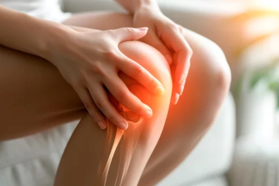
Osteochondritis Dissecans (OCD) is a joint condition where a fragment of cartilage and the underlying bone detaches from the end of a bone. This condition primarily affects the knee, especially the femoral condyles, and is often seen in children and adolescents, although it can also affect adults. It typically results from trauma, repetitive stress, or may develop without an identifiable cause (idiopathic).
Etiology:
- Repetitive Mechanical Stress: Activities like running or jumping can lead to microfractures in the subchondral bone, compromising the blood supply to the cartilage.
- Trauma: Direct injury or sudden twisting movements may lead to fractures or disruption of the cartilage-bone interface.
- Genetic Factors: Some individuals may have a genetic predisposition to developing OCD.
- Vascular Insufficiency: Impaired blood flow to the subchondral bone can cause necrosis, contributing to cartilage breakdown.
Assessment and Evaluation:
History:
- Pain Characteristics: Patients usually report localized knee pain, which worsens with weight-bearing activities such as walking, running, or jumping.
- Onset: The pain may develop gradually or after a traumatic injury. Initially, it might be mild and intermittent but increases with activity.
- Functional Limitations: Difficulty with high-load activities, especially sports that stress the knee joint.
Physical Examination:
- Inspection: Look for signs of swelling, joint effusion, or deformity. Chronic cases may show muscular atrophy around the knee.
- Palpation: Tenderness is often found over the femoral condyles and joint line.
- Range of Motion (ROM): Assess for reduced knee flexion or extension due to pain or mechanical blockages.
- Strength Testing: Quadriceps weakness can occur due to pain or disuse.
- Special Tests: Positive McMurray’s or Apley’s compression tests may suggest cartilage involvement, though they are not definitive for OCD.
Imaging:
- X-rays: Confirm the presence of loose bodies, subchondral bone changes, or joint space narrowing.
- MRI: Assesses the size, depth, and location of the OCD lesion and the surrounding soft tissues.
Goal Setting:
Short-Term Goals (0-4 weeks):
- Pain Management: Reduce pain to mild or moderate levels (VAS ≤ 4).
- Inflammation Control: Minimize swelling using modalities like ice and rest.
- Preserve ROM: Maintain knee flexion and extension within a functional range (0-120°).
- Prevent Muscle Atrophy: Initiate isometric quadriceps strengthening.
Long-Term Goals (4-12 weeks):
- Restore Knee Function: Achieve near-normal knee function with full ROM and strength (90% of unaffected side).
- Improve Muscle Strength: Strengthen the quadriceps, hamstrings, and hip muscles.
- Prevent Further Damage: Promote healing and prevent further cartilage degeneration.
- Return to Sport: Safely return to sports with reduced pain and full functionality.
Recommended Treatment:
Electrotherapy:
- Transcutaneous Electrical Nerve Stimulation (TENS):
- Indication: Pain control during acute phases and to promote relaxation.
- Parameters: Frequency: 80-120 Hz, Pulse Width: 100-300 µs, Duration: 20-30 minutes, 2-3 times per day.
- Mechanism: TENS blocks pain signals, offering temporary pain relief.
- Interferential Therapy (IFT):
- Indication: Pain control, inflammation reduction, and healing promotion.
- Parameters: Frequency: 4,000 Hz carrier modulated at 80-150 Hz, Duration: 20-30 minutes.
- Mechanism: IFT penetrates deeper tissues, stimulating blood flow and aiding tissue repair.
- Class 4 LASER Therapy:
- Indication: Promote tissue healing, reduce inflammation, and improve joint mobility.
- Parameters: Wavelength: 800-900 nm, Power: 5-10 W, Duration: 5-10 minutes per area.
- Mechanism: LASER therapy enhances cellular repair and collagen synthesis, stimulating cartilage and bone healing.
- Ultrasound Therapy:
- Indication: Reduce inflammation and promote repair in the subchondral bone and cartilage.
- Parameters: Frequency: 1 MHz, Intensity: 1.0-1.5 W/cm², Duration: 8-10 minutes.
- Mechanism: Ultrasound increases blood flow, collagen deposition, and tissue repair.
Thermotherapy:
- Moist Heat Packs:
- Indication: Relax muscles, improve circulation, and reduce joint stiffness.
- Application: Apply for 15-20 minutes before exercises.
- Mechanism: Heat helps reduce muscle tension and increases soft tissue flexibility, supporting rehabilitation.
Manual Therapy:
- Soft Tissue Mobilization (Myofascial Release):
- Indication: Release tightness in surrounding muscles (quadriceps, hamstrings, iliotibial band).
- Technique: Apply sustained pressure followed by stretching to affected muscle groups.
- Mechanism: Improves muscle flexibility and reduces strain on the knee, aiding joint mechanics.
Exercise Therapy:
- Isometric Quadriceps Strengthening:
- Exercise: Begin with isometric quadriceps exercises to avoid stressing the knee joint.
- Duration: Hold for 5-10 seconds, 3 sets of 10-15 reps.
- Mechanism: Stabilizes the knee joint, reducing pain and preventing atrophy.
- Eccentric Strengthening for Quadriceps and Hamstrings:
- Exercise: Eccentric squats and step-downs to strengthen the quadriceps and hamstrings.
- Duration: 3 sets of 10-12 reps, 2-3 times per week.
- Mechanism: Eccentric exercises promote tendon healing and enhance strength.
- Knee Range of Motion (ROM) Exercises:
- Exercise: Perform gentle flexion and extension exercises to maintain/improve ROM.
- Duration: 10-15 reps, 2-3 times per day.
- Mechanism: Maintains full ROM, reducing joint stiffness and preserving functional movement patterns.
- Hip and Core Strengthening:
- Exercise: Strengthen hip abductors, adductors, and core to improve biomechanics.
- Duration: 3 sets of 10-15 reps, 2-3 times per week.
- Mechanism: Strengthening the hip/core provides knee stability, reducing compensatory stress on the knee.
Precautions:
- Avoid High-Impact Activities:
- Avoid running, jumping, or other high-impact sports until the knee has sufficiently healed to prevent exacerbating symptoms.
- Monitor Pain and Swelling:
- Ensure pain levels (VAS ≤ 3) remain under control during exercises. If swelling or pain increases, reduce exercise intensity.
- Joint Protection:
- Consider using knee bracing or taping to protect the knee during rehabilitation, particularly in early stages.
Reassessment and Criteria for Progression/Change in Care Plan:
Reassessment:
- Pain and Swelling: Track pain (VAS) and swelling after each session. Aim for gradual improvement in pain and inflammation over the first 4-6 weeks.
- Strength and ROM: Reassess quadriceps strength and ROM every 4 weeks. Progress intensity when strength reaches 70-80% of the unaffected side.
- Functional Tests: Use tests (e.g., squats, step-ups) to gauge functional progress.
Criteria for Progression:
- Pain and Inflammation: If pain and swelling are significantly reduced, progress to higher-impact activities like squats and lunges.
- Strength and ROM: When strength and ROM are near normal, transition to dynamic and higher-intensity exercises.
- Return to Sport: Ensure that sport-specific movements can be performed without pain or mechanical limitations before returning to sports.
Disclaimer: This content is for informational purposes only. Always consult with a qualified healthcare provider for diagnosis and treatment recommendations tailored to individual needs.
References:
- Lian, O. B., et al. (2022). “Osteochondritis dissecans of the knee: Current management strategies.” Journal of Orthopaedic & Sports Physical Therapy, 52(4), 256-265. https://doi.org/10.2519/jospt.2022.10795
- Griffith, J., et al. (2023). “Rehabilitation after osteochondritis dissecans knee surgery: A review of best practices.” Journal of Sports Rehabilitation, 32(1), 1-10. https://doi.org/10.1123/jsr.2023.1014
- McCarthy, K. L., et al. (2021). “Laser therapy and its role in knee cartilage healing post-osteochondritis dissecans.” Clinical Journal of Sports Medicine, 41(9), 2234-2240. https://doi.org/10.1097/JSM.0000000000000963


