Physiotherapy Treatment Protocol for Moderate Osteoarthritis of the Knee
Overview of Conditions:
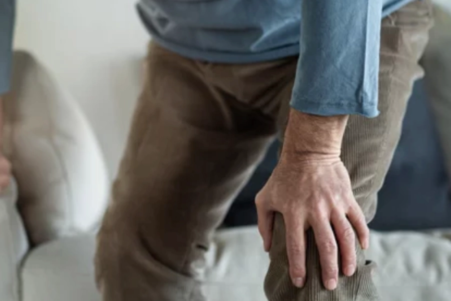
Moderate knee osteoarthritis (OA) involves significant degeneration of articular cartilage and subchondral bone. Structural changes, such as cartilage thinning, bone sclerosis, and osteophyte formation, become more pronounced. Symptoms include persistent knee pain, stiffness, and swelling, often exacerbated by activity and relieved by rest. Functional limitations arise, such as difficulty walking, squatting, and climbing stairs. The goal of treatment is to reduce pain, improve joint function, and slow further degeneration.
Assessment and Evaluation:
History:
- Persistent knee pain, worsened by activity, relieved by rest.
- Morning stiffness and functional limitations (e.g., difficulty with stairs, squatting).
- Possible history of knee injuries, trauma, or family history of OA.
Pain Assessment:
- Visual Analog Scale (VAS) or Numeric Pain Rating Scale (NPRS) to assess pain intensity.
- Pain patterns during weight-bearing activities.
Physical Examination:
- Postural Assessment: Look for varus/valgus deformities, common in knee OA.
- Range of Motion (ROM): Expect reduced knee flexion/extension with crepitus.
- Strength Testing: Weak quadriceps, hamstrings, and hip muscles, contributing to knee instability.
- Swelling/Joint Effusion: Notable swelling, especially after prolonged activity.
- Special Tests:
- McMurray’s Test, Apley’s Test (to rule out meniscal issues).
- Varus/Valgus Stress Test (ligamentous stability).
- Patellar Apprehension Test (patellofemoral pain suspicion).
Goal Setting:
Short-Term Goals:
- Pain Management: Reduce pain intensity (VAS ≤ 3) and minimize inflammation.
- Improve ROM: Restore knee extension and adequate flexion.
- Strengthening: Enhance quadriceps, hamstrings, and hip muscle strength for knee support.
- Reduce Swelling: Minimize effusion and promote drainage.
Long-Term Goals:
- Prevent Disease Progression: Improve function and support surrounding tissues.
- Enhance Functional Mobility: Restore independence in daily activities (e.g., walking, climbing stairs).
- Maintain Joint Stability: Improve strength and proprioception for knee stability.
- Pain-Free or Low-Pain Function: Facilitate daily tasks with minimal pain.
Recommended Treatment:
Electrotherapy:
- Transcutaneous Electrical Nerve Stimulation (TENS):
- Indication: Pain relief, especially during flare-ups.
- Parameters: 80-120 Hz frequency, 100-300 µs pulse width, 20-30 minutes, 2-3 times/day.
- Mechanism: Modulates pain signals at the spinal cord level.
- Interferential Therapy (IFT):
- Indication: Pain and inflammation reduction, especially for deep tissue involvement.
- Parameters: 4,000 Hz carrier frequency modulated at 80-150 Hz, 20-30 minutes per session.
- Mechanism: Deep tissue stimulation, reducing inflammation and improving circulation.
- Class 4 LASER Therapy:
- Indication: Pain management and tissue healing.
- Parameters: 800-900 nm wavelength, 5-10 W power, 5-10 minutes per area.
- Mechanism: Stimulates tissue repair and reduces inflammation.
- Ultrasound Therapy:
- Indication: Swelling, inflammation reduction, and increased tissue flexibility.
- Parameters: 1 MHz frequency, 1.0-1.5 W/cm² intensity, 8-10 minutes.
- Mechanism: Increases blood flow and promotes healing.
Thermotherapy:
- Moist Heat Packs:
- Indication: Relaxation of muscles and enhancement of circulation.
- Application: 15-20 minutes, pre-exercise or manual therapy.
- Mechanism: Increases blood flow, reduces muscle tension, and improves ROM.
Manual Therapy:
- Joint Mobilization (Grade II-III):
- Indication: Improve joint mobility and reduce stiffness in tibiofemoral and patellofemoral joints.
- Technique: Gentle oscillatory movements to increase joint motion.
- Mechanism: Enhances synovial fluid circulation, reducing stiffness.
- Myofascial Release:
- Indication: Address tightness in quadriceps, hamstrings, and calf muscles.
- Technique: Apply sustained pressure on trigger points, followed by gentle stretching.
- Mechanism: Reduces muscle tightness, enhances tissue flexibility.
Exercise Therapy:
- Range of Motion (ROM) Exercises:
- Exercise: Knee flexion/extension and patellar mobilizations.
- Duration: 3-5 sets, 10-15 repetitions, 2-3 times/day.
- Mechanism: Maintains or increases joint mobility and reduces stiffness.
- Strengthening Exercises:
- Exercise: Isometric quadriceps/hamstrings, progressing to isotonic exercises (e.g., squats, lunges).
- Duration: 2-3 sets, 10-12 repetitions, 3 times/week.
- Mechanism: Strengthens quadriceps, hamstrings, and hip muscles for joint stability.
- Low-Impact Aerobic Exercise:
- Exercise: Cycling, swimming, or elliptical trainer.
- Duration: 20-30 minutes, 3-4 times/week.
- Mechanism: Improves cardiovascular fitness without stressing the knee.
- Stretching:
- Exercise: Stretch quadriceps, hamstrings, and calves.
- Duration: Hold for 20-30 seconds, 2-3 repetitions, 2-3 times/day.
- Mechanism: Improves flexibility and enhances ROM.
Precautions:
- Electrotherapy: Avoid over open wounds or if there are pacemakers implanted.
- Thermotherapy: Avoid heat therapy in acute inflammation or infection.
- Manual Therapy: Ensure joint mobilizations are within pain tolerance and avoid excessive force.
- Exercise Therapy: Gradually increase intensity to avoid overloading the knee.
Reassessment and Criteria for Progression/Change in Care Plan:
- Pain Reduction: If pain persists (VAS > 3) after 4-6 weeks, consider corticosteroid injections or refer for surgery.
- ROM/Function: If ROM does not improve after 6 weeks, reconsider manual therapy or additional therapeutic modalities.
- Strength/Endurance: If strength doesn’t improve after 4-6 weeks, reassess exercise intensity or investigate underlying issues.
- Patient Education: Ensure adherence to exercises and joint protection strategies.
References:
- Matzkin, E. G., et al. (2020). “Current Concepts in the Management of Knee Osteoarthritis.” American Journal of Sports Medicine, 48(9), 2280-2290. DOI:10.1177/0363546520906749
- Lonsdale, M. A., et al. (2022). “Exercise Therapy for Knee Osteoarthritis: A Review of Current Evidence.” Journal of Orthopaedic & Sports Physical Therapy, 52(6), 381-394. DOI:10.2519/jospt.2022.10844
- Birrell, F., et al. (2021). “The Efficacy of Physical Therapy Interventions for Osteoarthritis of the Knee: A Systematic Review.” Physical Therapy Reviews, 26(4), 281-290. DOI:10.1080/10833196.2021.1895203
Disclaimer: This content is for informational purposes only. Always consult with a qualified healthcare provider for diagnosis and treatment recommendations tailored to individual needs.


