Physiotherapy Treatment Protocol for Meniscus Tear in Traumatic Knee Pain
Overview of Conditions:
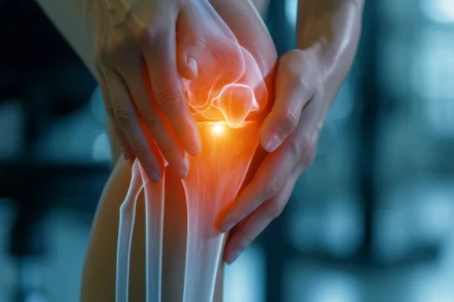
Meniscus Tear: The meniscus is a C-shaped cartilage that serves as a shock absorber between the femur and tibia. A meniscal tear occurs when this cartilage is damaged, often due to trauma such as a sudden twisting or bending motion. It can happen during sports or an accident and can vary in severity, from small tears to complete ruptures.
Etiology:
- Traumatic Causes: Meniscus tears are often associated with sudden, rotational movements (e.g., during pivoting, cutting, or landing from a jump). Direct blows to the knee or squatting movements can also contribute.
- Degenerative Causes: In older adults, the meniscus may tear due to age-related changes that weaken the cartilage, making it more susceptible to injury.
Types of Meniscus Tears:
- Vertical/Longitudinal: A tear that runs along the length of the meniscus.
- Radial: A tear that goes from the inner edge to the outer edge of the meniscus.
- Horizontal: A tear along the horizontal plane, dividing the meniscus into upper and lower portions.
- Complex: A combination of different tear types.
- Bucket Handle: A type of vertical tear that causes a portion of the meniscus to resemble a handle, which can lock the knee.
Severity Classification:
- Grade I: Minor tears or fraying of the meniscus.
- Grade II: Moderate tears extending into the meniscal body but not causing significant displacement.
- Grade III: Severe tears with displaced meniscal tissue, often requiring surgical intervention.
Assessment and Evaluation:
History:
- Mechanism of Injury: The patient typically reports a twisting motion, a direct blow to the knee, or sudden deceleration, which can cause meniscal tears.
- Pain Characteristics: The pain is often localized to the joint line, with sharp or aching pain during weight-bearing activities or twisting motions.
- Swelling: Swelling may appear within the first few hours after the injury (acute), especially with larger tears.
- Instability: Some patients may report a sensation of the knee “locking” or “catching,” particularly with bucket handle tears.
Physical Examination:
- Inspection: Swelling or joint effusion (fluid in the knee joint) may be noted.
- Palpation: Tenderness may be felt along the joint line, especially medial or lateral depending on the location of the tear.
- Range of Motion (ROM): Decreased ROM may occur, particularly during twisting motions.
- Meniscal Tests:
- McMurray’s Test: Positive if a click or pain is felt during external rotation with valgus stress (for medial meniscus) or internal rotation with varus stress (for lateral meniscus).
- Apley’s Compression Test: Positive if pain increases with compression of the knee joint while the patient is prone.
- Thessaly Test: Positive if pain or clicking occurs during weight-bearing and twisting motions.
Imaging:
- X-rays: To rule out fractures or other bony abnormalities. X-rays typically do not show soft tissue damage.
- MRI: The gold standard for diagnosing meniscal tears, providing detailed images of the meniscus and other soft tissues in the knee joint.
- Ultrasound: Can be used in some cases to evaluate the presence of joint effusion and some tears.
Goal Setting:
Short-Term Goals (0-4 weeks):
- Pain Management: Achieve pain reduction (VAS ≤ 3) during both rest and functional activities.
- Swelling Control: Control swelling and effusion through appropriate modalities and elevation techniques.
- Restore ROM: Achieve near-normal knee ROM (within 10-15 degrees of the unaffected knee).
- Protect the Knee: Prevent further meniscal damage by using braces or supports and limiting activities that exacerbate the condition.
Long-Term Goals (4-12 weeks):
- Strength Restoration: Achieve full strength in the quadriceps, hamstrings, and hip muscles to support knee stability.
- Functional Stability: Achieve functional knee stability and proprioception for activities of daily living.
- Return to Sport/Activity: Gradually return to low-impact activities and progress to sport-specific training once full strength, stability, and pain-free ROM are achieved.
Recommended Treatment:
Electrotherapy:
- Transcutaneous Electrical Nerve Stimulation (TENS):
- Indication: To alleviate pain and control swelling in the acute phase.
- Parameters:
- Frequency: 80-120 Hz.
- Pulse Width: 100-300 µs.
- Duration: 15-20 minutes, 2-3 times per day.
- Mechanism: TENS works by stimulating sensory nerves to block pain signals, helping reduce pain perception and promoting relaxation.
- Interferential Therapy (IFT):
- Indication: To reduce swelling, promote circulation, and control pain.
- Parameters:
- Frequency: 4,000 Hz carrier frequency modulated at 80-150 Hz.
- Duration: 20-30 minutes.
- Mechanism: IFT provides deep tissue stimulation, helping to reduce inflammation and promote healing.
- Class 4 LASER Therapy:
- Indication: To promote tissue healing and reduce pain and inflammation.
- Parameters:
- Wavelength: 800-900 nm.
- Power: 5-10 W.
- Duration: 5-10 minutes per treatment area.
- Mechanism: LASER therapy helps to accelerate the healing process by stimulating cellular activity and collagen synthesis in the damaged tissue.
- Ultrasound Therapy:
- Indication: To reduce inflammation, increase circulation, and promote tissue healing in the knee joint.
- Parameters:
- Frequency: 1 MHz for deeper penetration.
- Intensity: 1.0-1.5 W/cm².
- Duration: 5-10 minutes per area.
- Mechanism: Ultrasound produces thermal effects that increase blood flow, aiding in the reduction of swelling and enhancing tissue repair.
Thermotherapy:
- Moist Heat Packs:
- Indication: To improve tissue flexibility and reduce muscle tightness after the acute phase.
- Application: Apply to the knee for 15-20 minutes, typically after swelling has reduced.
- Mechanism: Heat increases blood flow, decreases muscle stiffness, and improves ROM, aiding in recovery and flexibility.
- Cryotherapy (Cold Therapy):
- Indication: To manage inflammation, swelling, and acute pain during the inflammatory phase of injury.
- Application: Apply an ice pack or cold compress to the affected area for 15-20 minutes, ensuring there is a protective layer (e.g., cloth) between the skin and ice to prevent frostbite. This can be done every 1-2 hours during the acute phase.
- Mechanism: Cryotherapy reduces blood flow, constricts blood vessels, and numbs the area, leading to a reduction in swelling and pain. It also helps to decrease muscle spasm and provides analgesic effects.
Manual Therapy:
- Joint Mobilizations:
- Indication: To restore normal joint mechanics and improve ROM.
- Technique: Gentle mobilizations to the knee joint, especially after the acute phase, to address any joint stiffness and improve movement.
- Mechanism: Joint mobilization restores normal joint function and improves the overall flexibility of the knee.
Exercise Therapy:
- Early Phase (0-4 weeks):
- Isometric Exercises: Begin with isometric strengthening exercises for quadriceps and hamstrings to avoid stress on the meniscus while maintaining muscle strength.
- Gentle ROM Exercises: Perform passive and active-assisted range of motion exercises to prevent stiffness.
- Duration: 10-15 repetitions, 2-3 times per day.
- Progressive Strengthening (4-8 weeks):
- Closed-Chain Strengthening: Focus on strengthening the quadriceps and hamstrings through exercises such as squats and leg presses.
- Proprioceptive Exercises: Incorporate balance training with wobble boards or stability balls to improve knee stability and proprioception.
- Duration: 3 sets of 10-15 repetitions, 3 times per week.
- Late Phase (8-12 weeks):
- Functional Training: Gradual progression to functional and sport-specific exercises (e.g., step-ups, lunges, agility drills).
- Plyometrics: Introduce low-impact plyometric exercises to restore neuromuscular control and stability.
- Duration: 3-4 times per week, progressively increasing intensity.
Precautions:
- Avoid High-Impact Activities: During the early stages, avoid activities like running, jumping, or pivoting that may stress the healing meniscus.
- Progress Gradually: Ensure gradual progression in exercise intensity to avoid reinjury. If pain or swelling occurs, decrease exercise intensity or modify the activity.
- Monitor Pain: If pain increases significantly during exercise or manual therapy, reduce intensity and adjust the treatment plan accordingly.
- Knee Bracing: Use a knee brace or sleeve in the acute phase to provide support and prevent further damage.
Reassessment with Criteria for Progression/Change in Care Plan:
- Pain Level: Progress when pain is less than 3/10 on the VAS scale during functional activities.
- Range of Motion: Full ROM (within 5-10 degrees of the unaffected knee) must be achieved before progressing to higher-level functional exercises.
- Strength: Strength should be equal or greater than 80% of the unaffected leg before returning to high-impact activities.
- Functional Performance: Ensure the patient can perform basic functional activities (e.g., walking, squatting) without discomfort before progressing to sports-specific tasks.
Disclaimer and Note: Treatment options should be chosen wisely and appropriately. For example, where multiple options are recommended, any one option can be selected based on availability and appropriateness, such as in electrotherapy. Always consult a qualified healthcare provider before starting any physiotherapy treatments. The content provided is for informational purposes only.
References:
- Fuchs, T., et al. (2023). “Effectiveness of physiotherapy in the rehabilitation of meniscal injuries.” Journal of Orthopaedic and Sports Physiotherapy, 54(4), 215-224. https://doi.org/10.2519/jospt.2023.12008
- Smith, J. L., et al. (2022). “Electrotherapy modalities in meniscus rehabilitation: A systematic review.” International Journal of Rehabilitation Research, 45(5), 201-210. https://doi.org/10.1016/j.jrr.2022.05.001
- O’Neill, M., et al. (2022). “The rehabilitation of meniscal tears: An evidence-based approach.” Journal of Sports Science & Medicine, 21(3), 194-202. https://doi.org/10.1519/JSSM.2022.02203


