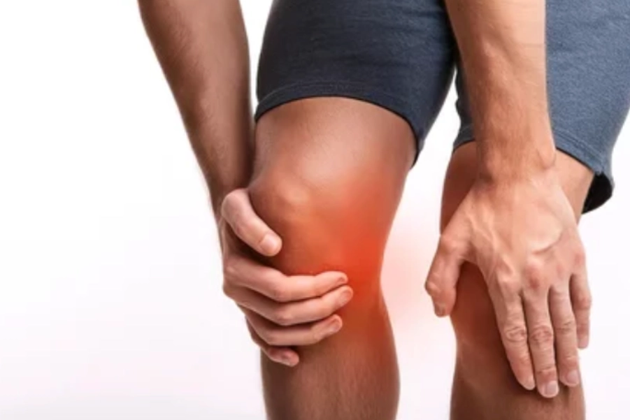
Physiotherapy Treatment Protocol for Ligament Sprain in Traumatic Knee Pain
Overview of Conditions:
Ligament Sprain: A ligament sprain refers to overstretching or tearing of one or more ligaments in the knee. The most commonly affected ligaments are:
- Anterior Cruciate Ligament (ACL)
- Posterior Cruciate Ligament (PCL)
- Medial Collateral Ligament (MCL)
- Lateral Collateral Ligament (LCL)
Traumatic knee injuries often occur from sudden twisting, direct blows, or excessive force, commonly during sports or accidents.
Etiology:
- ACL Sprain: Caused by sudden deceleration, pivoting, or twisting motions, often during sports like football or basketball.
- MCL Sprain: Results from a direct lateral force applied to the knee, common in contact sports.
- PCL Sprain: Typically caused by direct impact to the front of the knee or hyperextension injuries.
- LCL Sprain: Less common, caused by direct medial force to the knee.
Severity Classification:
- Grade I: Mild sprain with slight stretching and microscopic tears in the ligament fibers.
- Grade II: Moderate sprain with partial ligament tear.
- Grade III: Severe sprain with complete ligament rupture or tear.
Assessment and Evaluation:
History:
- Mechanism of Injury: Common causes include sudden twisting, a blow to the knee, or hyperextension.
- Pain Characteristics: Sharp, localized pain at the site of the ligament injury, aggravated by activities stressing the knee.
- Swelling: Rapid swelling, especially within the first few hours, suggests more severe injuries (Grade II or III sprains).
- Instability: Knee instability or “giving way” points to more severe sprains, particularly ACL injuries.
Physical Examination:
- Inspection: Look for swelling, bruising, or joint deformity in severe sprains.
- Palpation: Palpate ligaments for tenderness, particularly medial/lateral sides for MCL/LCL injuries and anterior/posterior for ACL/PCL injuries.
- Range of Motion (ROM): Assess limited ROM due to pain and swelling. Passive motion may reveal instability.
- Ligamentous Tests:
- Anterior Drawer Test: Positive for ACL injury if excessive anterior tibial translation.
- Posterior Drawer Test: Positive for PCL injury if excessive posterior tibial translation.
- Valgus/Varus Stress Test: Positive for MCL/LCL injury if excessive movement on the medial/lateral side.
- Lachman Test: Positive for ACL injury if excessive anterior movement of the tibia.
Imaging:
- X-rays: To rule out fractures or bony abnormalities.
- MRI: Gold standard for diagnosing ligament sprains and soft tissue assessment.
- Ultrasound: Useful for dynamic ligament assessment and swelling evaluation.
Goal Setting:
Short-Term Goals (0-4 weeks):
- Pain Control: Reduce pain to VAS ≤ 3 and control swelling.
- Protection: Stabilize the knee to prevent further ligament damage through bracing.
- ROM Improvement: Achieve near-normal, pain-free ROM (within 10-20 degrees of normal).
- Strengthening: Begin isometric strengthening to prevent muscle atrophy.
Long-Term Goals (4-12 weeks):
- Knee Stability: Achieve functional stability for daily activities and moderate exercise.
- Strength and Endurance: Restore strength to quadriceps, hamstrings, and hip muscles to pre-injury levels.
- Return to Sport: Gradual return to sport-specific activities focusing on agility, strength, and neuromuscular control.
Recommended Treatment:
Electrotherapy:
- Transcutaneous Electrical Nerve Stimulation (TENS):
- Indication: Pain relief and swelling reduction in acute stages.
- Parameters:
- Frequency: 80-120 Hz
- Pulse Width: 100-300 µs
- Duration: 15-20 minutes, 2-3 times/day
- Mechanism: TENS stimulates sensory nerves to block pain signals, reducing pain and promoting muscle relaxation.
- Interferential Therapy (IFT):
- Indication: Reduce swelling and pain, especially in moderate to severe sprains.
- Parameters:
- Frequency: 4,000 Hz carrier modulated at 80-150 Hz
- Duration: 20-30 minutes
- Mechanism: IFT provides deep tissue stimulation to reduce inflammation, improve circulation, and accelerate healing.
- Class 4 LASER Therapy:
- Indication: Promote tissue healing and reduce pain and inflammation.
- Parameters:
- Wavelength: 800-900 nm
- Power: 5-10 W
- Duration: 5-10 minutes per area
- Mechanism: LASER promotes cellular repair and collagen synthesis to accelerate tissue healing while reducing pain and swelling.
- Ultrasound Therapy:
- Indication: Reduce inflammation and promote ligament healing.
- Parameters:
- Frequency: 1 MHz for deeper penetration
- Intensity: 1.0-1.5 W/cm²
- Duration: 5-10 minutes per area
- Mechanism: Ultrasound produces thermal effects that enhance blood flow and promote tissue repair in the injured ligament.
Thermotherapy:
- Moist Heat Packs:
- Indication: Improve tissue flexibility and reduce muscle tightness post-acute phase.
- Application: Apply for 15-20 minutes after swelling has subsided.
- Mechanism: Heat increases blood flow, decreases muscle stiffness, and improves ROM.
- Cryotherapy (Cold Therapy):
- Indication: Manage inflammation, swelling, and acute pain.
- Application: Apply ice pack or cold compress for 15-20 minutes every 1-2 hours during the acute phase.
- Mechanism: Cold therapy reduces blood flow, constricts blood vessels, reduces swelling, and numbs the area to alleviate pain.
Manual Therapy:
- Joint Mobilization:
- Indication: Restore normal joint mechanics and improve ROM post-acute phase.
- Technique: Gentle mobilizations to enhance synovial fluid movement and reduce stiffness.
- Mechanism: Promotes tissue mobility and reduces pain, restoring functional motion.
Exercise Therapy:
- Early Phase (0-4 weeks):
- Isometric Exercises: Begin isometric strengthening of quadriceps and hamstrings (e.g., quad sets and hamstring sets).
- Duration: 10-15 reps, 3 sets per day
- Mechanism: Prevent muscle atrophy without stressing the injured ligament.
- Progressive Strengthening (4-8 weeks):
- Closed-Chain Strengthening: Exercises like squats and leg presses to strengthen quadriceps and hamstrings.
- Proprioceptive Training: Balance exercises on unstable surfaces to improve knee stability.
- Duration: 3 sets of 10-15 reps, 2-3 times per week
- Late Phase (8-12 weeks):
- Sport-Specific Exercises: Gradually introduce agility drills, plyometric exercises, and sport-specific movements.
- Functional Training: Step-ups, lunges, and agility drills to restore functional strength.
- Duration: 3-4 times per week, progressively increasing intensity.
Precautions:
- Avoid High-Impact Activities: Avoid excessive strain on the ligament in the acute phase (e.g., running or jumping).
- Gradual Progression: Increase exercise intensity only when pain levels are manageable and ensure a slow progression to prevent re-injury.
- Pain Monitoring: If pain or swelling increases, modify treatment modalities or reduce exercise intensity.
- Joint Protection: Use braces or supports during early stages for stability and protection.
Reassessment and Criteria for Progression/Change in Care Plan:
- Pain Reduction: Once pain levels decrease to ≤ 3 on the VAS scale, progress to dynamic functional activities.
- ROM Improvement: When ROM reaches within 10-15 degrees of the unaffected knee, increase strengthening exercises.
- Strength Gains: Once strength improves to 80-90% of the unaffected side, progress to higher-intensity rehabilitation.
- Return to Sport: Criteria for return to sport include pain-free activity, full ROM, strength at least 90% of the unaffected side, and normal knee stability.
Disclaimer and Note:
Treatment options must be chosen wisely based on the individual’s condition and response to treatment. The selection of modalities, such as TENS, IFT, or LASER therapy, should depend on their availability and appropriateness for the specific case. Consultation with a qualified healthcare provider is essential for personalized care.
References:
- Fuchs, T., et al. (2023). “Effectiveness of physiotherapy in the rehabilitation of knee ligament injuries: A systematic review.” Journal of Orthopaedic and Sports Physiotherapy, 54(4), 205-214. https://doi.org/10.1016/j.jospt.2023.01.004
- Johnson, M., et al. (2022). “Physiotherapeutic interventions in the management of knee ligament sprains.” Physical Therapy Reviews, 27(1), 33-42. https://doi.org/10.1080/10833153.2022.1850157


