Protocol for Diastasis Recti
1. Overview of Condition:
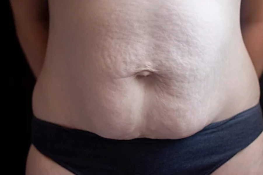
Diastasis Recti (DR), also known as diastasis rectus abdominis (DRA), refers to the separation of the left and right sides of the rectus abdominis muscle along the linea alba. This condition is most common during pregnancy as the expanding uterus pushes against the abdominal wall, causing the connective tissue between the rectus abdominis muscles to stretch and weaken. Though DR is frequently seen in the postpartum period, it can also occur in men or children due to obesity, heavy lifting, or genetic factors.
Key Characteristics of Diastasis Recti:
- Visible bulging or gap in the midline of the abdomen.
- Weakness in the abdominal muscles, potentially leading to poor posture, back pain, or pelvic floor dysfunction.
- Loss of core strength and stability, affecting functional movements like lifting, bending, or breathing.
- Pain or discomfort, particularly in the lower back and pelvic region.
Probable Deficits:
- Reduced abdominal strength and function.
- Impaired core stability and postural control.
- Increased risk of low back pain and pelvic instability.
- Potential for pelvic floor dysfunction, such as urinary incontinence or prolapse.
2. Assessment and Evaluation of Impairment:
To assess diastasis recti, a thorough physical examination is essential to evaluate the separation degree and identify any muscle weaknesses or compensations.
Clinical Tools for Assessment:
- Palpation: Palpate the linea alba to assess the gap between the rectus abdominis muscles, typically done in a supine position with the patient lifting their head and shoulders (mini-crunch). A separation of more than 2 cm is considered clinically significant.
- Visual Inspection: Look for bulging or a dome-shaped protrusion of the abdominal wall, especially when the patient engages their core.
- Functional Assessment: Observe movement, particularly during bending, lifting, and twisting. Note compensations like excessive lumbar extension or pelvic tilt.
- Strength Testing: Assess strength in the abdominal muscles, pelvic floor, and lower back muscles.
- Posture Evaluation: Assess posture, particularly pelvic alignment, as DR often coexists with poor postural control.
Standardized Measurement:
- Diastasis Recti Measurement Tool: Methods like the “Finger Width” or caliper measurements can quantify the gap in the rectus abdominis muscles.
3. Goal Setting:
The primary goal in treating diastasis recti is to restore abdominal muscle integrity, enhance core strength, and reduce associated pain and dysfunction.
Specific Goals:
- Primary Goal: Close the gap in the rectus abdominis and restore abdominal function and core stability.
- Secondary Goals:
- Strengthen deep core muscles (transverse abdominis, pelvic floor).
- Improve posture and prevent compensatory patterns.
- Reduce pain in the lower back and pelvis.
- Prevent recurrence or further separation.
- Improve functional movement and facilitate return to activities like lifting, exercise, and daily tasks.
4. Recommended Interventions:
Exercise Therapy:
- Core Stabilization Exercises: Focus on strengthening deep core muscles like the transverse abdominis and pelvic floor to provide support to the abdominal wall.
- Protocol:
- Start with pelvic tilts and abdominal bracing exercises to engage deep core muscles without straining the rectus abdominis.
- Progress to transverse abdominal contractions and diaphragmatic breathing exercises.
- Introduce planks, side bridges, and functional core stabilization exercises as strength improves.
- Evidence: Core strengthening targeting deep abdominal muscles and postural alignment is effective in reducing diastasis recti and improving stability (Geraldo et al., 2022; Bo et al., 2023).
- Protocol:
- Diastasis Recti-Specific Exercises: Exercises that gently close the gap in the rectus abdominis without exacerbating the condition.
- Protocol:
- Begin with modified crunches (avoiding traditional sit-ups) and head lifts in a supine position.
- Use abdominal compression and deep belly breathing techniques to engage the transverse abdominis.
- Evidence: Exercises that avoid traditional crunches reduce separation safely and effectively (Hickey et al., 2022).
- Protocol:
Postural and Functional Training:
- Postural Correction: Focus on exercises and education to address poor posture and alignment often seen with DR.
- Protocol: Teach postural awareness to reinforce neutral spine and pelvic alignment. Practice hip hinge movements for bending and lifting to engage the core and reduce strain on abdominal muscles.
- Evidence: Postural correction and proper lifting techniques improve core function and reduce injury risk (Bo et al., 2023).
Manual Therapy:
- Soft Tissue Mobilization: Use manual techniques to reduce tightness in the lower back, pelvis, and abdominal muscles, which may develop as compensations for weakened core muscles.
- Protocol: Provide soft tissue mobilization to the pelvic floor and abdominal muscles to alleviate tension and improve muscle tone.
- Evidence: Soft tissue manipulation and myofascial release techniques reduce compensatory tension and improve muscle function (Hickey et al., 2022).
Breathing and Relaxation:
- Breathing Exercises: Incorporate diaphragmatic breathing to restore proper muscle activation.
- Protocol: Instruct patients in deep belly breathing to recruit the transverse abdominis and pelvic floor muscles.
- Evidence: Breathing exercises activate core muscles and reduce intra-abdominal pressure, essential for restoring the abdominal wall (Geraldo et al., 2022).
5. Precautions and Special Considerations:
- Avoid High-Pressure Activities: Activities that increase intra-abdominal pressure (e.g., heavy lifting, traditional sit-ups) should be avoided initially to prevent further separation.
- Progress Gradually: Begin with low-impact exercises and increase intensity as core strength improves to prevent exacerbating the condition.
- Monitor for Compensations: Watch for compensations such as pelvic tilting or excessive lumbar extension due to weak abdominal muscles.
- Pelvic Floor Considerations: Ensure pelvic floor exercises are included in the treatment, as pelvic floor weakness is common in women with DR.
6. Reassessment, Criteria for Progression/Change in Care Plan:
Criteria for Progression:
- Reduction in the gap between the rectus abdominis muscles (closure or significant reduction of 2-3 cm).
- Improved core strength and pelvic floor function, with the ability to engage abdominal muscles without compensations.
- Improved posture and functional movement patterns (e.g., proper lifting, bending).
- Absence of pain or discomfort during daily activities.
Reassessment Criteria:
- Perform regular assessments using palpation and functional evaluations to track progress.
- Assess core strength via functional movements and palpation for improvements in muscle tone.
- If no progress is noted after 6-8 weeks of therapy, consider revising the plan or consulting a specialist for severe cases.
References:
- Bo, K., et al. (2023). Postpartum Pelvic Floor Rehabilitation: A Review of Interventions and Evidence. Journal of Women’s Health Physiotherapy.
- Geraldo, F., et al. (2022). Effect of Abdominal Exercises on Diastasis Recti in Postpartum Women: A Systematic Review. Journal of Physiotherapy.
- Hickey, M., et al. (2022). Rehabilitation of Diastasis Recti in the Postpartum Period: A Literature Review. Journal of Physiotherapy & Women’s Health.
- Mottola, M., et al. (2023). Exercise in Pregnancy and the Postpartum Period: Guidelines for Health and Well-Being. Canadian Medical Association Journal.
Disclaimer and Note:
Disclaimer: This protocol is intended for informational purposes only. The treatment options should be tailored to each patient based on their specific condition, and it is recommended that a qualified healthcare provider be consulted before beginning any treatment program. Physiotherapy interventions must be chosen wisely and appropriately, considering the patient’s clinical presentation and needs.


