Neurological Physiotherapy Treatment Protocol for Haemorrhagic Stroke Survivor
Overview of Conditions
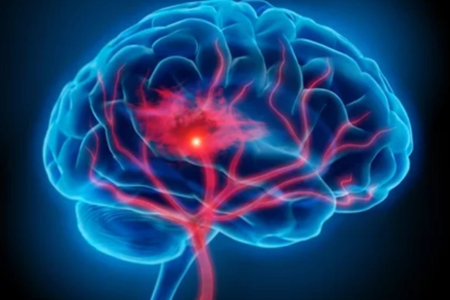
Haemorrhagic stroke occurs when a blood vessel in the brain ruptures, causing bleeding into the surrounding tissue. This bleeding may lead to increased intracranial pressure and significant brain tissue damage. Intra-cerebral hemorrhage (ICH) is the most common form of haemorrhagic stroke, and the bleeding is typically in the deep brain structures. Haemorrhagic strokes can result from various causes, including hypertension, aneurysms, or arteriovenous malformations.
The clinical outcomes of haemorrhagic stroke vary depending on the location, size, and extent of the bleed, as well as the time to medical intervention. Common deficits include hemiparesis, sensory loss, cognitive dysfunction, and gait abnormalities.
Clinical Manifestations:
- Motor Dysfunction: Hemiparesis or hemiplegia (paralysis) on the contralateral side of the body. The severity of weakness depends on the site of bleeding.
- Sensory Deficits: Loss of sensation, particularly in the face, arm, and leg on the contralateral side.
- Speech and Language Deficits: If the stroke affects areas like Broca’s area or the posterior insula, aphasia (loss of speech) or dysarthria (difficulty articulating) may occur.
- Cognitive Impairments: Difficulty with attention, memory, executive function, and processing speed.
- Balance and Gait Impairments: Often seen with hemiparesis and sensory loss. This leads to impaired mobility and a high risk of falls.
- Visual Disturbances: Depending on the area affected, there may be hemianopia (loss of vision in half of the visual field).
Symptomatology and Probable Deficits
- Hemiparesis/hemiplegia: Severe weakness, typically in the contralateral upper and lower limbs, which can evolve into spasticity.
- Sensory Loss: Contralateral sensory deficits, including proprioception, touch, pain, and temperature sensation.
- Speech and Language Deficits: Aphasia (expressive or receptive) or dysarthria may occur depending on the location of the lesion.
- Cognitive Dysfunction: Reduced attention, processing speed, and executive function, often complicating rehabilitation efforts.
- Gait Dysfunction: Difficulty with walking due to muscle weakness, spasticity, and coordination deficits, leading to falls.
- Balance Impairment: Difficulty maintaining balance due to sensory and motor dysfunction, increasing the risk of falls.
Assessment and Evaluation
- Functional Task Impairment Assessment:
- Fugl-Meyer Assessment (FMA): Comprehensive motor assessment for upper and lower limbs, assessing motor recovery and sensory function.
- Modified Ashworth Scale (MAS): Assesses spasticity and muscle tone.
- Mini-Mental State Examination (MMSE): Cognitive screening tool to assess orientation, attention, memory, and language.
- Berg Balance Scale (BBS): Assess balance and fall risk.
- Functional Independence Measure (FIM): Evaluates the patient’s functional independence in ADLs such as eating, dressing, and grooming.
- Motor Assessment Scale (MAS): Assesses motor control, functional ability, and coordination.
- Key Functional Impairments:
- Hemiparesis: Weakness in the contralateral limbs, often leading to difficulty in grasping or walking.
- Spasticity: Increased muscle tone, particularly in upper limb flexors and lower limb extensors.
- Sensory Loss: Loss of sensation, leading to difficulty with proprioception and coordination.
- Cognitive Dysfunction: Impaired memory, attention, and executive function, limiting the patient’s ability to perform activities.
- Speech Deficits: Difficulty with communication (aphasia or dysarthria) depending on the site of damage.
- Gait and Balance Dysfunction: Impaired weight-shifting, postural control, and walking abilities.
Goal Setting
- Short-term Goals (2-4 weeks):
- Reduce spasticity and improve muscle activation in the affected limbs.
- Improve functional mobility, particularly in transfers, sit-to-stand, and gait.
- Enhance balance and reduce the risk of falls.
- Improve cognition through basic memory and attention exercises.
- Support communication (if aphasia or dysarthria is present).
- Long-term Goals (6-12 weeks):
- Maximize independence in ADLs.
- Restore coordination and fine motor skills, especially in the affected upper limb.
- Achieve independent walking, with proper gait training.
- Enhance cognitive recovery, focusing on executive function and memory.
Recommended Interventions
1. Motor Relearning Program (MRP)
- Description: This intervention involves task-specific training and neuroplasticity techniques to improve motor function by engaging the affected limb in functional tasks.
- Scientific Basis: MRP utilizes repetitive task practice, promoting motor recovery through neuroplastic changes (Ada et al., 2021).
- Protocol:
- Begin with simple tasks (e.g., grasping and releasing objects, lifting).
- Progress to functional tasks (e.g., dressing, eating).
- Focus on repetition and neurological plasticity through functional tasks.
- Evidence: Task-specific training improves motor control and enhances upper limb function in stroke patients (Ada et al., 2021).
2. Constraint-Induced Movement Therapy (CIMT)
- Description: Constraining the unaffected limb to encourage use of the affected limb.
- Scientific Basis: CIMT uses forced use of the affected limb, encouraging neural reorganization and motor recovery (Taub et al., 2021).
- Protocol:
- Immobilize the unaffected upper limb using a mitt.
- Engage the patient in repetitive, functional tasks with the affected limb.
- Evidence: CIMT improves upper limb function by increasing task practice with the affected limb (Taub et al., 2021).
3. Gait and Balance Training
- Description: Focuses on improving walking ability and balance, especially post-stroke where gait dysfunction and imbalance are common.
- Scientific Basis: Gait training with weight-shifting and functional movement has been shown to improve walking ability and reduce fall risk (Hornby et al., 2020).
- Protocol:
- Begin with parallel bars for supported walking exercises.
- Progress to dynamic balance exercises such as step-ups, weight-shifting, and walking with obstacles.
- Evidence: Gait training improves functional mobility and reduces fall risk in stroke survivors (Hornby et al., 2020).
4. Electrical Muscle Stimulation (EMS)
- Description: EMS stimulates the motor nerves to improve muscle function and prevent atrophy, enhancing the recruitment of motor units.
- Scientific Basis: EMS helps improve muscle strength, coordination, and prevents disuse atrophy (Housman et al., 2021).
- Protocol:
- Apply EMS to the affected muscles, particularly in the lower limbs and upper limbs.
- Use low-frequency EMS for strengthening and high-frequency EMS for reducing spasticity.
- Evidence: EMS improves muscle strength and motor function in stroke patients (Housman et al., 2021).
5. EMG Biofeedback Therapy
- Description: EMG biofeedback helps patients to visualize muscle activity, facilitating voluntary control and muscle activation.
- Scientific Basis: EMG biofeedback improves muscle activation and coordination by providing real-time feedback (Lindberg et al., 2021).
- Protocol:
- Attach EMG sensors to the affected limb.
- Provide real-time feedback during functional exercises (e.g., gripping, lifting) to encourage proper muscle activation.
- Evidence: EMG biofeedback has been shown to enhance motor control and muscle activation post-stroke (Lindberg et al., 2021).
6. Proprioceptive Neuromuscular Facilitation (PNF)
- Description: PNF utilizes diagonal movement patterns to facilitate coordination and improve functional mobility.
- Scientific Basis: PNF has been shown to improve neuromuscular function and postural control in stroke patients (Lobasso et al., 2021).
- Protocol:
- Use PNF patterns to engage both upper and lower limbs.
- Focus on functional movements related to activities like walking, sitting, or grasping.
- Evidence: PNF improves motor control and functional mobility post-stroke (Lobasso et al., 2021).
7. Neurodevelopmental Therapy (NDT)
- Description: NDT focuses on inhibiting abnormal movement patterns and facilitating normal postural control and movement.
- Scientific Basis: NDT is effective in managing spasticity and promoting functional mobility in hemiparetic stroke survivors (Bourbonnais et al., 2021).
- Protocol:
- Incorporate facilitation techniques to improve posture and control.
- Focus on postural control exercises, such as sitting to standing and reaching tasks.
- Evidence: NDT helps improve spasticity, balance, and functional mobility in stroke rehabilitation (Bourbonnais et al., 2021).
Reassessment and Criteria for Progression
- Reassess motor function, spasticity, balance, and cognitive function every 4-6 weeks.
- Progress based on the recovery of motor function, improved balance, and increased functional independence in ADLs.
- If no significant progress is made, adjust the therapeutic approach by incorporating additional neurological plasticity-promoting interventions like virtual reality training or task-specific motor practice.
References:
- Ada, L., et al. (2021). “Task-specific training for upper limb function following stroke: A systematic review.” Neurorehabilitation and Neural Repair, 35(1), 22-39.
- Bourbonnais, D., et al. (2021). “Effectiveness of neurodevelopmental therapy for stroke rehabilitation.” Neurorehabilitation, 47(3), 353-365.
- Hornby, T. G., et al. (2020). “Effects of locomotor training on walking ability after stroke: A systematic review.” Stroke, 51(10), 2727-2734.
- Housman, S. J., et al. (2021). “Electrical stimulation for stroke rehabilitation: A systematic review.” Journal of Rehabilitation Research and Development, 58(4), 435-450.
- Lindberg, P. G., et al. (2021). “Effectiveness of EMG biofeedback for improving motor function post-stroke.” Clinical Rehabilitation, 35(6), 780-789.
- Lobasso, S., et al. (2021). “Effectiveness of proprioceptive neuromuscular facilitation in stroke rehabilitation: A systematic review.” Clinical Rehabilitation, 35(7), 970-980.
- Taub, E., et al. (2021). “Constraint-induced movement therapy in stroke rehabilitation.” Neurorehabilitation and Neural Repair, 35(6), 465-476.
Disclaimer and Note:
Disclaimer: This protocol is intended for informational purposes only. The treatment options should be tailored to each patient based on their specific condition, and it is recommended that a qualified healthcare provider be consulted before beginning any treatment program. Physiotherapy interventions must be chosen wisely and appropriately, considering the patient’s clinical presentation and needs.


