Evidence-based physiotherapy and rehabilitation resources
Editorial & review policy
Evidence-based physiotherapy and rehabilitation resources
Editorial & review policyHuman Anatomy · Upper limb
Everything above exists to position the hand. Twenty-seven bones, three nerves and more muscles than any comparable region — arranged so that the same structure can hold a hammer and thread a needle. Losing a little of it costs a great deal.
Part 2 · The upper limb
The carpal tunnel, three nerve territories, the arches, and the four grips
The hand contains 27 bones, 27 joints, 34 muscles and 3 major nerves in a segment weighing less than half a kilogram. It has the densest sensory innervation and the largest cortical representation of any body part relative to its mass. It is simultaneously the body’s most precise tool and its principal organ of exploration.
Two consequences follow for practice:
| Joint | Type | Surfaces | Range |
|---|---|---|---|
| Radiocarpal (wrist) | Synovial condyloid (ellipsoid), 2 DoF | Distal radius + TFCC ↔ scaphoid, lunate, triquetrum (the triquetrum contacts only in ulnar deviation). The ulna does not articulate with the carpus | Contributes ~60% of flexion, ~40% of extension |
| Midcarpal | Compound synovial | Proximal ↔ distal carpal rows | Contributes ~40% of flexion, ~60% of extension |
| Distal radio-ulnar | Synovial pivot | Ulnar notch of radius ↔ head of ulna, with the TFCC | Pronation–supination |
| Intercarpal | Plane synovial | Between adjacent carpals | Small gliding |
| Carpometacarpal 1 (thumb) | Saddle, 2 DoF (+ conjunct rotation) | Trapezium ↔ base of 1st metacarpal | Flexion–extension, abduction–adduction, opposition |
| Carpometacarpal 2–5 | Plane | Distal row ↔ metacarpal bases | 2nd and 3rd essentially fixed; 4th ~15°, 5th ~25–30° flexion |
| Metacarpophalangeal 2–5 | Condyloid, 2 DoF | Metacarpal head ↔ base of proximal phalanx | Flexion 90°, extension 30–45°, abduction/adduction ~20° (only in extension) |
| Interphalangeal | Hinge, 1 DoF | — | PIP flexion ~100–110°; DIP ~80–90° |
| Movement | Full range | Functional range for ADL |
|---|---|---|
| Flexion | 80–90° | ~10° |
| Extension | 70–80° | ~35° |
| Radial deviation | 15–20° | ~10° |
| Ulnar deviation | 30–45° | ~15° |
Ulnar deviation exceeds radial deviation because the radial styloid projects further distally, blocking radial deviation early. The “dart-thrower’s motion” — the oblique arc from radial extension to ulnar flexion — occurs largely at the midcarpal joint with minimal proximal-row motion, which is why it is the preferred early motion after scapholunate and proximal-row injury.
Close-packed position of the wrist: full extension with radial deviation. Resting position: neutral with slight ulnar deviation. Capsular pattern: flexion and extension equally limited.
Figure 1 · The arches of the hand
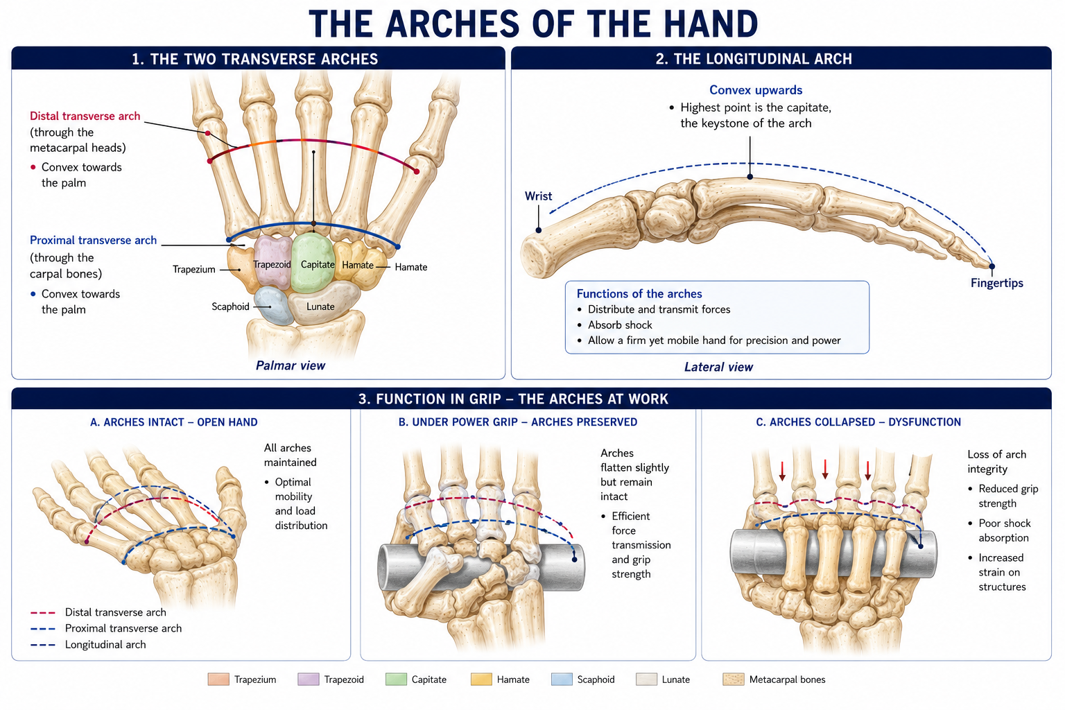
Three arches allow the flat hand to cup, conforming to whatever it holds. Their loss is one of the most functionally significant consequences of intrinsic muscle paralysis.
| Arch | Description | Keystone | Mobility |
|---|---|---|---|
| Proximal transverse | Across the distal carpal row | Capitate | Fixed |
| Distal transverse | Across the metacarpal heads | 2nd and 3rd metacarpal heads | Mobile — the 4th and 5th metacarpals flex around the fixed central pillar |
| Longitudinal | Along each ray, carpus → metacarpal → phalanges | MCP joints | Mobile |
The fixed central pillar (2nd and 3rd CMC joints and metacarpals, plus the distal carpal row) is the stable base against which the mobile units — the thumb, and the ring and little fingers — move. Understanding this explains why the thumb and ulnar two rays carry so much of the hand’s adaptability, and why a fixed, flattened hand is functionally crippled even with normal finger flexion.
Figure 2 · The carpal tunnel and the tunnels at the wrist
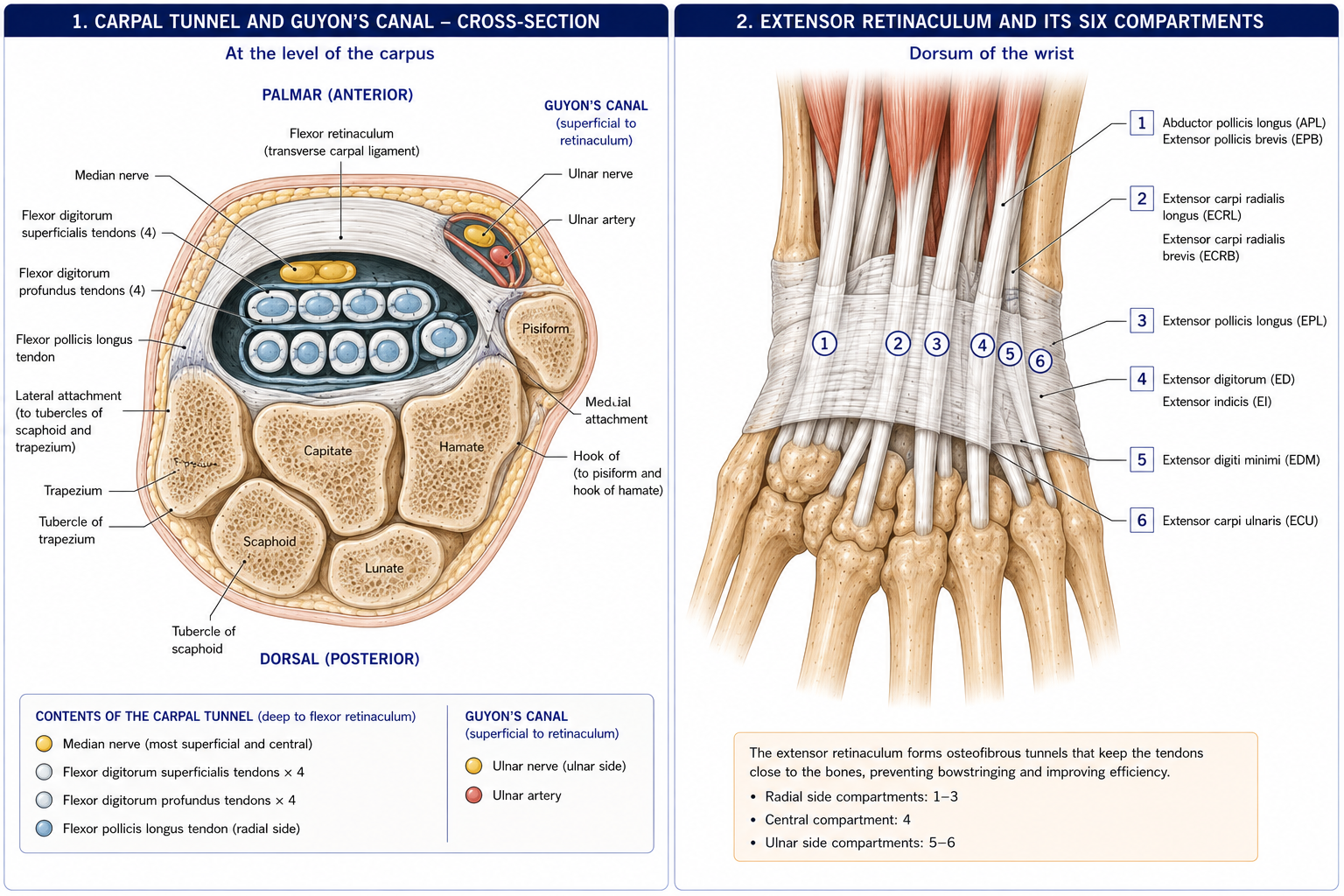
The long flexors and extensors are described in Chapter 11. Two structural features at the wrist matter here.
The flexor retinaculum is a strong fibrous band, ~2–3 cm wide, attached to:
Converting the carpal arch into the carpal tunnel, whose contents are ten structures:
Passing superficial to the retinaculum (not in the tunnel): the palmaris longus tendon, the ulnar nerve and artery (in Guyon’s canal), the palmar cutaneous branch of the median nerve, and the palmar cutaneous branch of the ulnar nerve. Also outside: flexor carpi radialis, which runs in its own groove in the trapezium within a split of the retinaculum, and flexor carpi ulnaris, which inserts on the pisiform.
Tendon sheaths: the ulnar bursa (common flexor sheath) surrounds the FDS and FDP tendons; the radial bursa surrounds FPL. The ulnar bursa communicates with the little finger’s digital sheath, and the radial bursa with the thumb’s — which is why infection in the little finger can track proximally into the palm and, across the potential communication between the bursae, into the thumb (a “horseshoe abscess”).
| Compartment | Contents | Clinical note |
|---|---|---|
| 1 | Abductor pollicis longus, extensor pollicis brevis | De Quervain’s tenosynovitis; Finkelstein’s test |
| 2 | ECRL, ECRB | Intersection syndrome where the first compartment crosses it |
| 3 | Extensor pollicis longus | Turns around Lister’s tubercle; ruptures after distal radius fracture or in rheumatoid disease |
| 4 | Extensor digitorum, extensor indicis | Posterior interosseous nerve terminates here (a target for denervation) |
| 5 | Extensor digiti minimi | Over the DRUJ; ruptures in rheumatoid arthritis (Vaughan-Jackson lesion begins here) |
| 6 | Extensor carpi ulnaris | ECU subsheath is part of the TFCC; subluxation causes ulnar-sided wrist pain |
Fibrous bands hold the flexor tendons against the phalanges, preventing bowstringing and preserving the moment arm.
Flexor tendon zones (Verdan) — zone II, from the distal palmar crease to the middle of the middle phalanx, is where FDS and FDP lie together within the sheath, and was historically called “no man’s land” because repairs there adhere so readily. Modern early controlled mobilisation protocols (Chapter 7) have transformed the outcome, but the zone still dictates the protocol.
Nineteen muscles wholly within the hand, in four groups. All are supplied by the ulnar nerve except the “LOAF” muscles, which are median.
| Group | Muscles | Nerve | Actions |
|---|---|---|---|
| Thenar | Abductor pollicis brevis, Flexor pollicis brevis (superficial head), Opponens pollicis | Recurrent (motor) branch of the median nerve (C8, T1) | Abduction, flexion and opposition of the thumb |
| Adductor pollicis (oblique and transverse heads) | Ulnar (deep branch) | Adducts the thumb; the key muscle of key (lateral) pinch | |
| Hypothenar | Abductor digiti minimi, flexor digiti minimi brevis, opponens digiti minimi (+ palmaris brevis, superficial) | Ulnar (deep branch) | Abduct, flex and oppose the little finger; cup the ulnar palm |
| Lumbricals | Four, arising from the FDP tendons (a rare muscle with both origin and insertion on tendon) and inserting into the radial side of the extensor expansion | 1st and 2nd (index, middle): median. 3rd and 4th (ring, little): ulnar | Flex the MCP and extend the IP joints; extraordinarily richly supplied with muscle spindles — proprioceptive fine-tuning of grip |
| Interossei | Four dorsal (D–AB: abduct); three palmar (P–AD: adduct) | Ulnar (deep branch) | Abduct/adduct the digits about the axis of the middle finger; also flex MCP and extend IP joints via the expansion |
Mnemonic: LOAF — Lateral two Lumbricals, Opponens pollicis, Abductor pollicis brevis, Flexor pollicis brevis (superficial head) — the median intrinsics. Everything else in the hand is ulnar.
They pass anterior to the MCP axis but posterior to the IP axes. One contraction therefore flexes the MCP joints while extending the IP joints — the position needed to flatten the hand around a large object, to hold a book, or to write.
Lose them (ulnar nerve palsy) and the long extensors act unopposed at the MCP joints while FDP acts unopposed at the IP joints — producing clawing (intrinsic-minus hand): MCP hyperextension with IP flexion. Lose the opposite (spasticity, Volkmann’s contracture) and you get the intrinsic-plus hand: MCP flexion with IP extension.
This is the reason the position of safe immobilisation (§12.10) puts the MCPs in flexion and the IPs in extension — it is the position in which the collateral ligaments are at maximum length and cannot shorten.
Over the dorsum of each finger, the extensor digitorum tendon flattens into the extensor expansion (dorsal hood), joined on each side by the lumbrical and interossei. It divides into:
Held in place by the sagittal bands at the MCP joint, the triangular ligament dorsally and the transverse retinacular ligaments laterally.
| Deformity | Lesion | Mechanism |
|---|---|---|
| Mallet finger | Rupture or avulsion of the terminal extensor tendon at the distal phalanx | DIP droops into flexion; cannot actively extend. Treated by continuous DIP extension splinting for 6–8 weeks — the joint must not be allowed to flex even momentarily, which patients must be taught explicitly |
| Swan-neck | PIP hyperextension with DIP flexion | Lateral bands migrate dorsally. Causes: untreated mallet, volar plate laxity, intrinsic tightness, rheumatoid arthritis |
| Boutonnière | PIP flexion with DIP hyperextension | Central slip rupture; the lateral bands slip volar to the PIP axis and become flexors of it. Elson’s test detects an acute central slip injury before the deformity appears — and detecting it then is the difference between a splint and a reconstruction |
| Claw hand (intrinsic-minus) | Ulnar (± median) palsy | MCP hyperextension, IP flexion |
| Trigger finger | A1 pulley stenosis | Catching, locking |
| Dupuytren’s contracture | Myofibroblastic proliferation in the palmar aponeurosis | Progressive MCP then PIP flexion contracture, usually ring and little fingers; nodules and cords; associated with northern European descent, diabetes, alcohol, epilepsy medication. Stretching does not work; treatment is surgical or by needle fasciotomy/collagenase, with post-operative therapy |
Figure 3 · Nerve supply of the hand
| Nerve | Palmar | Dorsal | Autonomous (reliable) zone |
|---|---|---|---|
| Median | Lateral 3½ digits + adjacent palm | Dorsum of the distal phalanges of the lateral 3½ digits | Pulp of the index finger |
| Ulnar | Medial 1½ digits + adjacent palm | Medial 1½ digits and the adjacent dorsal hand | Pulp of the little finger |
| Radial | — (no palmar territory) | Lateral dorsum of the hand and the proximal parts of the lateral 3½ digits | Dorsal first web space |
The autonomous zones are the small areas supplied by one nerve only, without overlap. Test there. Testing in overlap zones is the commonest reason a clinical examination fails to detect a nerve lesion.
| Lesion | Motor loss | Deformity | Functional consequence |
|---|---|---|---|
| Median (at the wrist) | Thenar muscles (LOAF); lateral two lumbricals | Ape (simian) hand — thenar wasting, thumb adducted and laterally rotated, cannot oppose | Loss of opposition and precision pinch — the single most disabling upper-limb nerve deficit, costing ~40–50% of hand function |
| Median (high, at the elbow) | Above plus FDS, lateral FDP, FPL, FCR, pronators | Hand of benediction on attempted fist-making | Loss of index/middle flexion and thumb IP flexion |
| Ulnar (at the wrist / Guyon’s) | All intrinsics except LOAF | Claw hand (ring and little), marked; first dorsal interosseous wasting is the earliest visible sign | Loss of grip power, finger abduction/adduction, and coordination. Froment’s sign — thumb IP flexes on key pinch, substituting FPL for the paralysed adductor pollicis. Wartenberg’s sign — the little finger drifts into abduction |
| Ulnar (high, at the elbow) | Above plus FCU and medial FDP | Less clawing (the ulnar paradox) | |
| Radial (at the wrist) | None (sensory only) | — | Numbness only |
| Radial (proximal) | Wrist and finger extensors | Wrist drop | Grip strength falls ~70% because the wrist cannot be stabilised (Chapter 6) |
The commonest peripheral entrapment neuropathy: prevalence ~3–5%, peak 40–60 years, more common in women.
Boundaries: floor = flexor retinaculum and hypothenar muscles; roof = palmar carpal ligament and palmaris brevis; medial wall = pisiform and FCU; lateral wall = hook of the hamate. Contents: the ulnar nerve and ulnar artery (no tendons).
Causes: ganglion (the commonest), hook of hamate fracture, ulnar artery thrombosis (hypothenar hammer syndrome), and prolonged handlebar pressure (cyclist’s palsy).
The localising sign: the dorsal cutaneous branch of the ulnar nerve leaves the nerve 5–7 cm proximal to the wrist, so dorsal ulnar hand sensation is preserved in a Guyon’s canal lesion but lost in a cubital tunnel lesion. That one finding separates the two, and it is worth having at your fingertips.
| Arch | Formed by | Level | Branches |
|---|---|---|---|
| Superficial palmar arch | Mainly the ulnar artery, completed by the superficial palmar branch of the radial | At the level of the distal border of the fully extended thumb | Three common palmar digital arteries + one to the medial little finger |
| Deep palmar arch | Mainly the radial artery, completed by the deep branch of the ulnar | ~1 cm proximal to the superficial arch, at the level of the proximal border | Palmar metacarpal arteries; princeps pollicis; radialis indicis |
Allen’s test: compress both radial and ulnar arteries, have the patient clench and open the hand until it blanches, then release one artery and time the return of colour (normally <5–7 seconds). It tests whether one artery alone can perfuse the hand — essential before radial artery harvest, cannulation, or in suspected hypothenar hammer syndrome.
Digital arteries run on the palmar-lateral aspect of each digit with the digital nerves, which is why digital blocks are placed there and why lacerations on the sides of a finger threaten both.
Figure 4 · The four grips
Napier’s classification remains the standard framework, with common clinical elaborations.
| Grip | Description | Key muscles | Example |
|---|---|---|---|
| Power (cylindrical) grip | The object is held between the flexed fingers and the palm, with the thumb wrapped around; the wrist is held in extension and slight ulnar deviation | Long flexors, with wrist extensors as essential synergists; hypothenar and interossei | Holding a hammer, a bar, a cup handle |
| Spherical grip | Fingers abducted around a rounded object | Interossei, long flexors | Holding a ball or a doorknob |
| Hook grip | Fingers flexed at IP joints; thumb not involved; the only grip preserved in median nerve palsy | Long flexors only | Carrying a bag or briefcase |
| Precision (pinch) grips | The object is held between the thumb and finger pulps, not touching the palm | Thenar muscles, lumbricals, interossei | — |
| — Tip (pulp-to-pulp) pinch | Thumb tip to index tip | FPL and FDP (both AIN) | Picking up a pin |
| — Tripod (three-jaw chuck) pinch | Thumb to index and middle pulps | Thenar + lumbricals | Holding a pen |
| — Key (lateral) pinch | Thumb pulp against the radial side of the index middle phalanx | Adductor pollicis (ulnar) and first dorsal interosseous | Turning a key; the grip tested by Froment’s sign |
Two things that are worth internalising:
| Position | Description | Use |
|---|---|---|
| Functional (position of function) | Wrist ~20–30° extension, slight ulnar deviation; MCP ~45°; PIP ~30–45°; DIP ~10–20°; thumb abducted and opposed | The position of readiness; the position for a hand that will be permanently stiff, since it retains most usable function |
| Position of safe immobilisation (intrinsic-plus / “safe” position) | Wrist ~20–30° extension; MCP 70–90° flexion; IP joints in full extension; thumb abducted and opposed | The position for splinting any injured hand. Each element has a reason (below) |
| Position of rest | Wrist slight flexion; fingers gently flexed with increasing flexion from index to little | The natural resting posture; the tenodesis cascade — disruption of the smooth cascade indicates a flexor tendon rupture, a finding visible before any test is performed |
Splinting a hand flat, which looks intuitively correct, produces exactly the wrong contractures at every joint. This is one of the highest-value pieces of applied anatomy in the whole subject.
Oedema management deserves a note of the same weight: hand oedema organises into fibrous tissue quickly, and once the dorsal skin and extensor mechanism are involved the hand stiffens into the intrinsic-minus posture. Elevation, active movement, compression and early mobilisation in the first days matter more than anything done later.
15 questions on this chapter. Tap one to see the answer and the reasoning.
Answer: (C) Four FDS, four FDP, FPL, and the median nerve.
Answer: (B)
Answer: (B)
Answer: (B) They pass palmar to the MCP axis and dorsal to the IP axes.
Answer: (C)
Answer: (B) The thumb IP flexes as FPL substitutes during key pinch.
Answer: (B) The lateral bands slip volar to the PIP axis.
Answer: (B) Their loss causes bowstringing.
Answer: (A) Strongly associated with diabetes.
Answer: (A)
Answer: (B) The dorsal cutaneous branch leaves 5–7 cm proximal to the wrist.
Answer: (B) The deep arch is mainly radial.
Answer: (B) Because MCP collaterals are taut in flexion and will shorten if held extended.
Answer: (C) It requires only the long flexors, and the thumb is not involved.
Answer: (C) The joint must not be allowed to flex at any point, including during splint changes.
Everything on this page, in one screen
| Source | Why it earns its place |
|---|---|
| Standring S (ed.) — Gray’s Anatomy, 42nd edn | Definitive anatomy of the wrist and hand |
| Palastanga N, Field D, Soames R — Anatomy and Human Movement | Functional anatomy of the hand |
| Neumann DA — Kinesiology of the Musculoskeletal System | Arches, extensor mechanism and grip mechanics |
| Skirven TM, Osterman AL, Fedorczyk JM, Amadio PC — Rehabilitation of the Hand and Upper Extremity | The definitive hand therapy reference |
| Green DP et al. — Green’s Operative Hand Surgery | The surgical reference; the source for zones, pulleys and deformities |
| Napier JR — “The prehensile movements of the human hand”, JBJS Br, 1956 | The original grip classification |
| Magee DJ — Orthopedic Physical Assessment | Wrist and hand tests and their accuracy |
| Kleinert HE; Duran RJ — early mobilisation protocols after flexor tendon repair | The protocols that changed zone II outcomes |
| Snell RS — Clinical Anatomy by Regions | Carpal tunnel, Guyon’s canal and nerve territories with clinical notes |
| Chaurasia BD — Human Anatomy, Vol 1 | Indian syllabus-matched descriptive account |
Chapter 12 of 24 · Human Anatomy · Physiotherapist India End of Part 2 — The upper limb. Next: Chapter 13 — Bones of the Lower Limb, opening Part 3.
Reviewed by the Physiotherapist India Team. · Human Anatomy contents