Evidence-based physiotherapy and rehabilitation resources
Editorial & review policy
Evidence-based physiotherapy and rehabilitation resources
Editorial & review policyHuman Anatomy · The trunk
A column of small bones that has to hold you upright all day, let you bend and turn, and carry the spinal cord safely through the middle of itself. Those demands do not sit together comfortably. Almost every problem you will treat in this region comes from the compromise between them.
Part 4 · The trunk
The curves, the disc, the facet joints and the back muscles
The vertebral column has to be a rigid mast that supports the head and transmits load to the pelvis, a flexible rod that permits movement in three planes, and an armoured tube that protects the spinal cord — all at once, in a structure that is loaded every waking hour.
It solves the problem by segmentation: 33 vertebrae, each moving only a few degrees, summing to a large total range. Segmentation also creates the vulnerability: 23 intervertebral discs, 46 facet joints, and dozens of ligaments and nerve roots, any one of which can become a symptom generator.
Low back pain is the leading cause of years lived with disability worldwide. Neck pain is not far behind. No other region in this book has a comparable clinical footprint, and none is more surrounded by outdated teaching. This chapter covers both the anatomy and the corrections.
Figure 1 · The five regions
| Region | Vertebrae | Notes |
|---|---|---|
| Cervical | 7 | Every mammal has seven, from the mouse to the giraffe |
| Thoracic | 12 | Articulate with ribs |
| Lumbar | 5 | Largest bodies |
| Sacral | 5, fused | Transmits load to the pelvis |
| Coccygeal | 4, fused | Vestigial tail |
| Total | 33 (24 presacral mobile) | Column length ~72 cm in men, ~61 cm in women; discs form ~20–25% of it |
| Curve | Direction | Type | Formed |
|---|---|---|---|
| Cervical lordosis | Convex anteriorly | Secondary (compensatory) | ~3–4 months, when the infant lifts the head |
| Thoracic kyphosis | Concave anteriorly | Primary | Present in fetal life |
| Lumbar lordosis | Convex anteriorly | Secondary | ~12–18 months, with standing and walking |
| Sacral kyphosis | Concave anteriorly | Primary | Fetal |
Primary curves follow the shape of the fetal C-curve and are due to vertebral body shape; secondary curves develop with postural milestones and are maintained largely by disc wedging.
Functional value of the curves: a curved column resists axial compression far better than a straight one. The classic engineering approximation is resistance ∝ (number of curves)² + 1 — giving a curved column roughly ten times the resistance of a straight rod. Whether or not the formula is exact, the principle is sound and it explains why the loss of lordosis under sustained flexion is mechanically costly.
Abnormal curvatures: excessive kyphosis (Scheuermann’s disease in adolescents, osteoporotic vertebral wedging in the elderly), excessive lordosis (hyperlordosis), scoliosis (§17.11) and gibbus (an angular kyphosis, classically from spinal tuberculosis — Pott’s disease — still relevant in India).
Figure 2 · A typical vertebra, and the ones that break the plan
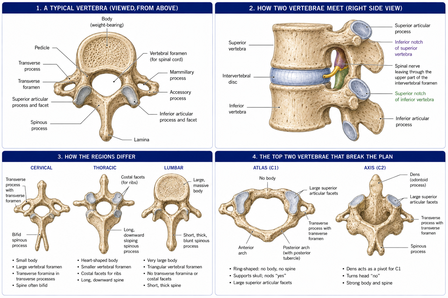
| Part | Detail |
|---|---|
| Body | Anterior weight-bearing mass, largely cancellous with a thin cortical shell and vertebral endplates of hyaline cartilage superiorly and inferiorly |
| Pedicles | Connect body to the posterior arch; form the superior and inferior vertebral notches which, with those of the adjacent vertebra, create the intervertebral foramen |
| Laminae | Complete the arch posteriorly |
| Spinous process | Posterior, midline |
| Transverse processes | Lateral, two |
| Articular processes | Two superior and two inferior, bearing the facets |
| Vertebral foramen | Encloses the cord (or cauda equina) |
| Feature | Cervical | Thoracic | Lumbar |
|---|---|---|---|
| Body | Small, wider transversely; uncinate processes on the superolateral margins | Heart-shaped; costal facets (demifacets) on the body | Large, kidney-shaped |
| Vertebral foramen | Large, triangular | Small, circular | Triangular |
| Transverse process | Foramen transversarium (transmits the vertebral artery, C6–C1 only), anterior and posterior tubercles | Costal facet for the rib tubercle (T1–T10) | Long and slender (the “costal process”); mamillary and accessory processes |
| Spinous process | Short and bifid (C2–C6); C7 is long (vertebra prominens) | Long, sloping steeply downwards (overlapping like roof tiles) | Short, broad, horizontal |
| Facet orientation | ~45° from horizontal, in the frontal plane | ~60° from horizontal, frontal plane | ~90°, in the sagittal plane |
| Movement permitted | All movements freely, especially rotation | Rotation (limited by ribs) | Flexion and extension; rotation minimal (~2° per segment) |
The facets act as bony guides. In the cervical spine their oblique frontal orientation permits flexion, extension, lateral flexion and free rotation. In the thoracic spine their frontal orientation permits rotation but the ribs restrict everything else. In the lumbar spine their sagittal orientation permits substantial flexion and extension but blocks rotation — total lumbar axial rotation is only about 10–13° across all five segments, and forcing rotation there loads the facets and the anulus in a way they are not designed for.
This single fact explains why lumbar rotation manipulations must be applied with care, why “rotate from the hips and thoracic spine” is sound advice for golfers and throwers, and why thoracic mobility deficits so often present as lumbar or cervical symptoms.
| Vertebra | Features |
|---|---|
| C1 (atlas) | No body and no spinous process — a ring of anterior and posterior arches with two lateral masses. Its “body” became the dens of C2. Anterior arch has a facet for the dens; the groove for the vertebral artery lies on the posterior arch |
| C2 (axis) | The dens (odontoid process) projecting superiorly — the pivot for head rotation. The dens is held by the transverse ligament of the atlas, whose failure (rheumatoid arthritis, Down syndrome, trauma) permits atlanto-axial instability and cord compression |
| C7 (vertebra prominens) | Long, non-bifid spinous process; foramen transversarium transmits only accessory veins, not the vertebral artery |
| T1 | Full costal facet for rib 1 and a demifacet for rib 2 |
| T11, T12 | Single full costal facets; transitional facet orientation |
| L5 | Largest body; deeply wedged anteriorly, contributing to the lumbosacral angle |
| Sacrum | Five fused vertebrae; auricular surface for the SI joint; four pairs of anterior and posterior sacral foramina; the sacral hiatus (the caudal opening of the sacral canal, between the sacral cornua) — the site of caudal epidural injection |
| Coccyx | Four fused rudimentary vertebrae; attachment for the pelvic floor and anococcygeal ligament |
Transitional anomalies are common and clinically relevant: sacralisation of L5 (fused to the sacrum) and lumbarisation of S1 (a free S1) occur in around 4–8% of people, and mis-numbering the segments is a well-documented source of wrong-level surgery.
| Joint | Type | Movement |
|---|---|---|
| Atlanto-occipital | Synovial condyloid (paired) | ~15–20° flexion–extension (“yes” nodding); slight lateral flexion; no rotation |
| Atlanto-axial | Median (pivot, between dens and atlas ring) + two lateral (plane) | ~45–50° rotation to each side — approximately half of all cervical rotation (“no”) occurs here |
The alar ligaments (dens to the occipital condyles) limit rotation and are tested by the alar ligament stress test. Upper cervical instability — from rheumatoid arthritis, Down syndrome, Marfan syndrome, os odontoideum, or trauma — is an absolute contraindication to cervical manipulation and must be screened for.
Figure 3 · The intervertebral disc
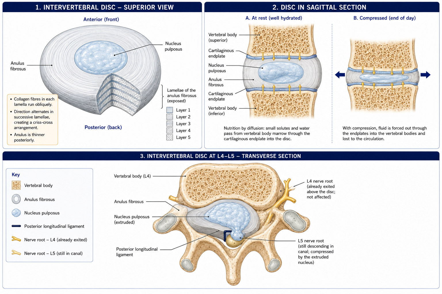
23 discs, from C2–C3 to L5–S1 (none between C1–C2 or within the sacrum). The largest avascular structure in the body.
| Component | Composition | Function |
|---|---|---|
| Nucleus pulposus | Notochord remnant; 70–90% water in youth; type II collagen in a loose network; high proteoglycan (aggrecan) content | A hydrostatic gel: converts axial compression into radial pressure against the anulus and endplates, distributing load evenly |
| Anulus fibrosus | 15–25 concentric lamellae of type I collagen (outer) grading to type II (inner); fibres run at ~65° from vertical (~30° from horizontal), alternating direction between adjacent lamellae | Contains the nucleus; resists tension, torsion and shear |
| Vertebral endplate | Hyaline cartilage, ~0.6–1 mm | The route of nutrition; the weakest link under pure compression |
The alternating fibre orientation matters: in any given rotation, only about half the lamellae are engaged — those oriented with the direction of twist. This halves the anulus’s torsional strength compared to its tensile strength, and is why rotation combined with flexion and compression is the most damaging loading pattern for the disc, and why lumbar segments (which cannot rotate much anyway) are damaged by it.
The adult disc has no blood supply of its own beyond the outer few millimetres of anulus. Nutrition reaches the nucleus by diffusion through the vertebral endplates (the dominant route) and through the outer anulus, driven by convection from cyclical loading exactly as in articular cartilage (Chapter 3).
Consequences:
Disc height falls through the day as fluid is expressed under load and recovers overnight. Adults lose 1–2 cm of stature over a day, most of it in the first few hours of being upright.
Clinically: disc pressure, bending stresses and the risk of anular strain are highest in the first hour or two after rising. Advising a patient with disc-related pain to avoid heavy lifting and end-range lumbar flexion first thing in the morning is anatomically sound advice, and it is one of the few genuinely mechanical pieces of advice worth giving.
Approximate intradiscal pressure relative to standing (=100%):
| Position | Relative pressure |
|---|---|
| Lying supine | ~25% |
| Side lying | ~75% |
| Standing | 100% |
| Standing, flexed forward | ~150% |
| Sitting unsupported | ~140% |
| Sitting, flexed forward with a weight | ~275% |
| Lifting with a flexed, rotated spine | Highest |
The classical teaching that “sitting is worse than standing” is broadly supported, though later in-vivo measurements (Wilke et al.) found smaller differences than Nachemson’s original figures. The clinically defensible message is not “sit correctly” but “change position regularly” — sustained loading in any posture is the problem.
Figure 4 · Why each region moves the way it does
Paired plane synovial joints between the superior articular process of one vertebra and the inferior articular process of the one above, with a capsule, synovium, and small intra-articular meniscoids (fibro-adipose folds implicated in acute locking).
Functions: 1. Guide and limit movement by their orientation (§17.3). 2. Share load: normally carrying ~16–20% of compressive load, but rising to ~40–70% in extension and in disc-degenerated segments — which is why extension-based pain suggests facet involvement. 3. Resist shear, particularly the anterior shear at L5–S1 imposed by the lumbosacral angle. 4. Proprioception — they are richly innervated.
Innervation: by the medial branch of the posterior (dorsal) ramus, with each joint supplied by the medial branches of two adjacent levels — the reason medial branch blocks and radiofrequency denervation must target two levels per joint.
The three-joint complex (Kirkaldy-Willis): each motion segment is a functional triad — one disc and two facet joints. Degeneration in any one loads the others. The degenerative cascade proceeds through dysfunction → instability → stabilisation (restabilisation by osteophytes and stiffening), which is why an elderly degenerate spine is often stiff but less painful than a middle-aged one in the instability phase.
Note: the uncovertebral joints (of Luschka) in the cervical spine (C3–C7) form between the uncinate processes and the vertebra above. They guide cervical motion, limit lateral translation, and — importantly — osteophytes here narrow the intervertebral foramen and are a common cause of cervical radiculopathy.
| Ligament | Extent | Function |
|---|---|---|
| Anterior longitudinal ligament (ALL) | Occiput/C1 to sacrum, along the anterior bodies; broad and strong; adherent to bodies and discs | Limits extension; the only ligament resisting hyperextension. Reinforces the disc anteriorly |
| Posterior longitudinal ligament (PLL) | Body of C2 to sacrum, within the vertebral canal; narrow, especially over the lumbar discs | Limits flexion. Its narrowness at the lumbar levels is why disc herniation is typically posterolateral, avoiding the ligament, and why it strikes the traversing nerve root |
| Ligamentum flavum | Between adjacent laminae | Up to 60–70% elastin — the most elastic tissue in the body. Pre-tensioned, so it resists flexion and recoils without buckling into the canal. Hypertrophies with age and buckles in extension, contributing to spinal stenosis |
| Interspinous | Between spinous processes | Limits flexion |
| Supraspinous | Tips of spinous processes, C7 to sacrum; continuous above as the ligamentum nuchae | Limits flexion |
| Intertransverse | Between transverse processes | Limits contralateral lateral flexion |
| Iliolumbar | L4–L5 transverse processes to the iliac crest | Stabilises the lumbosacral junction against shear |
| Sacroiliac complex | Anterior, interosseous (the strongest), posterior sacroiliac, sacrotuberous, sacrospinous | Resist sacral nutation and the enormous shear at the SI joint |
Craniovertebral ligaments: the transverse ligament of the atlas (the crucial one), the alar ligaments, the apical ligament, the tectorial membrane (the upward continuation of the PLL), and the atlanto-occipital membranes.
Figure 5 · The muscles of the back, layer by layer
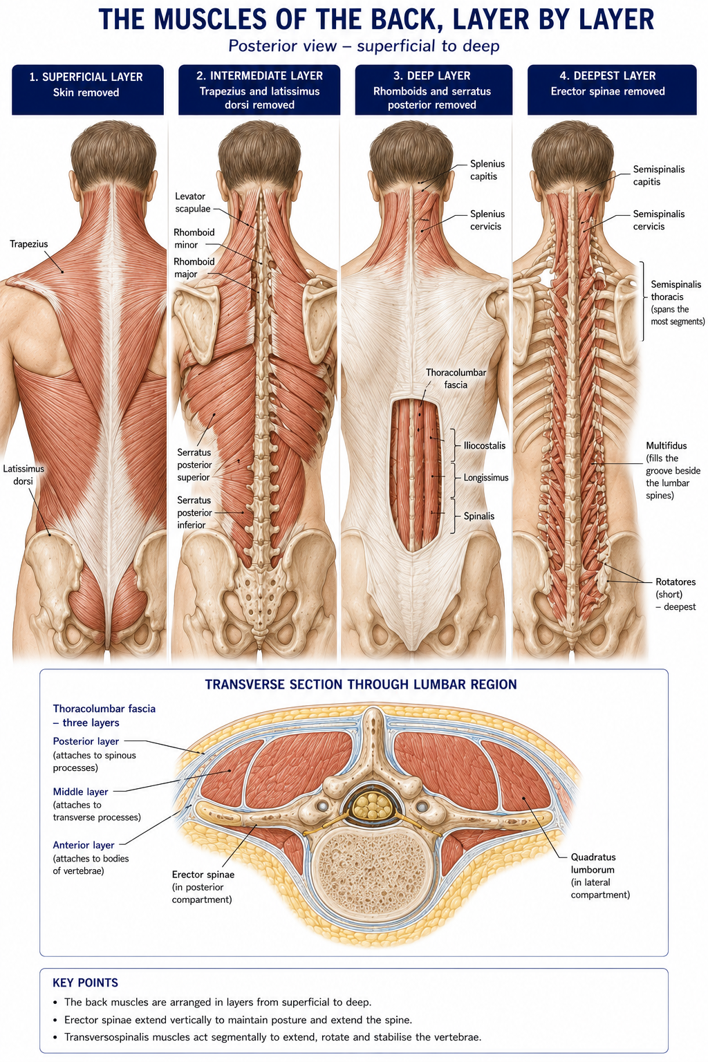
Organised in three groups. The key discriminator is nerve supply: the true (intrinsic, deep) back muscles are supplied by posterior (dorsal) rami; every other muscle in the back is an immigrant, supplied by anterior (ventral) rami.
| Muscle | Nerve | Action |
|---|---|---|
| Trapezius | Spinal accessory (CN XI) + C3, C4 | Scapular elevation, retraction, upward rotation |
| Latissimus dorsi | Thoracodorsal (C6–C8) | Shoulder extension, adduction, internal rotation |
| Levator scapulae | Dorsal scapular (C5) + C3, C4 | Scapular elevation |
| Rhomboid major and minor | Dorsal scapular (C5) | Retraction |
Serratus posterior superior (elevates ribs) and serratus posterior inferior (depresses ribs) — of doubtful respiratory significance, but they define the layer.
| Layer | Muscles | Function |
|---|---|---|
| Superficial | Splenius capitis and cervicis | Extension, ipsilateral lateral flexion and ipsilateral rotation of head and neck |
| Intermediate | Erector spinae — from lateral to medial: Iliocostalis, Longissimus, Spinalis (“I Long for Spinach”), arising from the common tendon on the sacrum, iliac crest and lumbar spinous processes | Bilateral: extension. Unilateral: ipsilateral lateral flexion. The main antigravity extensors; eccentrically control forward flexion |
| Deep (transversospinalis) | Semispinalis (spans 4–6 segments), multifidus (2–4 segments), rotatores (1–2 segments) | Extension, contralateral rotation, and above all segmental control and proprioception |
| Deepest (minor) | Interspinales, intertransversarii, levatores costarum | Segmental stabilisation and proprioception |
| Suboccipital | Rectus capitis posterior major and minor, obliquus capitis superior and inferior — bounding the suboccipital triangle (contents: vertebral artery and suboccipital nerve, C1) | Fine control of head position; extraordinarily high muscle spindle density (up to 200+ spindles per gram — among the highest in the body), making them a major source of cervicogenic proprioceptive input, and implicated in cervicogenic headache and dizziness |
Multifidus deserves separate mention. It is the largest and most medial of the lumbar transversospinalis group, contributing substantially to segmental stiffness. It shows rapid, segmental, side-specific atrophy after low back injury, with fatty infiltration that does not recover spontaneously with resolution of pain. This is a real and replicated finding. What the evidence does not support is the inference once drawn from it — that targeted multifidus/transversus retraining is superior to general exercise for chronic low back pain (Chapter 6).
Three layers enclosing erector spinae and quadratus lumborum. The posterior layer receives latissimus dorsi, the contralateral gluteus maximus, and the aponeuroses of transversus abdominis and internal oblique — forming the posterior oblique sling that transmits force diagonally across the body during gait and rotation. It is also densely innervated and is a plausible primary source of low back pain in its own right.
From outside in: epidural (extradural) space (fat, the internal vertebral venous plexus of Batson, and the target of epidural injection), dura mater, arachnoid, subarachnoid space with CSF, pia mater, and the spinal cord.
Key levels:
| Structure | Level |
|---|---|
| Spinal cord ends (conus medullaris) | L1–L2 in adults (L3 at birth, because the column grows faster than the cord) |
| Dural sac and subarachnoid space end | S2 — level of the PSIS |
| Filum terminale | Conus to the coccyx |
| Cauda equina | The lumbar and sacral roots below L1–L2 |
| Lumbar puncture | L3–L4 or L4–L5, below the conus, at the supracristal (L4) plane |
| Cord enlargements | Cervical (C4–T1) for the upper limb; lumbosacral (L1–S3) for the lower limb |
Formed in the intervertebral foramen by the union of the dorsal (sensory) root, bearing the dorsal root ganglion, and the ventral (motor) root. It divides almost immediately into:
Nerve root numbering — a perennial confusion, and worth stating precisely:
Anterior spinal artery syndrome therefore produces bilateral motor loss and loss of pain and temperature with preserved proprioception and vibration — a pattern that makes sense only if you know the vascular territories.
Figure 6 · What keeps a spine stable
| Region | Flexion | Extension | Lateral flexion (each) | Rotation (each) |
|---|---|---|---|---|
| Cervical | 45–50° | 45–70° | 40–45° | 80–90° (half at atlanto-axial) |
| Thoracic | 20–45° | 20–25° | 20–25° | 35–50° |
| Lumbar | 40–60° | 20–35° | 15–20° | 5–7° (total ~10–13°) |
Lateral flexion and axial rotation are mechanically linked, and the direction of coupling differs by region:
Forward bending is a coordinated sequence: the first ~40–60° comes from lumbar flexion, followed by progressive anterior pelvic rotation on the femoral heads, in a roughly 1:1 to 2:1 lumbar-to-hip ratio depending on the phase. Returning to upright reverses it, hips first.
The flexion–relaxation phenomenon: at full trunk flexion, the erector spinae become electrically silent, and load is borne passively by the posterior ligaments and the thoracolumbar fascia. This phenomenon is absent in most people with chronic low back pain, who maintain erector spinae activity throughout — a robust and reproducible finding, and a useful marker of guarding.
This section exists because the anatomy in this chapter is routinely used to justify practices the evidence does not support.
What is well established:
What this does not mean: anatomy is not irrelevant. It is what allows you to recognise the 10–15% who do have a specific, identifiable and sometimes serious cause, and it is what allows you to explain to a patient, accurately and reassuringly, what their structures actually are and are not doing.
Compression of the lumbosacral roots below the conus, most often by a large central disc herniation.
Features: bilateral (or alternating) leg pain, weakness or numbness; saddle (perineal) anaesthesia; bladder dysfunction — beginning as difficulty initiating or reduced sensation of flow, progressing to painless retention with overflow incontinence; bowel incontinence; and loss of anal tone and sexual dysfunction.
Ask about these directly in every acute low back presentation — patients do not volunteer them. Bladder symptoms are the ones that matter most, and reduced urinary sensation is an earlier and more useful question than frank incontinence.
This is a surgical emergency. Decompression within 24–48 hours substantially improves outcome. Do not arrange a follow-up appointment; arrange immediate hospital assessment.
Grading: bulge (circumferential, ≤25% of circumference) → protrusion (base wider than the dome) → extrusion (dome wider than the base) → sequestration (a free fragment).
Nerve root levels worth having automatic:
| Root | Motor | Reflex | Sensory |
|---|---|---|---|
| C5 | Shoulder abduction, elbow flexion | Biceps | Lateral arm |
| C6 | Elbow flexion, wrist extension | Brachioradialis | Thumb |
| C7 | Elbow extension, wrist flexion | Triceps | Middle finger |
| C8 | Finger flexion | — | Little finger |
| T1 | Finger abduction | — | Medial forearm |
| L2–L3 | Hip flexion | — | Anterior thigh |
| L4 | Knee extension, ankle dorsiflexion | Knee jerk | Medial malleolus |
| L5 | Great toe extension (EHL), hip abduction | — (medial hamstring) | Dorsum of foot, first web space |
| S1 | Plantarflexion, eversion | Ankle jerk | Lateral foot, sole |
Narrowing of the central canal, lateral recess or foramen, usually degenerative — from disc bulging, facet hypertrophy and ligamentum flavum buckling acting together.
Neurogenic claudication is the characteristic syndrome, and distinguishing it from vascular claudication is a core clinical skill:
| Neurogenic | Vascular | |
|---|---|---|
| Relieved by | Sitting or lumbar flexion (which opens the canal) | Standing still |
| Provoked by | Standing and walking, especially downhill/extension | Any exertion, reproducibly at a set distance |
| Walking uphill | Easier (flexed posture) | Harder |
| Cycling | Well tolerated (flexed) | Provokes symptoms |
| Pulses | Normal | Diminished; ABPI reduced |
The “shopping trolley sign” — relief when leaning on a trolley — is a classic and useful observation.
A three-dimensional deformity: lateral curvature with vertebral rotation, producing the rib hump seen on Adam’s forward bend test and quantified with a scoliometer.
An acceleration–deceleration injury of the neck. Quebec Task Force grades 0–IV. Structures implicated include the facet joints (the best-supported single source, on medial branch block studies), capsules, discs, muscles and ligaments.
The predictors of poor outcome are largely non-anatomical: high initial pain intensity, high initial disability, cold hyperalgesia, and post-traumatic stress symptoms. Early return to normal activity with advice and exercise outperforms immobilisation — soft collars are not recommended, and prolonged rest worsens outcomes.
15 questions on this chapter. Tap one to see the answer and the reasoning.
Answer: (B) They develop with head lifting and standing.
Answer: (C) Which permits flexion and extension but blocks rotation.
Answer: (B) Around 45–50° to each side.
Answer: (B) Only half the lamellae resist a given rotation, halving torsional strength.
Answer: (B) Driven by cyclical loading — movement is nutrition.
Answer: (B) It is pre-tensioned so it does not buckle into the canal; hypertrophy contributes to stenosis.
Answer: (B)
Answer: (B) The traversing root. A far-lateral herniation at the same level would compress L4.
Answer: (B)
Answer: (B) The dural sac ends at S2.
Answer: (B) A major proprioceptive source, implicated in cervicogenic headache and dizziness.
Answer: (C) Which is why imaging findings must be interpreted with great caution.
Answer: (B) Vascular claudication is relieved by standing still.
Answer: (B) From repetitive extension and rotation in adolescent athletes.
Answer: (B) Patients rarely volunteer it; it is a surgical emergency.
Everything on this page, in one screen
| Source | Why it earns its place |
|---|---|
| Standring S (ed.) — Gray’s Anatomy, 42nd edn | Definitive vertebral and back anatomy |
| Bogduk N — Clinical and Radiological Anatomy of the Lumbar Spine | The single best text on lumbar structure, innervation and pain sources |
| Neumann DA — Kinesiology of the Musculoskeletal System | Spinal mechanics, coupled motion and lumbopelvic rhythm |
| Palastanga N, Field D, Soames R — Anatomy and Human Movement | Functional anatomy of the trunk |
| Adams MA, Bogduk N, Burton K, Dolan P — The Biomechanics of Back Pain | Disc mechanics, nutrition and degeneration |
| Brinjikji W et al. — “Systematic literature review of imaging features of spinal degeneration in asymptomatic populations”, AJNR, 2015 | The prevalence data that must inform every conversation about imaging |
| Foster NE et al. — “Prevention and treatment of low back pain: evidence, challenges, and promising directions”, Lancet, 2018 (Low Back Pain Series) | The current evidence framework |
| NICE NG59 — Low back pain and sciatica in over 16s | Current guideline-level management |
| Weinstein SL et al. — “Effects of bracing in adolescents with idiopathic scoliosis (BrAIST)”, NEJM, 2013 | The bracing evidence |
| Chaurasia BD — Human Anatomy, Vol 3: Head, Neck and Brain | Indian syllabus-matched descriptive account |
Chapter 17 of 24 · Human Anatomy · Physiotherapist India Next: Chapter 18 — Thoracic Wall and Diaphragm: the cage, the intercostals and the mechanics of breathing.
Reviewed by the Physiotherapist India Team. · Human Anatomy contents