Evidence-based physiotherapy and rehabilitation resources
Editorial & review policy
Evidence-based physiotherapy and rehabilitation resources
Editorial & review policyHuman Anatomy · The trunk
The chest is the only part of the skeleton that has to change shape about twenty thousand times a day. It must be stiff enough to protect the heart and lungs, and mobile enough to pump air without you ever thinking about it. This chapter is about how a cage of bone manages to breathe.
Part 4 · The trunk
The cage, the intercostals and the mechanics of breathing
Cardiorespiratory physiotherapy is one of the three great domains of the profession, and it rests entirely on this chapter. Every technique — positioning, breathing exercises, manual hyperinflation, cough assist, inspiratory muscle training, chest wall mobilisation — is an intervention on the mechanics described here.
Two ideas run through everything:
Figure 1 · The thoracic cage and a typical rib
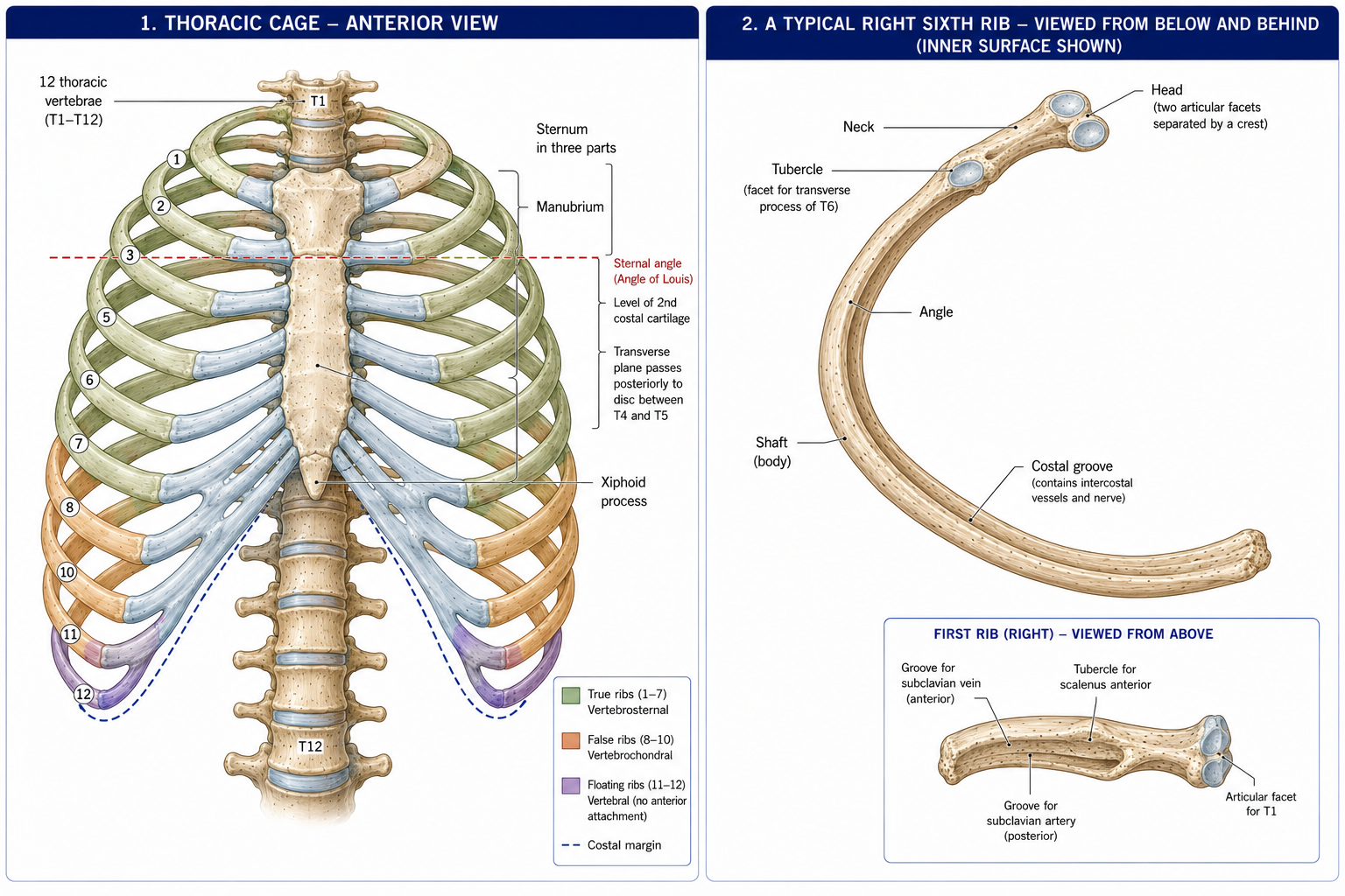
| Component | Detail |
|---|---|
| Posterior | 12 thoracic vertebrae and the intervertebral discs |
| Lateral | 12 pairs of ribs and 11 intercostal spaces |
| Anterior | Sternum and the costal cartilages |
| Superior aperture (thoracic inlet) | Bounded by T1, the first ribs and the manubrium; transmits the trachea, oesophagus, great vessels, nerves and the apices of the lungs. Small and rigid — hence “thoracic outlet syndrome” occurs at its margins |
| Inferior aperture (thoracic outlet) | Bounded by T12, ribs 11 and 12, the costal margin and the xiphisternum; closed by the diaphragm |
| Part | Features |
|---|---|
| Manubrium | Jugular (suprasternal) notch at the level of T2/T3; clavicular notches; facet for the first costal cartilage |
| Sternal angle (of Louis) | The manubriosternal joint — a palpable transverse ridge, and the single most important surface landmark in the thorax |
| Body | Demifacets for costal cartilages 2–7 |
| Xiphoid process | Cartilaginous until ~40 years; the landmark for the epigastric region and for hand placement in CPR |
Learn this one landmark thoroughly and half of thoracic surface anatomy follows.
| Classification | Ribs | Detail |
|---|---|---|
| True (vertebrosternal) | 1–7 | Attach directly to the sternum by their own costal cartilage |
| False (vertebrochondral) | 8–10 | Attach to the cartilage above, forming the costal margin |
| Floating (vertebral) | 11–12 | No anterior attachment |
| Typical | 3–9 | Have a head with two facets, neck, tubercle with an articular facet, angle, shaft and costal groove |
| Atypical | 1, 2, 10, 11, 12 | Rib 1: shortest, broadest, most curved; one facet; has the scalene tubercle with the subclavian groove behind (artery) and the subclavian vein in front; crossed by the lower trunk of the brachial plexus. Rib 2: tuberosity for serratus anterior. Ribs 10–12: single facets; 11 and 12 have no neck or tubercle |
The costal groove on the inferior internal border of each rib carries the intercostal vein, artery and nerve — VAN, from superior to inferior — which dictates where a needle or chest drain is placed (§18.4).
Figure 2 · Why the upper and lower ribs move differently
| Joint | Articulation | Type |
|---|---|---|
| Costovertebral (of the head) | Rib head with the demifacets of two adjacent vertebral bodies and the intervening disc (ribs 2–9); a single facet for ribs 1, 10, 11, 12 | Synovial plane, with an intra-articular ligament dividing it into two cavities |
| Costotransverse | Rib tubercle with the transverse process of the corresponding vertebra (absent for ribs 11 and 12) | Synovial plane, with lateral and superior costotransverse ligaments |
| Costochondral | Rib to its costal cartilage | Primary cartilaginous (synchondrosis) — no movement |
| Sternocostal | Costal cartilage to sternum | 1st is a synchondrosis; 2nd–7th are synovial plane |
| Interchondral | Between cartilages 6–10 | Synovial |
| Manubriosternal | — | Secondary cartilaginous (symphysis); may ossify after middle age |
The axis of rib movement runs through the costovertebral and costotransverse joints, and its orientation changes down the cage:
| Movement | Mechanism | Where it predominates | Effect |
|---|---|---|---|
| Pump-handle | The axis is nearly transverse, so the rib’s anterior end swings upwards and forwards, lifting the sternum | Upper ribs (1–6) | Increases the anteroposterior diameter |
| Bucket-handle | The axis is nearly anteroposterior, so the rib’s lateral part swings upwards and outwards | Lower ribs (7–10) | Increases the transverse diameter |
| Caliper | Lateral flaring | Ribs 11–12 | Small transverse increase |
Both mechanisms operate at every level; the terms describe which predominates. Together they can increase thoracic volume substantially, and their loss — from ankylosing spondylitis, kyphosis, prolonged mechanical ventilation, burns, or simply age — reduces vital capacity independently of lung disease.
Chest expansion measurement (at the axilla, the nipple line and the xiphisternum) quantifies this and is a standard, underused assessment. Normal is roughly 3–7 cm at the xiphoid level; less than 2.5 cm at the fourth intercostal space is a diagnostic criterion for ankylosing spondylitis.
Figure 3 · Inside an intercostal space
Eleven spaces, each containing three muscle layers and a neurovascular bundle.
| Layer | Fibre direction | Extent | Action |
|---|---|---|---|
| External intercostal | Downwards and forwards (“hands in pockets”) | From the tubercle to the costochondral junction, replaced anteriorly by the external intercostal membrane | Elevates the ribs — inspiratory |
| Internal intercostal | Downwards and backwards (at right angles to the external) | From the sternum to the angle of the rib, replaced posteriorly by the internal intercostal membrane | Interosseous part: depresses ribs — expiratory. Interchondral (parasternal) part: elevates ribs — inspiratory |
| Innermost intercostal | Same as internal | The middle of the space | With subcostalis and transversus thoracis, completes the deepest layer |
The neurovascular bundle — vein, artery, nerve from above downwards (VAN) — runs in the costal groove at the inferior border of the rib above, between the internal and innermost layers. A smaller collateral bundle runs along the superior border of the rib below, which is why no part of the space is entirely safe.
Insert immediately ABOVE the upper border of the rib below, in the middle of the intercostal space, to avoid the main neurovascular bundle in the costal groove above.
The “safe triangle” for intercostal drain insertion is bounded by the anterior border of latissimus dorsi, the lateral border of pectoralis major, a line superior to the horizontal level of the nipple (roughly the 5th intercostal space), and the apex below the axilla. It avoids the internal thoracic artery medially, the long thoracic nerve posteriorly, the diaphragm and liver/spleen inferiorly, and the thick musculature above.
A physiotherapist does not insert drains, but must know this to understand drain position, to mobilise a patient safely with a drain in situ, and to recognise a malpositioned one.
Arterial supply: posterior intercostal arteries — the upper two from the superior intercostal artery (from the costocervical trunk of the subclavian), the rest directly from the thoracic aorta; and anterior intercostal arteries from the internal thoracic artery and its musculophrenic branch.
Nerve supply:
Clinically: the intercostobrachial nerve (lateral cutaneous branch of T2) supplies the medial arm and axilla — routinely sacrificed in axillary dissection, producing predictable medial arm numbness. Herpes zoster classically follows a single intercostal dermatome. And the segmental innervation is why T4 = nipple and T10 = umbilicus are such reliable landmarks.
Figure 4 · The diaphragm seen from below
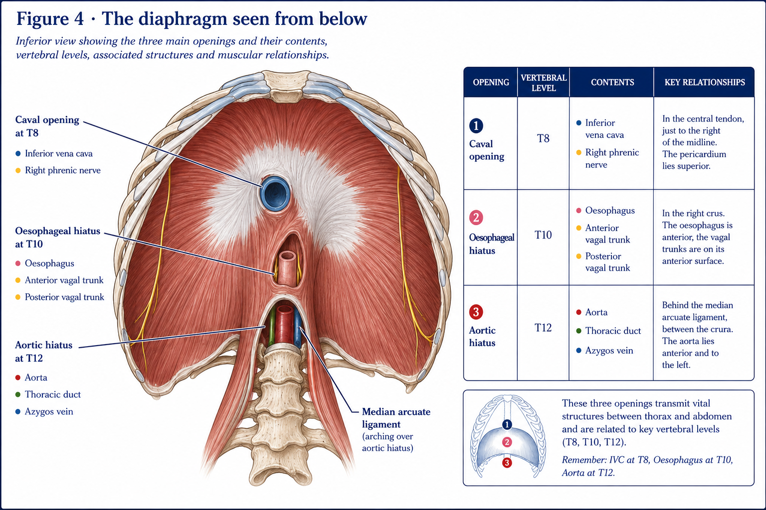
The principal muscle of respiration, a dome-shaped musculotendinous sheet separating thorax from abdomen.
| Part | Origin |
|---|---|
| Sternal | Two slips from the posterior xiphoid process |
| Costal | Inner surfaces of the lower six costal cartilages and ribs, interdigitating with transversus abdominis |
| Lumbar | Right crus from L1–L3 bodies (longer); left crus from L1–L2. Medial arcuate ligament over psoas major; lateral arcuate ligament over quadratus lumborum; median arcuate ligament joining the crura over the aorta |
| Insertion | The central tendon — a trefoil aponeurosis with no bony attachment, fused with the fibrous pericardium above |
The right dome is higher than the left (because of the liver), reaching approximately the 5th rib in expiration; the left reaches the 5th intercostal space.
| Opening | Level | Contents |
|---|---|---|
| Caval opening | T8 | Inferior vena cava, right phrenic nerve. In the central tendon, so it is held open and widens in inspiration — assisting venous return |
| Oesophageal hiatus | T10 | Oesophagus, anterior and posterior vagal trunks, oesophageal branches of the left gastric vessels. In the muscular right crus, which acts as a physiological sphincter — so it is compressed in inspiration. Weakness here permits hiatus hernia |
| Aortic hiatus | T12 | Aorta, thoracic duct, azygos vein. Behind the diaphragm (between the crura), so the aorta is not compressed during contraction |
Mnemonic: “I ate ten eggs at twelve” — IVC 8, Esophagus 10, Aorta 12. Or count the letters: VenaCava (T8, 3 letters ×… ) — most students find the sentence easier.
Minor structures pass elsewhere: the superior epigastric vessels behind the sternal slips; the splanchnic nerves pierce the crura; the sympathetic trunk passes behind the medial arcuate ligament; the hemiazygos vein pierces the left crus.
Motor supply is exclusively from the phrenic nerve (C3, C4, C5) — “C3, 4, 5 keeps the diaphragm alive.” Sensory supply to the central part is also phrenic; the peripheral part is supplied by the lower intercostal nerves (T7–T11).
Two consequences of enormous clinical importance:
On contraction, the dome descends — flattening and increasing the vertical dimension of the thorax — and, once the central tendon meets the resistance of the abdominal viscera, the zone of apposition (the cylindrical portion of the diaphragm applied to the inner chest wall) contracts to lift and evert the lower ribs, increasing the transverse diameter. It therefore does two jobs, and the second depends on abdominal wall tone providing a fulcrum.
Quiet inspiration: diaphragmatic descent of ~1.5 cm, contributing 70–80% of tidal volume. Deep inspiration: descent of ~6–10 cm.
A hyperinflated chest flattens the diaphragm. A flattened diaphragm has:
The COPD patient’s diaphragm is mechanically disadvantaged, not merely weak. That is why they recruit accessory muscles at rest, why they adopt forward-lean positions (which push the abdominal contents up, restoring the dome and the zone of apposition), and why pursed-lip breathing helps by reducing dynamic airway collapse and air trapping. Every one of those observations is anatomy applied.
Figure 5 · Which muscles breathe, and when
| Phase | Quiet breathing | Forced / increased demand |
|---|---|---|
| Inspiration | Diaphragm (70–80%), external intercostals, parasternal (interchondral) internal intercostals | Plus scalenes (active even in quiet breathing in many people), sternocleidomastoid, pectoralis major and minor, serratus anterior, latissimus dorsi, trapezius, erector spinae, levatores costarum, and alae nasi |
| Expiration | PASSIVE — elastic recoil of lung and chest wall | Abdominal muscles (rectus abdominis, external and internal oblique, transversus abdominis — the most important), internal intercostals (interosseous part), transversus thoracis, quadratus lumborum, latissimus dorsi |
Two points of clinical significance:
Accessory muscle use is a clinical sign. Sternocleidomastoid and scalene hypertrophy or visible activity at rest indicates chronically increased work of breathing.
A serous membrane in two continuous layers:
Between them, the pleural cavity — a potential space containing a few millilitres of serous fluid.
Recesses: the costodiaphragmatic recess (the deepest, where the lung does not reach in quiet breathing — the site of pleural effusion collection and thoracocentesis) and the costomediastinal recess.
The lung’s elastic recoil pulls inwards; the chest wall’s recoil pulls outwards. The fluid seal between the pleural layers prevents them separating, so the two opposing recoils generate a negative intrapleural pressure:
| Intrapleural pressure | Alveolar pressure | |
|---|---|---|
| End of quiet expiration | ~ −5 cmH₂O | 0 (atmospheric) |
| End of quiet inspiration | ~ −8 cmH₂O | 0 |
| During inspiration | Falls | Falls to ~−1, drawing air in |
| During expiration | Rises | Rises to ~+1, driving air out |
The sequence of a breath: 1. Inspiratory muscles contract → thoracic volume increases in all three dimensions (vertical by diaphragmatic descent, anteroposterior by pump-handle, transverse by bucket-handle). 2. Intrapleural pressure becomes more negative. 3. The lung, held to the wall by the fluid seal, expands (transpulmonary pressure rises). 4. Alveolar pressure falls below atmospheric. 5. Air flows in down the pressure gradient (Boyle’s law). 6. Expiration: muscles relax, elastic recoil restores volume, alveolar pressure exceeds atmospheric, air flows out.
Break the seal and the mechanism fails. In a pneumothorax, air enters the pleural cavity, intrapleural pressure rises toward atmospheric, and the lung collapses under its own recoil while the chest wall springs outwards. In a tension pneumothorax, a one-way valve effect raises intrapleural pressure above atmospheric, collapsing the lung completely and displacing the mediastinum — compressing the great veins and causing cardiovascular collapse. This is an immediate emergency: tracheal deviation away from the side, absent breath sounds, hyperresonance, distended neck veins and hypotension.
Compliance (Δvolume/Δpressure) falls in fibrosis, ARDS, pulmonary oedema, obesity and chest wall restriction; it rises in emphysema (destroyed elastic tissue), which is why emphysematous lungs inflate easily but empty poorly.
Surfactant, secreted by type II pneumocytes from ~24 weeks gestation, reduces alveolar surface tension, increases compliance, prevents alveolar collapse and equalises pressures between alveoli of different sizes (Laplace’s law). Its deficiency causes neonatal respiratory distress syndrome.
| Volume / capacity | Approximate value (70 kg adult male) | Definition |
|---|---|---|
| Tidal volume (TV) | ~500 mL | Volume per quiet breath (~350 mL reaches the alveoli; ~150 mL is anatomical dead space) |
| Inspiratory reserve volume (IRV) | ~3,000 mL | Extra volume that can be inspired above tidal |
| Expiratory reserve volume (ERV) | ~1,100 mL | Extra volume that can be expired below tidal |
| Residual volume (RV) | ~1,200 mL | Remains after maximal expiration — cannot be measured by spirometry |
| Inspiratory capacity (IC) | TV + IRV ≈ 3,500 mL | |
| Functional residual capacity (FRC) | ERV + RV ≈ 2,300 mL | The volume at the end of quiet expiration — the equilibrium point of lung and chest wall recoil. Falls in the supine position and after abdominal or thoracic surgery, the mechanism of post-operative atelectasis |
| Vital capacity (VC) | IRV + TV + ERV ≈ 4,600 mL | The most useful bedside measure in neuromuscular disease |
| Total lung capacity (TLC) | VC + RV ≈ 5,800 mL | |
| Closing volume | — | The volume at which dependent airways begin to close. Rises with age, supine position and smoking; when it exceeds FRC, dependent airways close during tidal breathing, causing shunt and hypoxaemia |
In neuromuscular disease, vital capacity is the number that matters. A VC below ~1 litre (or ~15 mL/kg) indicates impending respiratory failure; a fall of more than ~25% from sitting to supine indicates significant diaphragmatic weakness. Serial bedside VC in Guillain–Barré syndrome or motor neurone disease is a core physiotherapy and nursing measurement.
| Structure | Midclavicular line | Midaxillary line | Paravertebral line |
|---|---|---|---|
| Lower border of the LUNG | Rib 6 | Rib 8 | Rib 10 |
| Lower border of the PLEURA | Rib 8 | Rib 10 | Rib 12 |
The pleura extends two ribs below the lung at each line — the difference being the costodiaphragmatic recess. Both apices extend 2.5 cm above the medial third of the clavicle, into the neck — which is why a supraclavicular stab wound or a badly placed subclavian line causes a pneumothorax.
Fissures and lobes:
Auscultation implication: with the patient standing or sitting facing forward, you are listening mostly to upper lobes anteriorly and lower lobes posteriorly. The middle lobe and lingula are best heard in the axilla. Students who auscultate only the back systematically miss right middle lobe pathology.
This is where the anatomy becomes directly therapeutic.
Failure of any phase produces an ineffective cough:
| Impaired phase | Cause | Physiotherapy response |
|---|---|---|
| Inspiration | Inspiratory muscle weakness, pain, restrictive disease | Air stacking, glossopharyngeal breathing, lung volume recruitment bag |
| Compression | Bulbar weakness, tracheostomy, vocal cord palsy | Mechanical insufflation–exsufflation |
| Expulsion | Abdominal muscle paralysis (SCI, neuromuscular disease) | Manual assisted cough, abdominal binder, cough assist device |
Peak cough flow below ~270 L/min indicates an at-risk cough; below 160 L/min, cough is ineffective and airway clearance assistance is required.
| Condition | Anatomy | Implications |
|---|---|---|
| Rib fracture | Ribs 4–9 most commonly; ribs 1–2 imply high energy (check for vascular injury); ribs 9–12 imply liver, spleen or kidney injury | Pain causes splinting, hypoventilation, retained secretions, atelectasis and pneumonia. Analgesia is the enabling intervention; then deep breathing, supported cough and early mobilisation |
| Flail chest | ≥3 consecutive ribs fractured in ≥2 places | Paradoxical movement — the flail segment moves inward on inspiration. The main problem is the underlying pulmonary contusion, not the chest wall movement itself. Strapping is contraindicated |
| Pneumothorax | Air in the pleural space | Spontaneous (tall thin young men, apical blebs), traumatic, or iatrogenic. Tension pneumothorax is an emergency. Positive-pressure techniques and manual hyperinflation are contraindicated in an undrained pneumothorax |
| Pleural effusion | Fluid in the costodiaphragmatic recess | Stony dull percussion, absent breath sounds, reduced expansion; drained above the rib in the safe triangle |
| COPD | Hyperinflation → flattened diaphragm | Hoover’s sign; forward-lean positioning; pursed-lip breathing; pulmonary rehabilitation, which has among the strongest evidence bases in all of physiotherapy |
| Ankylosing spondylitis | Costovertebral and costotransverse ankylosis | Chest expansion <2.5 cm; the diaphragm becomes the sole ventilatory muscle; exercise is disease-modifying |
| Cervical spinal cord injury | Above C3 — no diaphragm, ventilator-dependent. C4–C5 — diaphragm preserved, intercostals and abdominals lost | VC falls to ~30% of predicted; VC improves in supine (the abdominal contents support the diaphragm) — the opposite of every other patient group, and a fact that surprises clinicians. Abdominal binders, assisted cough and inspiratory muscle training are core |
| Thoracic outlet syndrome | The costoclavicular space, the scalene triangle, and the pectoralis minor space; a cervical rib (present in ~0.5–1%) | Neurogenic (lower trunk, C8–T1 — true neurogenic TOS is rare), venous or arterial. Managed with posture, scalene and pectoralis minor management, and nerve gliding |
15 questions on this chapter. Tap one to see the answer and the reasoning.
Answer: (B) It also marks the carina, the aortic arch limits and the mediastinal division.
Answer: (B) They attach to the cartilage above, forming the costal margin.
Answer: (A)
Answer: (B)
Answer: (B) Within the right crus, which acts as a physiological sphincter.
Answer: (C) Which assists venous return.
Answer: (B) “C3, 4, 5 keeps the diaphragm alive.”
Answer: (B) Via C3–C5, because the diaphragm develops in the cervical region.
Answer: (C)
Answer: (B) Paradoxical inward movement of the lower costal margin on inspiration.
Answer: (B) The lung is at rib 8 there — the pleura extends two ribs lower at each line.
Answer: (B) In infants this is reversed.
Answer: (C) Hence ineffective cough in tetraplegia.
Answer: (B) The abdominal contents support the flaccid diaphragm — the opposite of most patient groups.
Answer: (B)
Everything on this page, in one screen
| Source | Why it earns its place |
|---|---|
| Standring S (ed.) — Gray’s Anatomy, 42nd edn | Definitive thoracic wall and diaphragm anatomy |
| Moore KL, Dalley AF, Agur AMR — Clinically Oriented Anatomy | The best clinical treatment of the intercostal space, safe triangle and surface anatomy |
| West JB — Respiratory Physiology: The Essentials | The reference for V/Q, zones and mechanics |
| Hough A — Physiotherapy in Respiratory and Cardiac Care | The physiotherapy-facing synthesis of everything in this chapter |
| Pryor JA, Prasad SA — Physiotherapy for Respiratory and Cardiac Problems | Positioning, airway clearance and assisted cough technique |
| De Troyer A, Boriek AM — “Mechanics of the respiratory muscles”, Compr Physiol, 2011 | Zone of apposition, diaphragm mechanics and hyperinflation |
| Guérin C et al. — “Prone positioning in severe acute respiratory distress syndrome (PROSEVA)”, NEJM, 2013 | The prone positioning evidence |
| Bott J et al. — BTS guideline on the physiotherapy management of the spontaneously breathing adult, Thorax, 2009 | Guideline-level practice |
| Bach JR — cough augmentation and peak cough flow literature | The thresholds and techniques for assisted cough |
| Chaurasia BD — Human Anatomy, Vol 1: Upper Limb and Thorax | Indian syllabus-matched descriptive account |
Chapter 18 of 24 · Human Anatomy · Physiotherapist India Next: Chapter 19 — Heart and Mediastinum: chambers, valves, the conducting system and the coronary supply.
Reviewed by the Physiotherapist India Team. · Human Anatomy contents