Evidence-based physiotherapy and rehabilitation resources
Editorial & review policy
Evidence-based physiotherapy and rehabilitation resources
Editorial & review policyHuman Anatomy · Upper limb
The most mobile joint in the body, and the most commonly dislocated. Its socket holds about a third of the head that sits in it. Nearly everything you will treat at the shoulder follows from that one fact, and from the four muscles that spend all day compensating for it.
Part 2 · The upper limb
Four joints, the rotator cuff, and why stability here is muscular rather than bony
Figure 1 · The four joints of the shoulder complex
It is a complex of four joints that must move in a coordinated sequence for the arm to reach overhead. Assess only the glenohumeral joint and you will miss most shoulder pathology.
| Joint | Type | Contribution |
|---|---|---|
| Glenohumeral | Synovial ball-and-socket, 3 DoF | The largest share of elevation; the most mobile joint in the body |
| Acromioclavicular | Synovial plane, with a fibrocartilaginous disc | Small rotations permitting the scapula to conform to the thorax |
| Sternoclavicular | Synovial saddle, with a complete intra-articular disc | The only bony link to the axial skeleton; elevation, protraction and axial rotation of the clavicle |
| Scapulothoracic | A functional (physiological) articulation — not a true joint; two muscular gliding planes | Upward rotation, protraction, elevation and posterior tilt during elevation |
Two further “pseudo-joints” are worth naming because pathology occurs in them: the subacromial (suprahumeral) space and the bicipital groove.
| Feature | Detail |
|---|---|
| Type | Synovial, saddle in shape but functioning as a ball-and-socket with 3 DoF |
| Surfaces | Sternal end of the clavicle with the clavicular notch of the manubrium and the first costal cartilage. The bony fit is poor — less than half the clavicular surface is in contact |
| Disc | A complete fibrocartilaginous disc dividing the joint into two cavities; it prevents medial displacement of the clavicle and is a principal stabiliser |
| Ligaments | Anterior and posterior sternoclavicular ligaments (posterior is the strongest); interclavicular ligament; costoclavicular (rhomboid) ligament — the strongest and the true axis of movement |
| Movements | Elevation/depression (~45°/10°), protraction/retraction (~15–20° each), and posterior axial rotation of ~30–50° during full arm elevation |
Clinical. Because the ligaments are so strong, force is usually transmitted past the joint and the clavicle fractures rather than the joint dislocating. When dislocation does occur, posterior dislocation is an emergency — the clavicle can compress the trachea, oesophagus, great vessels or brachial plexus. In adolescents, an apparent dislocation is usually a medial physeal separation (the medial epiphysis fuses at 22–25 years).
The joint’s axial rotation is obligatory for full elevation: fusing or restricting it limits arm elevation to roughly 110°.
| Feature | Detail |
|---|---|
| Type | Synovial plane joint |
| Surfaces | Lateral end of the clavicle with the medial acromion; often with an incomplete fibrocartilaginous disc that degenerates from the third decade |
| Ligaments | Acromioclavicular ligament (superior and inferior; the superior is reinforced by deltoid and trapezius) — resists horizontal (anteroposterior) translation. Coracoclavicular ligament in two parts: conoid (posteromedial) and trapezoid (anterolateral) — resists vertical displacement and suspends the scapula from the clavicle |
| Movements | Small: ~5–8° of rotation and tilting, allowing the scapula to follow the curve of the thorax during elevation |
| Type | Injury | Clinical |
|---|---|---|
| I | AC ligament sprain only | Tender, no deformity |
| II | AC ligaments torn, CC intact | Slight step, some vertical instability |
| III | Both AC and CC torn | Obvious step, clavicle rides high (in reality the scapula and limb drop). Usually non-operative initially |
| IV–VI | Posterior, superior (>100%) or inferior displacement | Surgical |
Assessment. Localised tenderness over the joint; pain in the high (>120°) painful arc; positive cross-body adduction (scarf) test; and pain on O’Brien’s active compression test. Degenerative AC change is nearly universal on imaging after 40 and correlates poorly with symptoms — treat the patient, not the report.
Figure 2 · The glenohumeral joint
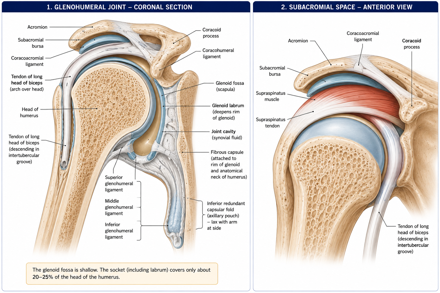
A large hemispherical humeral head (retroverted ~30°, inclined ~130–140°) against a small, shallow, pear-shaped glenoid cavity with ~5° retroversion and slight upward tilt. The surface area ratio is roughly 3:1 or 4:1; only about one-third of the head contacts the glenoid at any time.
The glenoid labrum — a fibrocartilaginous rim — deepens the socket by around 50%, roughly doubles its depth, increases the contact area, acts as the attachment for the capsule and the long head of biceps superiorly and the inferior glenohumeral ligament inferiorly, and creates a suction seal.
The capsule is lax and voluminous — roughly twice the volume of the humeral head — and is redundant inferiorly (the axillary recess), which is what permits the range. It attaches to the glenoid margin (beyond the labrum) and to the anatomical neck of the humerus, except medially where it extends ~1 cm down the shaft.
| Ligament | Course | Function |
|---|---|---|
| Superior GHJ ligament (SGHL) | Supraglenoid tubercle to lesser tubercle | Restrains inferior translation of the adducted arm; part of the rotator interval sling with the coracohumeral ligament |
| Middle GHJ ligament (MGHL) | Anterior labrum to lesser tubercle | Restrains anterior translation at ~45° abduction and in external rotation |
| Inferior GHJ ligament (IGHL) | A hammock-like complex — anterior band, axillary pouch, posterior band | The principal static restraint in the abducted, externally rotated (apprehension) position. The anterior band is what avulses in a Bankart lesion |
| Coracohumeral ligament | Coracoid to both tubercles, bridging the rotator interval | Restrains inferior translation and external rotation in adduction; contracts in adhesive capsulitis, which is why external rotation is lost first |
| Coracoacromial ligament | Coracoid to acromion | With the acromion and coracoid, forms the coracoacromial arch — the roof of the subacromial space, preventing superior dislocation of the head |
| Transverse humeral ligament | Across the intertubercular groove | Retains the long head of biceps tendon |
Bursae: the subacromial–subdeltoid bursa (the largest in the body, and continuous in most people), the subscapular bursa (communicating with the joint through the foramen of Weitbrecht), the subcoracoid bursa, and the infraspinatus bursa.
Static restraints (bony congruence, labrum, capsule, ligaments, negative intra-articular pressure) are important at end range but lax through the mid-range, where most function occurs. Mid-range stability comes from:
| Movement | Approximate range (glenohumeral + total) |
|---|---|
| Flexion | 180° total |
| Extension | 45–60° |
| Abduction | 180° total (~120° glenohumeral) |
| Adduction | 45° across the body |
| External rotation | 90° (arm abducted) |
| Internal rotation | 70–90° |
| Horizontal abduction/adduction | 45° / 135° |
Close-packed position: full abduction with external rotation. Resting position: ~55° abduction, 30° horizontal adduction. Capsular pattern: external rotation > abduction > internal rotation.
Figure 3 · The rotator cuff
Four muscles whose tendons blend with the capsule to form a continuous cuff over the head, deficient only anteroinferiorly (the rotator interval, and the site through which most dislocations occur).
| Muscle | Origin | Insertion | Nerve | Actions |
|---|---|---|---|---|
| Supraspinatus | Supraspinous fossa | Superior facet, greater tubercle | Suprascapular (C5, C6) | Initiates abduction (first ~15–30°); compresses and stabilises the head throughout; a major stabiliser rather than a major mover |
| Infraspinatus | Infraspinous fossa | Middle facet | Suprascapular (C5, C6) | External rotation (the principal external rotator, ~60% of ER torque); posterior stability |
| Teres minor | Upper lateral border of scapula | Inferior facet | Axillary (C5, C6) | External rotation; inferior stability. The only cuff muscle supplied by the axillary nerve — hence spared in suprascapular nerve lesions |
| Subscapularis | Subscapular fossa | Lesser tubercle | Upper and lower subscapular (C5–C7) | Internal rotation; the sole anterior cuff, and the principal anterior restraint |
The single most important cuff concept: the cuff’s primary role is not to move the arm but to hold the humeral head centred on the glenoid so that the large muscles can move it. Deltoid’s line of pull in early abduction is almost vertical, and would translate the head superiorly into the acromion; the inferior cuff generates an opposing inferior and compressive force so that the resultant is a rotation, not a translation. Cuff failure allows superior migration of the humeral head, visible radiographically as a reduced acromiohumeral interval (<7 mm), and eventually cuff tear arthropathy.
| Muscle | Origin | Insertion | Nerve | Action |
|---|---|---|---|---|
| Deltoid | Lateral clavicle, acromion, spine of scapula | Deltoid tuberosity | Axillary (C5, C6) | Anterior: flexion, internal rotation, horizontal adduction. Middle: abduction (the prime mover from ~15–90°). Posterior: extension, external rotation, horizontal abduction |
| Pectoralis major | Clavicular head; sternocostal head | Lateral lip of intertubercular groove (with a characteristic fibre twist) | Lateral and medial pectoral (C5–T1) | Adduction, internal rotation, flexion (clavicular head), extension from flexion (sternocostal head) |
| Latissimus dorsi | Thoracolumbar fascia, T7–sacrum, iliac crest, lower 3–4 ribs, inferior angle of scapula | Floor of intertubercular groove | Thoracodorsal (C6–C8) | Extension, adduction, internal rotation — “the handcuff muscle”; a powerful climbing and crutch-walking muscle |
| Teres major | Inferior angle / lower lateral border | Medial lip of intertubercular groove | Lower subscapular (C5–C6) | Adduction, internal rotation, extension. Not part of the cuff |
| Coracobrachialis | Coracoid | Mid-medial humerus | Musculocutaneous (C5–C7) — which pierces it | Flexion, adduction |
| Trapezius | Occiput, ligamentum nuchae, C7–T12 spines | Lateral clavicle, acromion, scapular spine | Spinal accessory (CN XI) + C3, C4 proprioceptive | Upper: elevation, upward rotation. Middle: retraction. Lower: depression, upward rotation |
| Serratus anterior | Outer surfaces of ribs 1–8/9 | Costal surface of the medial border | Long thoracic (C5, C6, C7) — “C5–6–7 keeps the wing from heaven” | Protraction and upward rotation; holds the medial border to the thorax |
| Levator scapulae | Transverse processes C1–C4 | Superior angle to root of spine | Dorsal scapular (C5) + C3, C4 | Elevation, downward rotation |
| Rhomboid major and minor | C7–T5 spines | Medial border | Dorsal scapular (C5) | Retraction, elevation, downward rotation |
| Pectoralis minor | Ribs 3–5 | Coracoid | Medial pectoral (C8, T1) | Protraction, depression, anterior tilt — tightness here is a common contributor to scapular dyskinesis |
| Subclavius | First rib | Subclavian groove | Nerve to subclavius (C5, C6) | Depresses and stabilises the clavicle; protects the neurovascular bundle |
Figure 4 · How the joint and the scapula share the work
Full arm elevation to 180° requires the coordinated contribution of all four joints.
| Contribution | Amount | Notes |
|---|---|---|
| Glenohumeral elevation | ~120° | |
| Scapular upward rotation | ~60° | |
| Overall ratio | ~2:1 (GH:ST) | An average across the whole range |
| Setting phase (0–30°) | Highly variable; scapula stabilises, ratio may be 4:1 or greater | Why the ratio should not be applied to small ranges |
| After ~30° | Approximately 2:1 and more consistent |
Accompanying obligatory movements:
Clinically: ask a patient to elevate with the arm held in internal rotation and watch it stop early. That is a demonstration, not a trick, and it explains why restoring external rotation is a prerequisite for restoring elevation.
Scapular dyskinesis is best described by pattern (inferior angle prominence, medial border prominence, excessive superior translation) and is best treated by addressing what causes it — pain, stiffness (especially pectoralis minor and posterior capsule), and weakness or mistiming of the serratus–trapezius force couple. It is common in asymptomatic athletes, so its presence alone is not a diagnosis.
Roof: the coracoacromial arch — acromion, coracoacromial ligament, coracoid process, and the undersurface of the acromioclavicular joint. Floor: the humeral head and greater tubercle. Contents: the supraspinatus tendon, the upper part of infraspinatus, the subacromial–subdeltoid bursa, the long head of biceps tendon, and the superior capsule.
The space measures ~7–14 mm with the arm at the side and narrows during elevation, particularly between 60° and 120° — the painful arc.
The Neer model — that a hooked acromion mechanically abrades the cuff, causing progressive bursitis, tendinitis and tearing, treatable by acromioplasty — dominated shoulder practice for thirty years. It has not survived scrutiny:
The current framing is subacromial (or rotator cuff related) shoulder pain: a clinical syndrome with contributions from tendon pathology, load intolerance, muscle dysfunction, scapular and postural factors, and central pain mechanisms. The primary treatment is progressive, graded exercise, with corticosteroid injection offering short-term relief and surgery reserved for the minority.
This is worth teaching explicitly because the older model is still widely taught, and it leads clinicians toward passive treatment and unnecessary surgery.
Figure 5 · The axilla and the brachial plexus
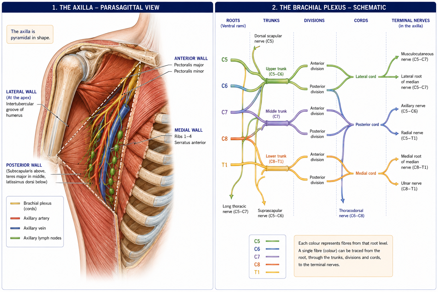
A pyramidal space between the upper limb and the thoracic wall — the neurovascular gateway.
| Boundary | Structures |
|---|---|
| Apex (cervico-axillary canal) | Clavicle, first rib, superior border of scapula |
| Base | Axillary fascia and skin |
| Anterior wall | Pectoralis major, pectoralis minor, subclavius, clavipectoral fascia |
| Posterior wall | Subscapularis, teres major, latissimus dorsi |
| Medial wall | Serratus anterior over ribs 1–4 |
| Lateral wall | Intertubercular groove of the humerus |
Contents: the axillary artery and vein, the cords and branches of the brachial plexus, axillary lymph nodes (five groups: pectoral/anterior, subscapular/posterior, humeral/lateral, central, apical), the long thoracic and thoracodorsal nerves on the walls, the intercostobrachial nerve (T2), the axillary tail of the breast, and fat.
Clinical. The axillary nodes drain the breast, upper limb and adjacent trunk — hence axillary clearance in breast cancer, and hence the physiotherapist’s role in post-surgical shoulder range and lymphoedema management. The long thoracic nerve is vulnerable during axillary dissection (producing winging) and the intercostobrachial nerve is routinely sacrificed, producing medial arm numbness that patients should be warned about.
Divided by pectoralis minor: the first part has one branch (superior thoracic), the second two (thoracoacromial, lateral thoracic), and the third three (subscapular — the largest, anterior and posterior circumflex humeral).
Roots C5–T1 → trunks (upper C5–6, middle C7, lower C8–T1) → divisions (anterior and posterior) → cords (lateral, posterior, medial, named for their relation to the axillary artery) → branches.
Mnemonic: Really Tired? Drink Coffee Before. The plexus is treated in detail in Chapter 24; for now know:
| Cord | Terminal branches |
|---|---|
| Lateral | Musculocutaneous; lateral root of median |
| Medial | Ulnar; medial root of median; medial cutaneous nerves of arm and forearm |
| Posterior | Axillary and radial (plus upper/lower subscapular and thoracodorsal) |
Erb’s palsy (C5–6, upper trunk — birth traction or a fall on the shoulder): loss of deltoid, supraspinatus, infraspinatus, biceps, brachialis, brachioradialis and supinator → the arm hangs adducted, internally rotated, elbow extended, forearm pronated — the “waiter’s tip” posture. Klumpke’s palsy (C8–T1, lower trunk — traction with the arm above the head): intrinsic hand muscle paralysis → claw hand; may include Horner’s syndrome if T1 sympathetic fibres are involved.
| Space | Boundaries | Contents |
|---|---|---|
| Quadrangular | Teres minor above, teres major below, long head of triceps medially, humerus laterally | Axillary nerve and posterior circumflex humeral artery |
| Triangular (upper) | Teres minor, teres major, long head of triceps | Circumflex scapular artery |
| Triangular interval (lower) | Teres major, long head of triceps, humerus | Radial nerve and profunda brachii |
~95–97% of shoulder dislocations. Mechanism: abduction, extension and external rotation — the arm forced back and out, as in a tackle or a fall on an outstretched abducted arm.
| Structure | Injury |
|---|---|
| Anteroinferior labrum and IGHL | Bankart lesion (labral avulsion); bony Bankart if the glenoid rim fractures |
| Posterolateral humeral head | Hill–Sachs lesion — an impaction fracture against the glenoid rim |
| Axillary nerve | Injured in 5–35% of cases — test deltoid function and lateral shoulder sensation before and after reduction, and document it |
| Rotator cuff | Tears common in patients over 40, in whom cuff rupture is the more likely lesion than labral avulsion |
Clinical picture:
Posterior dislocation is rare (~2–4%) but classically missed: it follows seizures, electric shock and posterior blows, and the arm is held in internal rotation with no external rotation possible. The AP radiograph can look almost normal — the “light bulb sign” — so an axillary or scapular-Y view is mandatory.
Inferior dislocation (luxatio erecta) presents with the arm fixed overhead and has a high rate of neurovascular injury.
A spectrum from reactive tendinopathy through partial-thickness to full-thickness tearing, with age the dominant risk factor. Supraspinatus is most commonly involved.
Assessment uses a battery rather than a single test, because individual special tests have poor diagnostic accuracy in isolation:
| Test | Targets |
|---|---|
| Empty can / full can (Jobe) | Supraspinatus |
| External rotation lag sign; resisted ER | Infraspinatus |
| Lift-off; belly-press; bear-hug | Subscapularis |
| Hornblower’s sign | Teres minor |
| Neer, Hawkins–Kennedy | Subacromial pain provocation (high sensitivity, poor specificity) |
| Drop arm sign | Large/massive tear |
| Painful arc 60–120° | Subacromial; >120° suggests AC joint |
Management is progressive loading — isometrics initially where irritable, then graded cuff and scapular strengthening with restoration of range and control — with surgery reserved for acute traumatic tears in younger patients, and for those who fail a genuine course of rehabilitation.
A fibrotic contracture of the capsule, particularly the rotator interval and coracohumeral ligament, with reduced joint volume.
| Nerve | Cause | Signs |
|---|---|---|
| Axillary (C5–6) | Surgical neck fracture, anterior dislocation, quadrangular space syndrome | Deltoid weakness; regimental badge sensory loss |
| Suprascapular (C5–6) | Suprascapular notch (both muscles) or spinoglenoid notch (infraspinatus only — as with a ganglion from a labral tear) | Weak abduction and external rotation; infraspinatus wasting visible |
| Long thoracic (C5–7) | Traction, backpack, axillary dissection, viral | Medial border winging, worse on wall push-up; difficulty elevating above 90° |
| Spinal accessory (CN XI) | Posterior triangle surgery (lymph node biopsy) — a classic iatrogenic injury | Trapezius wasting, drooping shoulder, lateral winging, inability to elevate fully |
| Musculocutaneous (C5–7) | Rare; surgery, dislocation | Weak elbow flexion and supination; lateral forearm sensory loss |
| Parsonage–Turner syndrome (neuralgic amyotrophy) | Idiopathic/post-viral brachial neuritis | Severe acute shoulder pain for days to weeks, followed by patchy weakness and wasting (often suprascapular and long thoracic). Often misdiagnosed as cuff pathology — the sequence of severe pain then weakness is the clue |
15 questions on this chapter. Tap one to see the answer and the reasoning.
Answer: (B)
Answer: (B) It resists vertical displacement; the AC ligament resists horizontal translation.
Answer: (C) Its anterior band avulses as a Bankart lesion.
Answer: (B) The only cuff muscle not supplied by the suprascapular or subscapular nerves.
Answer: (B) 120° GH to 60° ST, though the setting phase is variable.
Answer: (B) To clear the greater tubercle from the coracoacromial arch.
Answer: (C) Spinal accessory injury produces lateral winging with a drooping shoulder.
Answer: (B)
Answer: (B) The Bankart lesion is the labral avulsion.
Answer: (B) Always test and document before reduction.
Answer: (B) The AP film may look normal — obtain an axillary view.
Answer: (B)
Answer: (C) Reflecting coracohumeral ligament and rotator interval contracture.
Answer: (C) Which reframes subacromial pain as a syndrome managed primarily with exercise.
Answer: (C) The pain-then-weakness sequence is characteristic.
Everything on this page, in one screen
| Source | Why it earns its place |
|---|---|
| Standring S (ed.) — Gray’s Anatomy, 42nd edn | Definitive regional anatomy |
| Neumann DA — Kinesiology of the Musculoskeletal System | The best account of scapulohumeral rhythm, force couples and shoulder mechanics |
| Palastanga N, Field D, Soames R — Anatomy and Human Movement | Functional anatomy of the shoulder complex |
| Magee DJ — Orthopedic Physical Assessment | The special tests and their reported accuracy |
| Beard DJ et al. — “Arthroscopic subacromial decompression for subacromial shoulder pain (CSAW)”, Lancet, 2018 | The placebo-controlled trial that changed practice |
| Paavola M et al. — “Subacromial decompression versus diagnostic arthroscopy (FIMPACT)”, BMJ, 2018 | The confirmatory Finnish trial |
| Lewis J — “Rotator cuff related shoulder pain: assessment, management and uncertainties”, Man Ther, 2016 | The contemporary clinical framework |
| Kibler WB et al. — “Scapular summit” consensus statements, Br J Sports Med | Scapular dyskinesis: definition, assessment and its limits |
| Neviaser AS, Neviaser RJ — “Adhesive capsulitis of the shoulder”, J Am Acad Orthop Surg, 2011 | Phases, associations and management |
| Chaurasia BD — Human Anatomy, Vol 1 | Indian syllabus-matched descriptive account |
Chapter 10 of 24 · Human Anatomy · Physiotherapist India Next: Chapter 11 — Arm, Elbow and Forearm: compartments, the cubital fossa, pronation and supination, and the two epicondyles.
Reviewed by the Physiotherapist India Team. · Human Anatomy contents