Evidence-based physiotherapy and rehabilitation resources
Editorial & review policy
Evidence-based physiotherapy and rehabilitation resources
Editorial & review policyHuman Anatomy · The nervous system
Every other chapter in this book describes something the nervous system moves, feels or controls. This one is the control system itself. Learn how it is organised before you learn any of its parts, because almost every neurological sign a physiotherapist meets is a question about <em>where</em> the problem is rather than what it is.
Part 5 · Head, neck and the nervous system
Brain, spinal cord, tracts, cranial nerves, and the art of localisation
Neurological physiotherapy rests on one skill: taking a pattern of deficits and deducing where the lesion is. Weakness plus loss of pain and temperature on one side, with loss of proprioception on the other, is a hemisection of the cord. Weakness of the face, arm and leg on one side with the forehead spared is a lesion above the facial nucleus. Ataxia with intention tremor and dysdiadochokinesia is cerebellar, and the side of the signs tells you the side of the lesion.
None of that requires a scanner. It requires knowing where the tracts run, where they cross, and what each region does. That is what this chapter is for.
Figure 1 · How the nervous system is divided
| Division | Components |
|---|---|
| Central nervous system | Brain and spinal cord |
| Peripheral nervous system | 12 cranial nerves, 31 pairs of spinal nerves, ganglia, and peripheral receptors |
| Somatic | Voluntary motor and conscious sensory |
| Autonomic | Sympathetic (thoracolumbar, T1–L2), parasympathetic (craniosacral, CN III, VII, IX, X and S2–S4), enteric |
| Cell | Location | Function |
|---|---|---|
| Neuron | Both | Cell body (soma), dendrites, axon; myelinated axons conduct by saltatory conduction between nodes of Ranvier |
| Astrocyte | CNS | Metabolic support, potassium buffering, the blood–brain barrier, glial scar formation (which impedes CNS regeneration) |
| Oligodendrocyte | CNS | Myelinates up to 50 axons each; does not support regeneration |
| Microglia | CNS | Resident macrophages; central to neuroinflammation and to central sensitisation |
| Ependymal cells | CNS | Line ventricles; choroid plexus produces CSF |
| Schwann cell | PNS | Myelinates ONE internode of ONE axon; forms the bands of Büngner that guide regenerating axons |
| Satellite cells | PNS ganglia | Support |
Why peripheral nerves regenerate and central axons do not: Schwann cells and the endoneurial tube provide a permissive, guiding environment with growth factors, allowing regrowth at roughly 1 mm per day. In the CNS, oligodendrocytes and the astrocytic glial scar produce inhibitory molecules (Nogo-A, MAG, chondroitin sulphate proteoglycans), and there is no equivalent guidance tube. This is the fundamental reason a peripheral nerve injury recovers and a spinal cord injury does not — and the reason CNS rehabilitation depends on plasticity and reorganisation rather than regeneration.
Fibre types (Erlanger–Gasser), because they determine what you can test and what modalities affect:
| Type | Diameter / myelin | Conducts | Function |
|---|---|---|---|
| Aα (Ia, Ib) | Large, heavily myelinated | Fastest (~70–120 m/s) | Muscle spindle primary, Golgi tendon organ; alpha motor |
| Aβ (II) | Large myelinated | Fast | Touch, vibration, pressure, spindle secondary — the fibres stimulated by TENS and by touch, and the basis of gate control |
| Aγ | Medium | — | Gamma motor to intrafusal fibres |
| Aδ (III) | Small, thinly myelinated | ~5–30 m/s | Fast, sharp, well-localised pain; cold |
| C (IV) | Unmyelinated | Slowest (~0.5–2 m/s) | Slow, dull, burning pain; warmth; itch; postganglionic sympathetic |
Large fibres are the first affected by compression and the last affected by ischaemia; small fibres the reverse — which is why compressive neuropathy loses vibration and light touch first while ischaemia and diabetic small-fibre neuropathy lose pain and temperature first.
Figure 2 · The brain
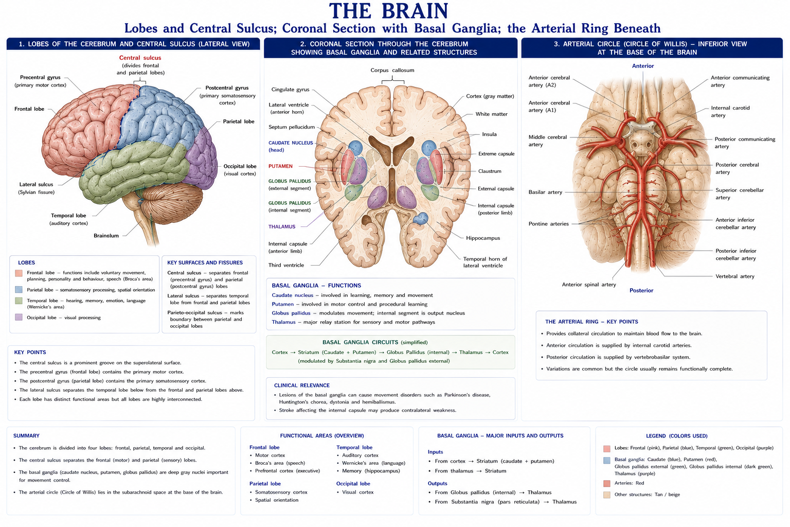
| Lobe | Key areas | Function | Lesion effects |
|---|---|---|---|
| Frontal | Precentral gyrus (primary motor, area 4); premotor and supplementary motor (area 6); Broca’s area (44, 45), dominant hemisphere; frontal eye field (8); prefrontal cortex | Voluntary movement, motor planning, expressive language, executive function, personality, behavioural inhibition | Contralateral spastic weakness; Broca’s (expressive) aphasia; apraxia; disinhibition, perseveration, poor planning; primitive reflexes |
| Parietal | Postcentral gyrus (primary somatosensory, areas 3, 1, 2); somatosensory association | Sensation; spatial awareness; body schema | Contralateral sensory loss, astereognosis, agraphaesthesia, two-point discrimination loss; non-dominant lesion → hemispatial neglect and anosognosia; dominant → Gerstmann syndrome |
| Temporal | Primary auditory (41, 42); Wernicke’s area (22), dominant; hippocampus and amygdala | Hearing, receptive language, memory, emotion | Wernicke’s (receptive) aphasia — fluent but meaningless speech with impaired comprehension; memory deficits; superior quadrantanopia (“pie in the sky”) |
| Occipital | Primary visual (17) and association (18, 19) | Vision | Contralateral homonymous hemianopia with macular sparing; visual agnosia |
| Insula | Deep to the lateral sulcus | Interoception, autonomic, pain affect, taste | Contributes to the affective dimension of pain |
| Limbic system | Cingulate gyrus, hippocampus, amygdala, fornix | Emotion, memory, motivation | Central to the affective component of chronic pain |
The homunculus. Both the motor and sensory cortices are somatotopically mapped, with representation proportional to precision of control or sensory density, not to body size. The hand, face and tongue occupy disproportionately large areas; the trunk very little. Two clinical consequences:
Arcuate fasciculus connects Wernicke’s to Broca’s area; its lesion produces conduction aphasia — fluent speech with good comprehension but impaired repetition.
Corpus callosum — the largest commissure, connecting the hemispheres.
Internal capsule — the compact white matter funnel between the basal ganglia and thalamus, carrying the corticospinal and corticobulbar fibres in the posterior limb and genu. Because everything is packed together, a small lacunar infarct here produces a dense complete contralateral hemiplegia — the classic “pure motor stroke”, disproportionate to lesion size.
Caudate nucleus + putamen (= striatum), globus pallidus (= lentiform with putamen), subthalamic nucleus, substantia nigra.
Function: they do not initiate movement; they modulate it — selecting desired movement and suppressing unwanted movement, through the direct (facilitatory) and indirect (inhibitory) pathways, both modulated by dopamine from the substantia nigra pars compacta.
| Disorder | Lesion | Features |
|---|---|---|
| Parkinson’s disease | Loss of dopaminergic neurons in the substantia nigra pars compacta | Bradykinesia (obligatory for diagnosis), resting “pill-rolling” tremor (4–6 Hz), rigidity (lead-pipe or cogwheel), postural instability; hypokinetic gait with festination and freezing; hypomimia, micrographia, hypophonia |
| Huntington’s disease | Striatal (caudate) degeneration; autosomal dominant CAG repeat | Chorea, cognitive decline, psychiatric change |
| Hemiballismus | Contralateral subthalamic nucleus | Violent, flinging proximal limb movements |
| Dystonia, athetosis | Various | Sustained or writhing involuntary posturing |
Basal ganglia lesions produce hypokinetic or hyperkinetic movement disorders — NOT weakness, and NOT the classical UMN signs. Rigidity is not spasticity: rigidity is velocity-independent and present in both directions; spasticity is velocity-dependent with a clasp-knife quality. Confusing the two is one of the commonest errors in neurological assessment.
Rehabilitation implication: the basal ganglia deficit is in internally generated, automatic, sequential movement. External cues bypass the deficit by routing movement through cortical–cerebellar pathways — which is precisely why auditory cueing (metronome), visual cues (floor lines), attentional strategies and large-amplitude training (LSVT BIG) work in Parkinson’s disease. That is anatomy generating a treatment.
The great sensory relay — all ascending sensory pathways except olfaction synapse here before reaching the cortex. Key nuclei: VPL (body sensation), VPM (face), LGB (vision), MGB (hearing), VA/VL (motor relay from basal ganglia and cerebellum).
Thalamic lesion → contralateral sensory loss and, in a proportion, Dejerine–Roussy syndrome: severe, intractable central post-stroke pain with allodynia and hyperpathia in the anaesthetic territory.
Hypothalamus — autonomic control, temperature regulation, endocrine control via the pituitary, hunger, thirst, circadian rhythm.
Three functional divisions:
| Division | Input | Function | Lesion |
|---|---|---|---|
| Vestibulocerebellum (flocculonodular lobe) | Vestibular | Balance, eye movement | Truncal ataxia, nystagmus, wide-based gait |
| Spinocerebellum (vermis and paravermis) | Spinal proprioceptive | Posture and gait | Gait and truncal ataxia — classically alcohol-related anterior vermis degeneration |
| Cerebrocerebellum (lateral hemispheres) | Cortex via pontine nuclei | Planning, coordination and timing of skilled limb movement; motor learning | Limb ataxia, intention tremor, dysmetria, dysdiadochokinesia |
The cardinal rule: cerebellar signs are IPSILATERAL — because the cerebellar output decussates in the superior cerebellar peduncle and then the corticospinal tract decussates again, a double crossing that returns the influence to the same side.
Signs (DANISH): Dysdiadochokinesia, Ataxia, Nystagmus, Intention tremor, Scanning (staccato) speech, Hypotonia. Plus dysmetria (past-pointing), rebound phenomenon, and impaired heel-shin testing.
Peduncles: superior (mainly output, to the red nucleus and thalamus), middle (input from the pontine nuclei), inferior (input from the spinal cord and vestibular nuclei).
| Part | Contains | Cranial nerve nuclei |
|---|---|---|
| Midbrain | Cerebral peduncles, substantia nigra, red nucleus, superior and inferior colliculi | III, IV |
| Pons | Pontine nuclei, middle cerebellar peduncle | V, VI, VII, VIII |
| Medulla | Pyramids (and the pyramidal decussation), olives, respiratory and cardiovascular centres | IX, X, XI, XII |
Mnemonic: “2 above 2, 4 in the pons, 4 in the medulla” — CN I and II are not brainstem; III and IV in the midbrain; V–VIII pontine; IX–XII medullary.
Reticular formation — running through the whole brainstem; the ascending reticular activating system governs consciousness and arousal; descending components modulate muscle tone, pain (descending inhibition) and autonomic function.
Figure 3 · Grey and white swap places
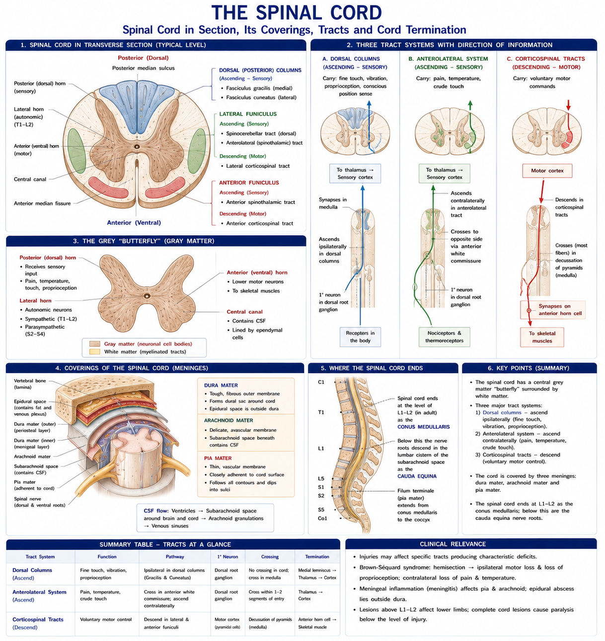
Grey matter centrally (H-shaped: dorsal, lateral and ventral horns), white matter peripherally in dorsal, lateral and ventral columns. Rexed laminae I–X organise the grey matter; laminae I, II (substantia gelatinosa) and V are the key nociceptive laminae and the site of gate control.
The lateral horn (T1–L2) contains preganglionic sympathetic neurons; S2–S4 contains the parasympathetic equivalent.
| Tract | Modality | First-order | Decussation | Third-order | Clinical |
|---|---|---|---|---|---|
| Dorsal column–medial lemniscus (gracile and cuneate) | Fine touch, vibration, proprioception, two-point discrimination | DRG → ascends ipsilaterally in the dorsal columns to the gracile/cuneate nuclei | In the MEDULLA (internal arcuate fibres) | Thalamus (VPL) → cortex | Ipsilateral loss below a cord lesion; positive Romberg; sensory ataxia. Damaged in tabes dorsalis and B12 deficiency |
| Lateral spinothalamic | Pain and temperature | DRG → synapses in the dorsal horn | IMMEDIATELY, in the cord, via the anterior white commissure — ascending 1–2 segments before crossing | Thalamus → cortex | Contralateral loss beginning 1–2 segments below the lesion. Damaged in syringomyelia (cape-like loss) |
| Anterior spinothalamic | Crude touch, pressure | As above | In the cord | Thalamus | Considerable overlap, so rarely lost in isolation |
| Spinocerebellar (posterior and anterior) | Unconscious proprioception | — | Posterior uncrossed; anterior crosses twice | Cerebellum | Ataxia |
The decussation difference is the single most useful fact in spinal cord localisation. Dorsal columns cross high, in the medulla; spinothalamic crosses low, at the level of entry. Therefore in a cord hemisection you get ipsilateral proprioceptive loss and contralateral pain and temperature loss — the dissociated sensory loss of Brown-Séquard syndrome.
| Tract | Origin | Decussation | Function |
|---|---|---|---|
| Lateral corticospinal | Motor cortex (~30% area 4, ~30% area 6, ~40% parietal) | ~85–90% at the pyramidal decussation in the medulla | Skilled voluntary movement, especially of the distal limbs |
| Anterior corticospinal | Motor cortex | The remaining ~10–15%, crossing at segmental level | Axial and proximal control |
| Corticobulbar | Motor cortex | To cranial nerve nuclei; mostly bilateral — hence the forehead rule (Chapter 22) and why unilateral lesions spare most bulbar function | Head and neck motor |
| Rubrospinal | Red nucleus | Midbrain | Flexor tone in the upper limb; minor in humans |
| Reticulospinal (pontine and medullary) | Reticular formation | Largely uncrossed | Posture, gait, gross axial and proximal movement; a major contributor to spasticity when descending inhibition is lost |
| Vestibulospinal | Vestibular nuclei | Uncrossed | Extensor tone, balance, antigravity posture |
| Tectospinal | Superior colliculus | Midbrain | Reflex head turning to visual and auditory stimuli |
The reticulospinal and vestibulospinal tracts matter more in rehabilitation than their textbook billing suggests: much of the recovery of gross proximal and trunk function after corticospinal damage is mediated by these surviving pathways, and much of the tone abnormality after stroke arises from their release from cortical inhibition.
| Feature | Upper motor neuron | Lower motor neuron |
|---|---|---|
| Tone | Increased — spasticity (velocity-dependent, clasp-knife) | Decreased — flaccidity |
| Reflexes | Exaggerated, clonus | Reduced or absent |
| Plantar response | Extensor (Babinski positive) | Flexor or absent |
| Wasting | Late, from disuse | Early and marked |
| Fasciculation | Absent | Present |
| Weakness pattern | Pyramidal distribution — upper limb extensors and lower limb flexors weaker | Segmental / peripheral nerve distribution |
| Examples | Stroke, SCI, MS, cerebral palsy | Poliomyelitis, peripheral nerve injury, radiculopathy, Guillain–Barré |
Motor neurone disease (ALS) characteristically produces mixed upper and lower motor neuron signs — brisk reflexes in a wasted, fasciculating limb — which is diagnostically distinctive.
A crucial caveat for acute practice: in the first hours to weeks after an acute UMN lesion, there is a period of SPINAL SHOCK or CEREBRAL SHOCK — flaccidity and areflexia, which resemble a lower motor neuron picture. Spasticity develops later. Judging a lesion level from tone in the first days is unreliable.
| Syndrome | Lesion | Presentation | Prognosis |
|---|---|---|---|
| Complete transection | Whole cord | Total loss of motor and all sensation below the level; bladder, bowel and sexual dysfunction | Poor for motor recovery |
| Brown-Séquard (hemisection) | Half the cord | Ipsilateral: motor loss, proprioception and vibration loss (dorsal columns cross high). Contralateral: pain and temperature loss, from 1–2 segments below | The best prognosis of the incomplete syndromes |
| Central cord | Central cord, typically hyperextension in an older spondylotic neck | Upper limbs affected more than lower limbs (medial corticospinal fibres serve the arm), with sacral sparing and variable bladder involvement | The commonest incomplete SCI; often good ambulatory recovery, poorer hand function |
| Anterior cord | Anterior spinal artery territory | Complete motor loss with loss of pain and temperature; proprioception and vibration PRESERVED | Poorest prognosis of the incomplete syndromes |
| Posterior cord | Dorsal columns | Loss of proprioception and vibration with preserved motor and pain/temperature; sensory ataxia | Rare |
| Conus medullaris | L1–L2 cord segment | Early, symmetrical bladder and bowel dysfunction, saddle anaesthesia, mixed UMN and LMN signs; less radicular pain | — |
| Cauda equina | Lumbosacral roots | Asymmetrical, LMN, severe radicular pain, saddle anaesthesia, later bladder involvement. A surgical emergency (Chapter 17) | Depends on decompression timing |
The International Standards for Neurological Classification of Spinal Cord Injury:
Autonomic dysreflexia must be flagged here because it is a physiotherapy emergency: in lesions at or above T6, a noxious stimulus below the level (typically a blocked catheter, bowel distension, pressure injury, or an ingrowing toenail) triggers unopposed sympathetic outflow → severe hypertension, pounding headache, flushing and sweating above the level, pallor below, and reflex bradycardia. Sit the patient up, loosen constrictive clothing, and find and remove the stimulus. Untreated, it causes stroke, seizure and death.
| Nerve | Name | Type | Function | Test | Lesion |
|---|---|---|---|---|---|
| I | Olfactory | S | Smell | Identify odours | Anosmia (head injury shearing at the cribriform plate) |
| II | Optic | S | Vision | Acuity, fields, pupillary light reflex (afferent) | Field defects localise along the pathway |
| III | Oculomotor | M | All extraocular muscles except SO and LR; levator palpebrae; parasympathetic to sphincter pupillae and ciliary | Eye movements, pupil | Ptosis, “down and out” eye, dilated unreactive pupil. A surgical third (pupil-involving) suggests compression — uncal herniation or aneurysm |
| IV | Trochlear | M | Superior oblique | Look down and in | Vertical diplopia worse on descending stairs; head tilt away. The only nerve to exit dorsally and the longest intracranial course |
| V | Trigeminal | B | Facial sensation (V₁ ophthalmic, V₂ maxillary, V₃ mandibular); muscles of mastication (V₃); corneal reflex afferent | Light touch in three divisions; clench and open the jaw | Trigeminal neuralgia — brief, severe, electric-shock pain, usually V₂/V₃ |
| VI | Abducens | M | Lateral rectus | Lateral gaze | Failure of abduction, horizontal diplopia; a false localising sign in raised ICP because of its long course |
| VII | Facial | B | Muscles of facial expression; taste anterior ⅔ tongue; lacrimal and salivary secretomotor; stapedius | Raise eyebrows, close eyes tightly, smile, puff cheeks | The forehead rule (Chapter 22) |
| VIII | Vestibulocochlear | S | Hearing and balance | Whisper test, Rinne, Weber; Dix–Hallpike, head impulse | Sensorineural loss, vertigo, nystagmus |
| IX | Glossopharyngeal | B | Taste posterior ⅓; sensation from pharynx, middle ear, carotid sinus/body; stylopharyngeus | Gag reflex (afferent) | Rarely isolated |
| X | Vagus | B | Palate, pharynx and larynx motor; parasympathetic to thorax and abdomen; sensory | Say “ah” — uvula deviates AWAY from the lesion; hoarseness; gag efferent | Dysphagia, dysphonia, aspiration risk |
| XI | Accessory | M | Sternocleidomastoid and trapezius | Shrug against resistance; turn head against resistance | Drooping shoulder, lateral winging |
| XII | Hypoglossal | M | Tongue muscles | Protrude the tongue — it deviates TOWARDS the lesion (“the lick wound”) | Wasting and fasciculation in LMN lesions |
Mnemonic for names: Oh Oh Oh To Touch And Feel Very Good Velvet, Ah Heaven. For type (S = sensory, M = motor, B = both): Some Say Marry Money But My Brother Says Big Brains Matter More.
Two deviation rules that get confused constantly:
Bulbar versus pseudobulbar palsy — a distinction with direct swallowing implications:
| Bulbar (LMN) | Pseudobulbar (UMN, bilateral) | |
|---|---|---|
| Tongue | Wasted, fasciculating | Spastic, small, immobile |
| Speech | Nasal, flaccid dysarthria | Slow, strained, “hot potato” |
| Jaw jerk | Absent or normal | Exaggerated |
| Gag | Absent | Exaggerated |
| Emotion | Normal | Emotional lability (pathological crying/laughing) |
Figure 4 · The spinal cord in section, and its coverings
| Layer | Features |
|---|---|
| Dura mater | Tough; two layers in the cranium (periosteal and meningeal), separating to form the dural venous sinuses. Reflections: falx cerebri, tentorium cerebelli, falx cerebelli, diaphragma sellae. Innervated by the trigeminal nerve — hence its role in headache |
| Arachnoid mater | Avascular; arachnoid granulations return CSF to the superior sagittal sinus |
| Pia mater | Adherent to the brain surface; forms the denticulate ligaments and filum terminale in the cord |
Space
Contents
Haemorrhage
Extradural
Potential
Middle meningeal artery at the pterion; lucid interval, biconvex (lens) shape on CT
Subdural
Potential
Bridging veins; crescent-shaped; chronic in the elderly and alcoholic after minor trauma — often presenting weeks later with confusion or a fluctuating hemiparesis, and easily missed
Subarachnoid
CSF, major vessels
Berry aneurysm rupture — “thunderclap” worst-ever headache, neck stiffness, photophobia
Two lateral ventricles → interventricular foramina (of Monro) → third ventricle → cerebral aqueduct (of Sylvius) → fourth ventricle → foramina of Luschka (lateral) and Magendie (median) → subarachnoid space → arachnoid granulations → superior sagittal sinus.
CSF: ~150 mL total, produced at ~500 mL/day by the choroid plexus — so it turns over three to four times daily. Clear, with glucose ~⅔ of plasma, protein 15–45 mg/dL, and fewer than 5 cells/mm³. Functions: buoyancy (reducing the brain’s effective weight from ~1400 g to ~50 g), cushioning, and metabolic exchange.
Hydrocephalus: communicating (impaired absorption — post-haemorrhagic, post-meningitic) or non-communicating/obstructive (aqueduct stenosis, tumour). Normal pressure hydrocephalus presents with the triad of gait apraxia (“magnetic” gait), urinary incontinence and dementia — “wet, wacky and wobbly” — and is important because the gait disturbance is often the presenting complaint to a physiotherapist and it is treatable.
Formed by:
| Artery | Territory | Syndrome |
|---|---|---|
| Anterior cerebral (ACA) | Medial surface — the leg area of the homunculus | Contralateral weakness and sensory loss worse in the LEG than the arm; urinary incontinence; abulia and personality change |
| Middle cerebral (MCA) | Lateral surface — face and arm areas, Broca’s and Wernicke’s areas | Contralateral weakness and sensory loss worse in the FACE and ARM; aphasia if dominant; neglect if non-dominant; homonymous hemianopia; gaze preference towards the lesion. The commonest stroke |
| Posterior cerebral (PCA) | Occipital and inferomedial temporal lobes | Contralateral homonymous hemianopia with macular sparing; visual agnosia; memory impairment |
| Vertebrobasilar | Brainstem and cerebellum | Crossed signs: ipsilateral cranial nerve palsy with contralateral limb signs. Vertigo, diplopia, dysarthria, dysphagia, ataxia. Locked-in syndrome from basilar occlusion (ventral pons) |
| PICA — lateral medullary (Wallenberg) syndrome | Lateral medulla | Ipsilateral: facial pain/temperature loss (spinal V), Horner’s, ataxia, dysphagia and hoarseness (nucleus ambiguus). Contralateral: body pain and temperature loss. Motor power preserved — a beautifully instructive lesion |
| Lacunar (perforating vessels) | Internal capsule, basal ganglia, pons | Pure motor, pure sensory, ataxic hemiparesis, or dysarthria–clumsy hand — without cortical signs (no aphasia, no neglect) |
Stroke recognition: FAST (Face, Arms, Speech, Time) for public use; BE-FAST adds Balance and Eyes, capturing posterior circulation strokes that FAST misses.
The physiotherapy imperative: thrombolysis and thrombectomy are time-critical, and very early mobilisation must be calibrated rather than maximal — the AVERT trial found that very early, high-dose out-of-bed activity within 24 hours produced worse outcomes than usual care, while shorter, more frequent sessions were favourable. The message is early and frequent, not early and aggressive.
Recovery after CNS injury depends on reorganisation, not regeneration. The mechanisms:
| Mechanism | Detail |
|---|---|
| Resolution of diaschisis | Recovery of function in structurally intact but functionally depressed regions remote from the lesion — much of the fastest early recovery |
| Unmasking of latent pathways | Existing but normally suppressed connections become functional |
| Synaptic plasticity (LTP/LTD) | Strengthening or weakening of synapses with use — “cells that fire together wire together” |
| Cortical map reorganisation | Somatotopic maps expand or contract with use, disuse or training |
| Axonal sprouting and dendritic arborisation | Local structural remodelling |
| Ipsilateral and contralesional recruitment | Variable, and not always beneficial — excessive contralesional activity can be maladaptive |
Kleim and Jones’s ten principles of experience-dependent plasticity are the most directly usable framework in neurological rehabilitation, and they translate this anatomy into prescription:
Constraint-induced movement therapy is the clearest clinical application — it directly targets learned non-use, and produces measurable cortical map reorganisation alongside functional gain.
The uncomfortable honest point: typical clinical practice delivers a small fraction of the repetitions used in the animal and trial protocols that established these effects. Studies of stroke rehabilitation sessions routinely count tens of upper limb repetitions where the evidence base suggests several hundred. Knowing the anatomy of plasticity means knowing that dose is the variable most often underdelivered.
15 questions on this chapter. Tap one to see the answer and the reasoning.
Answer: (B) Spinothalamic fibres cross at cord level.
Answer: (B) Which is why contralateral sensory loss begins 1–2 segments below a lesion.
Answer: (B)
Answer: (C) Typically hyperextension in an older spondylotic neck, with arms worse than legs.
Answer: (B) Because of the double decussation.
Answer: (B)
Answer: (C) Subthalamic lesion causes hemiballismus.
Answer: (B)
Answer: (B) The uvula, by contrast, deviates away.
Answer: (C) The leg area lies on the medial surface.
Answer: (B) It produces crossed sensory loss with ipsilateral Horner’s, ataxia and dysphagia.
Answer: (B) Bridging veins cause subdural haematoma.
Answer: (B) It is treatable, and the gait often presents to physiotherapy first.
Answer: (B) Sit the patient up and find the noxious stimulus.
Answer: (C)
Everything on this page, in one screen
| Source | Why it earns its place |
|---|---|
| Snell RS — Clinical Neuroanatomy | The best single neuroanatomy text for clinical students |
| Blumenfeld H — Neuroanatomy through Clinical Cases | Localisation taught the way clinicians actually reason |
| Standring S (ed.) — Gray’s Anatomy, 42nd edn | Reference detail |
| Vishram Singh — Textbook of Clinical Neuroanatomy | Indian syllabus-matched, widely used |
| Lundy-Ekman L — Neuroscience: Fundamentals for Rehabilitation | The physiotherapy-facing synthesis — the best fit for this chapter |
| Shumway-Cook A, Woollacott MH — Motor Control: Translating Research into Clinical Practice | Motor control and its clinical application |
| Kleim JA, Jones TA — “Principles of experience-dependent neural plasticity”, J Speech Lang Hear Res, 2008 | The ten principles |
| AVERT Trial Collaboration — “Efficacy and safety of very early mobilisation within 24 h of stroke onset”, Lancet, 2015 | The mobilisation dose evidence |
| ASIA — International Standards for Neurological Classification of Spinal Cord Injury | The classification you will use |
| Carr JH, Shepherd RB — Neurological Rehabilitation: Optimizing Motor Performance | Task-oriented practice grounded in this anatomy |
Chapter 23 of 24 · Human Anatomy · Physiotherapist India Next: Chapter 24 — Spinal and Peripheral Nerves: the plexuses, dermatomes and myotomes, and nerve injury.
Reviewed by the Physiotherapist India Team. · Human Anatomy contents