Evidence-based physiotherapy and rehabilitation resources
Editorial & review policy
Evidence-based physiotherapy and rehabilitation resources
Editorial & review policyHuman Anatomy · The nervous system
This is the chapter that lets you turn a patient's description into a location. Numbness in a strip down the arm, weakness of one movement, a reflex that has gone quiet — each of those is a statement about a particular root or a particular nerve, and the anatomy here is what lets you read it.
Part 5 · Head, neck and the nervous system
The plexuses, dermatomes and myotomes, and nerve injury and repair
Everything in this book converges here. The bone chapters told you where nerves are tethered and compressed. The muscle chapters told you what each nerve does. The regional chapters gave you the courses. This chapter gives you the system: how the 31 pairs of spinal nerves are organised into plexuses, how to map a lesion from the pattern of loss, and what happens to a nerve when it is injured.
The clinical question this chapter answers is the one you will ask most often in practice:
Is this a nerve root, a plexus, or a peripheral nerve — and how do I tell?
The connective tissue architecture mirrors that of muscle (Chapter 5), and it determines everything about injury and repair.
| Layer | Invests | Function |
|---|---|---|
| Endoneurium | Each individual axon with its Schwann cell sheath | Forms the endoneurial tube — the scaffold that guides regenerating axons. Its survival is the single most important prognostic factor in nerve injury |
| Perineurium | Each fascicle | Dense, mechanically strong, and the site of the blood–nerve barrier. It maintains endoneurial fluid pressure |
| Epineurium | The whole nerve trunk; interfascicular epineurium separates fascicles | Cushions, permits gliding, carries the vasa nervorum |
| Mesoneurium | Loose areolar tissue outside the epineurium | Allows the nerve to glide relative to surrounding tissue — the anatomical basis of neurodynamics |
Nerves are designed to move. A peripheral nerve must accommodate up to 10–20% strain and several millimetres to centimetres of excursion during limb movement (the median nerve moves ~7 mm at the elbow, the sciatic nerve ~10 mm at the hip). It does this by unfolding its undulating course, then unfolding the fascicles’ own undulation, and finally by gliding. Only after all three are exhausted does the nerve stretch.
Nerves are also metabolically demanding and vulnerable to ischaemia. Intraneural blood flow is impaired at as little as 20–30 mmHg of external pressure, and at 50 mmHg conduction begins to fail. This — not “pinching” — is the mechanism of most entrapment neuropathies, and it explains why symptoms are often worse at night (when limb positions are sustained and blood pressure falls) and why they resolve so quickly on position change.
Figure 1 · A typical spinal nerve
31 pairs: 8 cervical, 12 thoracic, 5 lumbar, 5 sacral, 1 coccygeal.
Formed in the intervertebral foramen by the union of:
It divides almost immediately into:
| Branch | Destination |
|---|---|
| Posterior (dorsal) ramus | The true back muscles and the skin of the back (Chapter 17) |
| Anterior (ventral) ramus | Everything else — limbs and the anterolateral trunk; forms the plexuses |
| Meningeal (sinuvertebral) branch | Re-enters the canal for the PLL, outer anulus, dura and periosteum |
| Rami communicantes | White (myelinated, preganglionic, only T1–L2) to the sympathetic chain; grey (unmyelinated, postganglionic, at every level) back to the spinal nerve |
Exit rule (Chapter 17, repeated because it is examined every year): C1–C7 exit ABOVE their corresponding vertebra; C8 exits between C7 and T1; from T1 down, nerves exit BELOW their corresponding vertebra.
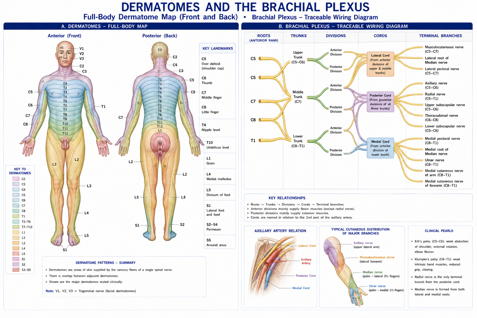
Figure 2 · Dermatomes, and the brachial plexus
Lies deep to sternocleidomastoid, on scalenus medius and levator scapulae.
| Type | Branches |
|---|---|
| Cutaneous (emerging at Erb’s point, mid-posterior border of SCM) | Lesser occipital (C2), great auricular (C2, C3), transverse cervical (C2, C3), supraclavicular (C3, C4) |
| Muscular | Ansa cervicalis (C1–C3) → infrahyoid muscles; branches to prevertebral muscles; C1 fibres travel with the hypoglossal nerve |
| Phrenic (C3, C4, C5) | The diaphragm — “C3, 4, 5 keeps the diaphragm alive” |
The supraclavicular nerves (C3–C4) supply skin down to the second rib — which is why shoulder-tip pain refers from the diaphragm (Chapters 8 and 18), and why a patient may point to the clavicle when the problem is subphrenic.
Roots → Trunks → Divisions → Cords → Branches (Really Tired? Drink Coffee Before).
| Level | Components |
|---|---|
| Roots | Anterior rami C5–T1, emerging between anterior and middle scalene |
| Trunks | Upper (C5, C6), Middle (C7), Lower (C8, T1) — in the posterior triangle |
| Divisions | Anterior and posterior of each trunk — behind the clavicle |
| Cords | Lateral, Posterior, Medial — named for their relation to the axillary artery, in the axilla |
| Branches | Terminal |
Branches worth knowing by level, because the level of a branch localises the lesion:
| Origin | Nerve | Roots |
|---|---|---|
| Roots | Dorsal scapular (rhomboids, levator scapulae) | C5 |
| Long thoracic (serratus anterior) | C5, C6, C7 — “C5, 6, 7 keeps the wing from heaven” | |
| Upper trunk | Suprascapular (supraspinatus, infraspinatus) | C5, C6 |
| Nerve to subclavius | C5, C6 | |
| Lateral cord | Musculocutaneous | C5–C7 |
| Lateral pectoral | C5–C7 | |
| Lateral root of median | C5–C7 | |
| Posterior cord | Upper and lower subscapular; thoracodorsal | C5–C8 |
| Axillary | C5, C6 | |
| Radial | C5–T1 | |
| Medial cord | Medial pectoral; medial cutaneous nerves of arm and forearm | C8, T1 |
| Ulnar | C8, T1 | |
| Medial root of median | C8, T1 |
Mnemonic for posterior cord branches: ULTRA — Upper subscapular, Lower subscapular, Thoracodorsal, Radial, Axillary.
Plexus injury patterns:
| Injury | Level | Presentation |
|---|---|---|
| Erb–Duchenne palsy | Upper trunk (C5–C6) — birth traction, fall on the shoulder, motorcycle injury | “Waiter’s tip”: arm adducted, internally rotated, elbow extended, forearm pronated. Loss of deltoid, supraspinatus, infraspinatus, biceps, brachialis, brachioradialis, supinator |
| Klumpke palsy | Lower trunk (C8–T1) — traction with the arm overhead | Claw hand from intrinsic paralysis; Horner’s syndrome if T1 sympathetic fibres are involved |
| Posterior cord | — | Radial + axillary loss |
| Neurogenic thoracic outlet | Lower trunk at the scalene triangle | Medial forearm and hand symptoms, thenar wasting |
| Preganglionic (root avulsion) | Proximal to the DRG | Poor prognosis, not surgically repairable directly. Clues: Horner’s syndrome, winged scapula (dorsal scapular/long thoracic involvement), preserved sensory nerve action potentials despite anaesthesia, and severe deafferentation pain |
Within psoas major (Chapter 20).
| Nerve | Roots | Supplies |
|---|---|---|
| Iliohypogastric | L1 | Lower abdominal wall |
| Ilioinguinal | L1 | Groin, upper medial thigh, genitalia |
| Genitofemoral | L1, L2 | Cremaster; femoral triangle skin |
| Lateral femoral cutaneous | L2, L3 | Anterolateral thigh — meralgia paraesthetica |
| Femoral | L2–L4 | Anterior thigh; saphenous nerve |
| Obturator | L2–L4 | Adductors; medial thigh; hip and knee articular branches |
| Lumbosacral trunk | L4, L5 | To the sacral plexus |
Mnemonic: I, I Get Laid On Fridays — Iliohypogastric, Ilioinguinal, Genitofemoral, Lateral femoral cutaneous, Obturator, Femoral.
On the posterior pelvic wall, anterior to piriformis.
| Nerve | Roots | Supplies |
|---|---|---|
| Superior gluteal | L4, L5, S1 | Gluteus medius and minimus, TFL. Injury → Trendelenburg |
| Inferior gluteal | L5, S1, S2 | Gluteus maximus |
| Sciatic | L4–S3 | Hamstrings and everything below the knee. Tibial + common fibular divisions |
| Posterior femoral cutaneous | S1–S3 | Posterior thigh, buttock |
| Pudendal | S2–S4 | Perineum, external sphincters |
| Nerve to quadratus femoris, nerve to obturator internus, nerve to piriformis | L4–S2 | Short rotators |
Figure 3 · Dermatome and myotome
Maps disagree because adjacent dermatomes overlap substantially (Chapter 8). Learn the reliable single points — the ASIA key sensory points — rather than trying to memorise boundaries.
| Level | Landmark |
|---|---|
| C2 | Occipital protuberance |
| C3 | Supraclavicular fossa |
| C4 | Acromioclavicular joint / top of shoulder |
| C5 | Lateral antecubital fossa / lateral elbow |
| C6 | Thumb |
| C7 | Middle finger |
| C8 | Little finger |
| T1 | Medial antecubital fossa |
| T4 | Nipple |
| T6 | Xiphoid |
| T10 | Umbilicus |
| T12/L1 | Inguinal region |
| L2 | Mid-anterior thigh |
| L3 | Medial femoral condyle |
| L4 | Medial malleolus |
| L5 | Dorsum of foot, first web space |
| S1 | Lateral heel |
| S2 | Popliteal fossa |
| S3, S4–S5 | Perianal / saddle — the critical points for cauda equina and for SCI completeness |
| Level | Movement |
|---|---|
| C1–C2 | Neck flexion |
| C3 | Neck lateral flexion |
| C4 | Shoulder elevation |
| C5 | Shoulder abduction, elbow flexion |
| C6 | Elbow flexion, wrist extension |
| C7 | Elbow extension, wrist flexion |
| C8 | Finger flexion, thumb extension |
| T1 | Finger abduction and adduction |
| L2 | Hip flexion |
| L3 | Knee extension |
| L4 | Ankle dorsiflexion |
| L5 | Great toe extension (EHL), hip abduction |
| S1 | Ankle plantarflexion, eversion, hip extension |
| S2 | Knee flexion |
| S4–S5 | Anal sphincter, bladder |
| Reflex | Root |
|---|---|
| Biceps | C5, C6 |
| Brachioradialis | C6 |
| Triceps | C7 |
| Knee (patellar) | L3, L4 |
| Ankle (Achilles) | S1, S2 |
| Plantar (Babinski) | UMN sign |
Note that sclerotomes (Chapter 8) explain the deep, aching, poorly localised bone pain that follows neither a dermatome nor a peripheral nerve territory — a common source of confusion.
This table is the practical heart of the chapter.
| Feature | Radiculopathy (root) | Plexopathy | Peripheral neuropathy (single nerve) |
|---|---|---|---|
| Sensory pattern | Dermatomal, with overlap; often incomplete | Multiple dermatomes and multiple nerve territories | Peripheral nerve territory — respects the autonomous zone |
| Motor pattern | Myotomal — muscles sharing a root but supplied by different peripheral nerves | Multiple nerves | Muscles supplied by that one nerve, below the lesion only |
| Reflexes | Segmental reflex reduced | Multiple reflexes | Only the reflex mediated by that nerve |
| Pain | Radicular — sharp, lancinating, in a band | Variable, often severe | Localised, with distal paraesthesia |
| Provocation | Spurling’s test, straight leg raise, slump — worse with neck/spine movement, cough, sneeze, Valsalva | Positional; supraclavicular tenderness | Tinel’s at the site; local compression |
| Key discriminator | A muscle supplied by the same root but a DIFFERENT peripheral nerve is also weak | Both root and nerve patterns violated | A muscle supplied by the same root but a different nerve is NORMAL |
| Electrodiagnosis | Sensory nerve action potential PRESERVED (lesion is proximal to the DRG); denervation in paraspinals | SNAP reduced; paraspinals normal | SNAP reduced across the lesion; conduction block or slowing |
A patient has weak wrist extension. Is it a C7 radiculopathy or a radial nerve palsy?
Test a muscle that shares the root but not the nerve. That one manoeuvre separates root from nerve more reliably than any special test.
Similarly, weak ankle dorsiflexion: L5 radiculopathy also weakens hip abduction (superior gluteal nerve) and inversion (tibialis posterior, tibial nerve); common fibular nerve palsy spares both (Chapter 15).
| Seddon | Sunderland | Lesion | Wallerian degeneration | Recovery |
|---|---|---|---|---|
| Neurapraxia | I | Focal demyelination; axon intact | No | Complete, days to 12 weeks; conduction block on NCS with normal distal conduction |
| Axonotmesis | II | Axon severed; endoneurium intact | Yes | Complete, by regeneration at ~1 mm/day along the intact tube |
| III | Axon and endoneurium disrupted; perineurium intact | Yes | Incomplete — misdirection within the fascicle | |
| IV | Axon, endoneurium and perineurium disrupted; epineurium intact (neuroma-in-continuity) | Yes | Poor; usually requires surgery | |
| Neurotmesis | V | Complete transection of the whole nerve | Yes | None without surgical repair |
| — | VI (Mackinnon) | Mixed pattern across fascicles | Variable | Variable |
Wallerian degeneration begins within 24–48 hours distal to the lesion: axonal and myelin breakdown, macrophage clearance, and Schwann cell proliferation into bands of Büngner within the surviving endoneurial tubes. The cell body undergoes chromatolysis and shifts to a regenerative phenotype.
Axonal sprouts advance at approximately 1 mm per day (~1 inch per month), after an initial delay of 2–4 weeks at the injury site.
Practical prediction: a radial nerve injury at the spiral groove is roughly 30 cm from the wrist extensors → around 10–12 months to reinnervation. This is not an academic calculation; it is what you tell the patient, what determines the splinting plan, and what tells you when to become concerned that recovery is not happening.
The advancing front is tracked by a progressing Tinel’s sign — the point of maximal paraesthesia on percussion moves distally as regeneration proceeds. A Tinel’s that fails to advance over successive months indicates that repair is not occurring and warrants surgical review.
The limiting factor is time, not distance. Denervated muscle loses its capacity to be reinnervated after roughly 12–18 months, as motor endplates degrade and the muscle is replaced by fibrofatty tissue. This is why proximal injuries have a worse prognosis than distal ones — the axons simply cannot arrive in time — and why nerve transfer surgery (bringing a donor nerve close to the target) has transformed the management of proximal brachial plexus injury.
Sensory recovery sequence: pain and temperature → vibration → moving touch → static touch → two-point discrimination. Sensory re-education is delivered along this sequence.
| Phase | Priorities |
|---|---|
| Acute / denervated | Prevent deformity and contracture (splinting in the anti-deformity position); maintain full passive range; protect insensate skin; oedema management; educate about the timeline |
| Reinnervation | Track with Tinel’s sign and manual muscle testing; begin active movement as soon as flicker appears, in gravity-eliminated positions first; avoid fatiguing the newly reinnervated muscle |
| Recovery | Progressive strengthening; sensory re-education; retraining functional patterns; addressing compensations |
| Throughout | Neurodynamic mobilisation where indicated; management of neuropathic pain |
A note on electrical stimulation of denervated muscle: the evidence is genuinely equivocal. It may retard atrophy, but there is also evidence it can impair reinnervation, and it is uncomfortable and time-consuming. It is not a substitute for range maintenance and splinting.
| Nerve | Site | Syndrome |
|---|---|---|
| Median | Carpal tunnel | Nocturnal paraesthesia, thenar sparing of the palmar cutaneous branch |
| Pronator teres | Pronator syndrome — with palmar cutaneous sensory change | |
| Anterior interosseous | Purely motor — “OK sign” failure | |
| Ulnar | Cubital tunnel | Clawing, Froment’s; dorsal cutaneous branch affected |
| Guyon’s canal | Dorsal ulnar sensation spared | |
| Radial | Spiral groove | Wrist drop |
| Arcade of Frohse (PIN) | Finger drop, no sensory loss | |
| Superficial branch | Wartenberg’s syndrome | |
| Suprascapular | Suprascapular / spinoglenoid notch | Weak abduction and external rotation |
| Lateral femoral cutaneous | Inguinal ligament | Meralgia paraesthetica |
| Common fibular | Fibular neck | Foot drop with preserved inversion |
| Tibial | Tarsal tunnel | Burning sole; heel spared |
| Interdigital | Third web space | Morton’s neuroma |
| Pudendal | Alcock’s canal | Perineal pain worse on sitting |
Upton and McComas (1973) proposed that a proximal lesion of an axon makes it more vulnerable to a second, distal compression, because axonal transport is impaired along its whole length. Clinically it is invoked for the coexistence of cervical radiculopathy and carpal tunnel syndrome, which is more common than chance would predict.
Honest appraisal: the epidemiological association is real; the causal mechanism remains contested and the concept has been over-applied. Use it as a prompt to examine the whole neural pathway rather than as a diagnosis in itself — the practical value is that it stops you treating a wrist while ignoring a neck.
Peripheral nerves must glide, elongate and tolerate compression during movement. When that mechanical or physiological capacity is impaired — by adhesion, oedema, inflammation or ischaemia — movements that load the nerve reproduce symptoms.
| Test | Nerve | Sensitising manoeuvre |
|---|---|---|
| ULNT 1 | Median | Shoulder depression, abduction, external rotation, elbow extension, forearm supination, wrist and finger extension; contralateral cervical lateral flexion |
| ULNT 2a | Median (with shoulder depression bias) | — |
| ULNT 2b | Radial | Shoulder depression, internal rotation, elbow extension, forearm pronation, wrist flexion and ulnar deviation |
| ULNT 3 | Ulnar | Wrist extension, forearm pronation, elbow flexion, shoulder abduction and external rotation — the “hand to ear” position |
| Straight leg raise | Sciatic (tibial) | Ankle dorsiflexion (Bragard’s); adduction and internal rotation |
| Slump test | Whole neuraxis | Cervical flexion; knee extension; ankle dorsiflexion |
| Prone knee bend / femoral slump | Femoral, mid-lumbar roots | Hip extension |
Interpretation requires care. A test is positive only if it reproduces the patient’s symptoms, and if those symptoms change with a structurally differentiating movement at a distant joint (adding cervical lateral flexion changes arm symptoms; adding ankle dorsiflexion changes posterior thigh symptoms). Discomfort at end range is normal — most asymptomatic people feel a stretch on ULNT 1 and slump.
Treatment uses sliders (tensioning at one end while releasing at the other, producing excursion with minimal strain — the gentler option for irritable presentations) and tensioners (loading both ends, increasing strain — used later for capacity). The evidence supports them as an adjunct within a broader programme, particularly in carpal tunnel syndrome and in nerve-related leg pain, not as a stand-alone treatment.
Figure 4 · The autonomic outflow
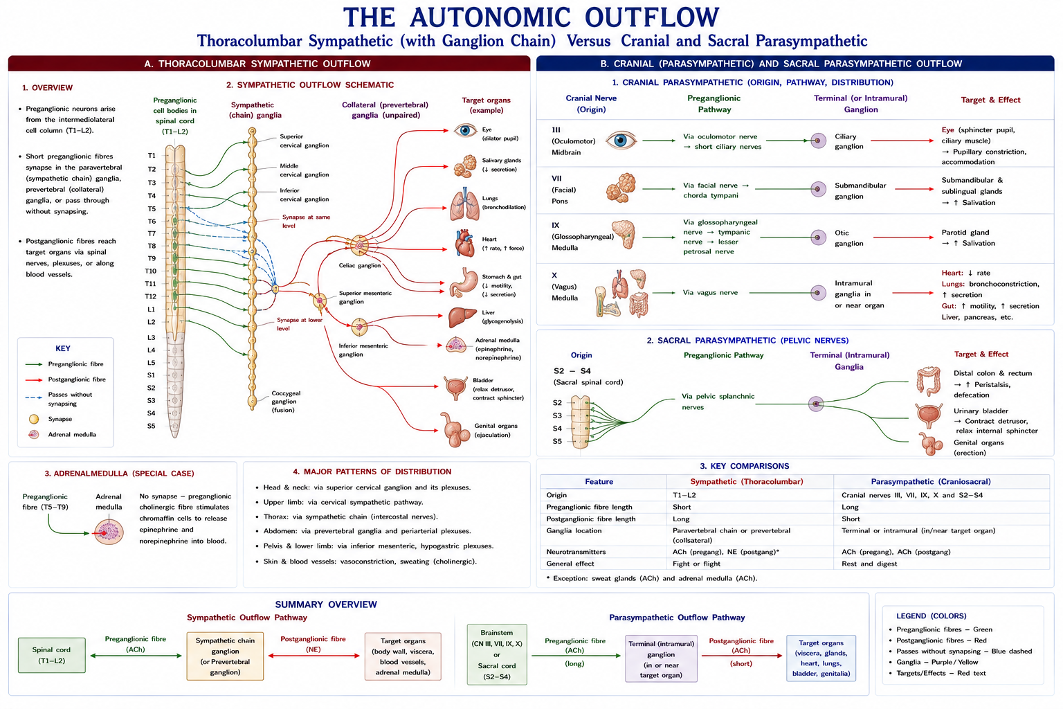
| Sympathetic | Parasympathetic | |
|---|---|---|
| Outflow | Thoracolumbar, T1–L2 | Craniosacral — CN III, VII, IX, X and S2–S4 |
| Ganglia | Paravertebral chain and prevertebral (coeliac, mesenteric) — close to the cord | In or near the target organ |
| Preganglionic fibre | Short | Long |
| Postganglionic fibre | Long | Short |
| Transmitter (postganglionic) | Noradrenaline (except sweat glands — acetylcholine) | Acetylcholine |
| Function | Fight or flight: ↑ HR and contractility, bronchodilatation, pupil dilatation, vasoconstriction (skin and gut), ↑ sweating, glycogenolysis, inhibits digestion | Rest and digest: ↓ HR, bronchoconstriction, pupil constriction, ↑ digestion and secretion, bladder emptying |
Referred visceral pain travels with the sympathetics, which is the anatomical basis of the referral patterns in Chapters 8, 19 and 20.
Clinical syndromes:
| Syndrome | Features |
|---|---|
| Horner’s syndrome | Ptosis, miosis, anhidrosis (± enophthalmos), from interruption of the sympathetic supply to the head — at the cord (T1), the apical lung (Pancoast tumour), the lower brachial plexus (Klumpke), the carotid sheath (dissection), or the brainstem (Wallenberg). Its presence in a brachial plexus injury indicates preganglionic root avulsion — a prognostically decisive sign |
| Autonomic dysreflexia | SCI at or above T6 (Chapter 23) — a physiotherapy emergency |
| Complex regional pain syndrome | Disproportionate pain with sensory, vasomotor, sudomotor and motor/trophic changes, diagnosed by the Budapest criteria. Treated with graded motor imagery, mirror therapy, desensitisation and graded exposure to movement — early mobilisation and education are the mainstays, and immobilisation worsens it |
| Orthostatic hypotension | Common in SCI, diabetes and Parkinson’s disease; a mobilisation planning issue |
| Autonomic neuropathy | Diabetes (Chapter 21) — blunted heart rate response means exercise must be prescribed by RPE |
| Condition | Pattern | Physiotherapy relevance |
|---|---|---|
| Diabetic polyneuropathy | Distal symmetrical, “glove and stocking”, sensory > motor, length-dependent | Foot protection, balance training, falls prevention (Chapter 16) |
| Guillain–Barré syndrome | Acute, ascending, symmetrical, predominantly motor, areflexic; often post-infective | Monitor vital capacity — respiratory failure is the danger; positioning, range, graded strengthening avoiding overwork fatigue, and a long rehabilitation |
| CIDP | Chronic, relapsing version | Similar, over a longer course |
| Charcot–Marie–Tooth (HMSN) | Hereditary; distal wasting (“inverted champagne bottle” legs), pes cavus, claw toes, foot drop | Orthoses (AFOs), balance, energy conservation, avoid overwork weakness |
| Alcoholic / nutritional (B12, thiamine) | Distal sensory, sometimes with dorsal column signs | Balance, sensory strategies |
| Chemotherapy-induced (platinum, taxanes, vinca) | Distal sensory, often painful | Increasingly common; balance and falls risk |
| Leprosy | Thickened nerves with patchy sensory and motor loss (ulnar, median, common fibular, facial) | Still a significant cause of disability in India; protective sensory loss management, splinting and deformity prevention |
A shared principle: in most polyneuropathies and in post-polio syndrome, overwork weakness is a real phenomenon — excessive eccentric or maximal loading of a partially denervated muscle can produce lasting loss. Prescribe moderate-intensity, submaximal, well-spaced work with careful monitoring, not maximal strengthening.
Twenty-four chapters, five parts. If you take four things from all of it:
Chapter 24 of 24 · Human Anatomy · Physiotherapist India End of Part 5 — and of the series.
15 questions on this chapter. Tap one to see the answer and the reasoning.
Answer: (C) Its survival distinguishes Sunderland II from III.
Answer: (B)
Answer: (B) Being outside the cord, it explains preserved SNAPs in radiculopathy.
Answer: (C)
Answer: (C) It may include Horner’s syndrome.
Answer: (B) Prognostically decisive and not directly repairable.
Answer: (B) For example, flexor carpi radialis (median, C7).
Answer: (A) Recovery is complete within days to 12 weeks, without Wallerian degeneration.
Answer: (A) Roughly an inch a month, after a 2–4 week initial delay.
Answer: (C) Which is why proximal injuries have the worse prognosis.
Answer: (B)
Answer: (C) With forearm pronation and wrist flexion.
Answer: (B)
Answer: (B) The classic exception.
Answer: (B) Prescribe moderate, submaximal, well-spaced loading.
Everything on this page, in one screen
| Source | Why it earns its place |
|---|---|
| Standring S (ed.) — Gray’s Anatomy, 42nd edn | Definitive plexus and peripheral nerve anatomy |
| Snell RS — Clinical Neuroanatomy | Roots, plexuses and localisation |
| Lundy-Ekman L — Neuroscience: Fundamentals for Rehabilitation | The physiotherapy-facing synthesis |
| Butler DS — The Sensitive Nervous System; Mobilisation of the Nervous System | The reference for neurodynamics |
| Shacklock M — Clinical Neurodynamics | Testing, differentiation, sliders and tensioners |
| Seddon HJ (1943); Sunderland S (1951) | The original injury classifications |
| Mackinnon SE, Dellon AL — Surgery of the Peripheral Nerve | Repair, transfers and the reinnervation timeline |
| Upton ARM, McComas AJ — “The double crush in nerve-entrapment syndromes”, Lancet, 1973 | The original hypothesis — read alongside its critiques |
| Harden RN et al. — Budapest criteria for CRPS, Pain, 2010 | The diagnostic standard |
| ASIA — International Standards for Neurological Classification of Spinal Cord Injury | Key sensory points and myotomes |
| Chaurasia BD — Human Anatomy, all volumes | Indian syllabus-matched descriptive account |
Reviewed by the Physiotherapist India Team. · Human Anatomy contents