Evidence-based physiotherapy and rehabilitation resources
Editorial & review policy
Evidence-based physiotherapy and rehabilitation resources
Editorial & review policyHuman Anatomy · Head and neck
The neck has to hold up a five-kilogram head, let you turn it to look behind you, and carry everything running between the head and the body through a space you can close your hand around. It is the most crowded region in the body, and almost nothing in it is protected by bone.
Part 5 · Head, neck and the nervous system
The skull, the TMJ, the muscles of facial expression, and the triangles of the neck
Three caseloads live in this chapter.
Facial palsy. Bell’s palsy has an annual incidence of around 20–30 per 100,000 and most cases recover — but the ones that do not need facial retraining, and the difference between an upper and a lower motor neuron lesion is decided by a single anatomical fact about the forehead.
Temporomandibular disorders. They affect around 5–12% of the population, are the second commonest musculoskeletal pain condition after low back pain, and respond well to conservative management — yet most physiotherapists feel unqualified to treat them because the anatomy was taught as dentistry.
The neck. Cervicogenic headache, cervicogenic dizziness, whiplash, thoracic outlet syndrome, torticollis and post-neck-dissection shoulder dysfunction all require the anatomy of the triangles, the fascial planes and the cranial nerves that traverse them. So does the cervical arterial screening every clinician must perform before treating a neck.
Figure 1 · The skull
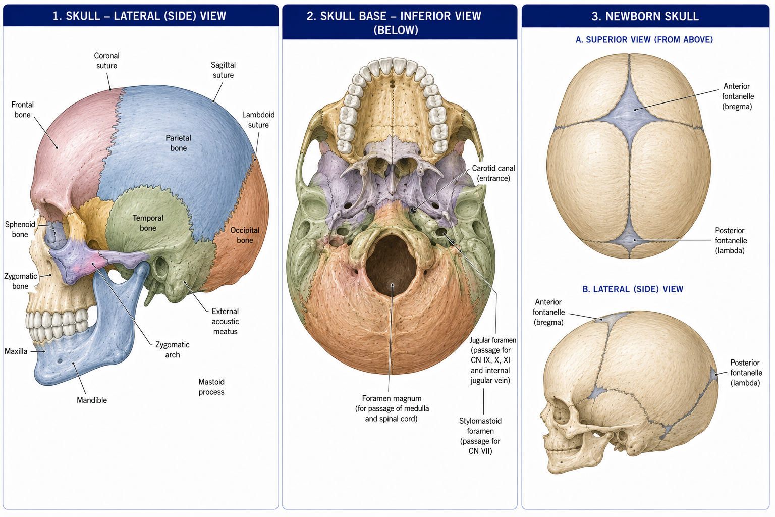
22 bones (excluding the auditory ossicles), in two functional groups.
| Group | Bones |
|---|---|
| Neurocranium (8) | Frontal, two parietal, two temporal, occipital, sphenoid, ethmoid |
| Viscerocranium / facial (14) | Two maxillae, two zygomatic, two nasal, two lacrimal, two palatine, two inferior nasal conchae, vomer, mandible |
Developmental origin: the vault and most facial bones ossify in membrane (from neural crest ectomesenchyme for the face); the skull base ossifies in cartilage (chondrocranium). This is why the vault can mould during birth and why base growth follows synchondroses.
| Suture | Between |
|---|---|
| Coronal | Frontal and parietals |
| Sagittal | The two parietals |
| Lambdoid | Parietals and occipital |
| Squamous | Parietal and temporal |
| Metopic | The two frontal halves — normally fused by 2 years |
Fontanelle
Position
Closes
Anterior (bregma)
Coronal + sagittal, diamond-shaped, the largest
~18 months
Posterior (lambda)
Sagittal + lambdoid, triangular
~2–3 months
Sphenoidal and mastoid
Lateral
~6 months and ~1–2 years
Clinically: the anterior fontanelle is a window for ultrasound and a bedside indicator — bulging in raised intracranial pressure, sunken in dehydration. Premature sutural fusion is craniosynostosis, producing a characteristic skull shape depending on which suture fuses.
The pterion — the H-shaped junction of the frontal, parietal, temporal and greater wing of sphenoid, on the temple about 4 cm above the zygomatic arch — is the thinnest part of the skull and overlies the anterior division of the middle meningeal artery. A blow here causes an extradural haematoma, classically with a lucid interval followed by rapid deterioration. It is the single most examined point of skull surface anatomy.
| Fossa | Floor | Major foramina and contents |
|---|---|---|
| Anterior | Frontal, ethmoid (cribriform plate), lesser wing of sphenoid | Cribriform plate — olfactory nerves (CN I) |
| Middle | Greater wing of sphenoid, temporal | Optic canal — CN II, ophthalmic artery. Superior orbital fissure — CN III, IV, V₁, VI, ophthalmic veins. Foramen rotundum — V₂. Foramen ovale — V₃ and the accessory meningeal artery. Foramen spinosum — middle meningeal artery. Foramen lacerum — traversed by the internal carotid |
| Posterior | Occipital, petrous temporal | Internal acoustic meatus — CN VII, VIII. Jugular foramen — CN IX, X, XI and the internal jugular vein. Hypoglossal canal — CN XII. Foramen magnum — medulla, vertebral arteries, spinal roots of CN XI |
Mnemonic for the foramen ovale contents: OVALE — Otic ganglion above, V3, Accessory meningeal artery, Lesser petrosal nerve, Emissary veins.
The mandible is the only mobile skull bone. Its condylar process articulates at the TMJ; its coronoid process takes temporalis; the angle takes masseter laterally and medial pterygoid medially; the mandibular foramen on the medial ramus transmits the inferior alveolar nerve (the dental block site), which emerges at the mental foramen. The mental, infraorbital and supraorbital foramina lie roughly in a vertical line — the three exit points of the trigeminal cutaneous branches, and the standard palpation points for trigeminal sensory testing.
A synovial joint with unique features: it is bilateral and must move as a pair, its articular surfaces are covered by fibrocartilage rather than hyaline cartilage (because the bones develop in membrane), and it is divided into two functional compartments by a disc.
| Feature | Detail |
|---|---|
| Surfaces | Head (condyle) of the mandible with the mandibular fossa and articular eminence of the temporal bone |
| Disc | A biconcave fibrocartilaginous disc dividing the joint into an upper (discotemporal) and a lower (discomandibular) compartment. It is attached to the joint capsule, to the condylar poles medially and laterally, and posteriorly to the bilaminar (retrodiscal) zone — vascular and richly innervated, and the principal pain source in TMD |
| Ligaments | Lateral (temporomandibular) — the main restraint; sphenomandibular (a remnant of Meckel’s cartilage, the “swinging hinge” fulcrum); stylomandibular |
Lower compartment: rotation. Upper compartment: translation.
Normal opening is 40–50 mm (approximately three of the patient’s own fingers vertically); lateral excursion and protrusion are ~10 mm each. Opening under 35 mm is restricted.
Movements: depression, elevation, protrusion, retrusion, and lateral excursion. Lateral excursion is asymmetrical — the ipsilateral condyle rotates while the contralateral condyle translates forward and medially, which is why unilateral pathology produces deviation.
All four are supplied by the mandibular division of the trigeminal nerve (V₃).
| Muscle | Attachments | Action |
|---|---|---|
| Temporalis | Temporal fossa → coronoid process and anterior ramus | Elevation; posterior fibres retract |
| Masseter | Zygomatic arch → lateral ramus and angle | Elevation — the most powerful; some protrusion |
| Medial pterygoid | Lateral pterygoid plate, tuberosity of maxilla → medial angle | Elevation, protrusion, contralateral excursion. With masseter forms a sling around the angle |
| Lateral pterygoid | Greater wing of sphenoid and lateral pterygoid plate → condylar neck and the disc | The odd one out: it DEPRESSES (opens) the jaw, protrudes, and produces contralateral excursion. Its superior head attaches to the disc and controls disc position during closing |
Also assisting depression: the suprahyoid muscles (digastric, mylohyoid, geniohyoid, stylohyoid) when the hyoid is fixed by the infrahyoids — which is why jaw, hyoid, tongue and cervical posture are mechanically coupled, and why forward head posture alters mandibular resting position.
| Category | Features |
|---|---|
| Myogenous (masticatory muscle disorder) | The commonest; muscle tenderness, pain on function, often with parafunction (bruxism, clenching) |
| Disc displacement with reduction | The disc sits anteriorly at rest; the condyle clicks onto it during opening and clicks off during closing — a reciprocal click. Common and often asymptomatic |
| Disc displacement without reduction | Closed lock — the condyle cannot translate onto the displaced disc; opening limited to ~25–30 mm with deviation towards the affected side, and no click |
| Degenerative joint disease | Crepitus, load-related pain |
| Subluxation / dislocation | The condyle translates anterior to the eminence and cannot return — an open lock |
Assessment: measure opening, note deviation (towards the restricted side) or deflection, palpate masseter, temporalis and the lateral pterygoid region, auscultate for click or crepitus, and always examine the cervical spine — the upper cervical spine and the TMJ share trigeminocervical convergence (§22.7) and their symptoms overlap constantly.
Management is conservative first-line and effective: education and reassurance, parafunction awareness, jaw rest within a comfortable range, controlled active exercise and manual therapy, postural and cervical management, and stress and sleep management. Occlusal splints have a role, and irreversible occlusal or surgical intervention should follow, not precede, conservative care.
Figure 2 · Why a stroke spares the forehead
All are supplied by the facial nerve (CN VII) and develop from the second pharyngeal arch. They are unusual in inserting into skin rather than bone, which is what allows expression.
| Muscle | Action |
|---|---|
| Occipitofrontalis (frontal belly) | Raises eyebrows; wrinkles the forehead |
| Orbicularis oculi | Closes the eye — palpebral part gently, orbital part forcibly |
| Orbicularis oris | Closes and purses the lips |
| Buccinator | Presses the cheek against the teeth — keeps food between the teeth during chewing and is the muscle of blowing. Pierced by the parotid duct |
| Zygomaticus major and minor, levator labii superioris, levator anguli oris | Elevate the angle of the mouth — smiling |
| Depressor anguli oris, depressor labii inferioris | Depress the mouth |
| Platysma | Tenses the neck skin; depresses the mandible and the corners of the mouth |
| Risorius, mentalis, nasalis, corrugator supercilii, procerus | Various |
Course: brainstem → internal acoustic meatus → facial canal in the petrous temporal bone (giving the greater petrosal nerve, the nerve to stapedius, and the chorda tympani) → stylomastoid foramen → through the parotid gland, where it divides into five terminal branches.
Terminal branches: Temporal, Zygomatic, Buccal, Marginal mandibular, Cervical — “Ten Zebras Bit My Curls” or “Two Zulus Buggered My Cat” (both are in circulation; use whichever you can say in an exam).
Functions: motor to the muscles of facial expression, stapedius, stylohyoid and the posterior belly of digastric; taste from the anterior two-thirds of the tongue (chorda tympani); parasympathetic secretomotor to the lacrimal, submandibular and sublingual glands; and a small sensory patch at the external acoustic meatus.
The part of the facial nucleus supplying the upper face (frontalis and orbicularis oculi) receives BILATERAL corticobulbar input; the part supplying the lower face receives only CONTRALATERAL input.
Therefore:
| Upper motor neuron lesion (stroke, above the nucleus) | Lower motor neuron lesion (Bell’s palsy, nerve trunk) | |
|---|---|---|
| Forehead | SPARED — the patient can still raise the eyebrow and wrinkle the forehead | PARALYSED — cannot raise the eyebrow |
| Lower face | Contralateral droop | Ipsilateral droop |
| Eye closure | Preserved | Lost — corneal exposure risk |
| Other features | Often with limb signs; emotional (spontaneous) smiling may be preserved | May include hyperacusis (stapedius), loss of taste, reduced lacrimation, depending on the level |
This single sign — forehead sparing — distinguishes a stroke from a Bell’s palsy at the bedside, and a physiotherapist must be able to make that distinction. A patient presenting with a facial droop and an intact forehead requires urgent stroke assessment, not facial exercises.
Bell’s palsy — idiopathic (probably viral, HSV-1 reactivation) lower motor neuron facial palsy, of acute onset over hours. Around 70–85% recover fully, most within 3 weeks to 3 months. Corticosteroids started within 72 hours improve outcome (Sullivan et al., NEJM 2007); antivirals add little.
Physiotherapy role:
Figure 3 · The triangles of the neck
Sternocleidomastoid divides each side into an anterior and a posterior triangle.
| Triangle | Boundaries | Contents |
|---|---|---|
| ANTERIOR | Midline, anterior border of SCM, mandible | Subdivided into four |
| — Submental | Anterior bellies of digastric, hyoid | Submental nodes |
| — Submandibular (digastric) | Two bellies of digastric, mandible | Submandibular gland, facial artery and vein, hypoglossal nerve, nodes |
| — Carotid | Superior belly of omohyoid, posterior belly of digastric, SCM | Common carotid and its bifurcation (at the upper border of the thyroid cartilage, C4), internal jugular vein, vagus nerve, hypoglossal nerve, ansa cervicalis |
| — Muscular | Superior belly of omohyoid, SCM, midline | Infrahyoid (strap) muscles, thyroid, parathyroids, trachea, oesophagus |
| POSTERIOR | Posterior border of SCM, anterior border of trapezius, middle third of clavicle | Subdivided by the inferior belly of omohyoid |
| — Occipital | Above omohyoid | Spinal accessory nerve (CN XI), cutaneous branches of the cervical plexus, roots and trunks of the brachial plexus, lymph nodes |
| — Supraclavicular (subclavian) | Below omohyoid | Third part of the subclavian artery, subclavian vein, suprascapular artery, brachial plexus trunks |
1. The spinal accessory nerve is extremely superficial. It crosses the posterior triangle in the investing fascia, roughly from the junction of the upper and middle thirds of the posterior border of SCM to a point 5 cm above the clavicle on trapezius. It is the nerve most commonly injured by minor surgery in the neck — a lymph node biopsy in the posterior triangle is a classic cause. The result is trapezius palsy: a drooping shoulder, lateral winging, and inability to elevate the arm fully (Chapter 10). Physiotherapists see these patients, and the history of a small neck operation is the clue.
2. Erb’s point — where the four cutaneous branches of the cervical plexus (lesser occipital, great auricular, transverse cervical, supraclavicular) emerge around the middle of the posterior border of SCM. It is the landmark for a superficial cervical plexus block. (The name is also used for a different point over the upper trunk of the brachial plexus — context matters.)
| Muscle | Attachments | Nerve | Action |
|---|---|---|---|
| Sternocleidomastoid | Manubrium and medial clavicle → mastoid process and superior nuchal line | Spinal accessory (CN XI) + C2, C3 proprioceptive | Unilateral: ipsilateral lateral flexion with CONTRALATERAL rotation. Bilateral: flexion of the lower cervical spine with extension of the upper; accessory inspiratory muscle |
| Trapezius (upper) | See Chapter 10 | Spinal accessory + C3, C4 | Elevation, upward rotation |
| Scalenes (anterior, middle, posterior) | Cervical transverse processes → ribs 1 and 2 | Anterior rami C3–C8 | Lateral flexion; accessory inspiratory muscles. The scalene triangle (anterior and middle scalene with the first rib) transmits the brachial plexus and subclavian artery — the subclavian vein passes anterior to anterior scalene |
| Longus colli and capitis | Anterior vertebral bodies | Anterior rami C1–C6 | The deep cervical flexors — segmental control and craniocervical flexion; impaired in neck pain and whiplash, and the target of the craniocervical flexion test and retraining |
| Suprahyoid and infrahyoid | Hyoid to mandible/skull; hyoid to sternum/scapula/thyroid | Various (CN V, VII, XII; ansa cervicalis C1–C3) | Swallowing, jaw depression, hyoid fixation |
The deep cervical flexors deserve emphasis. The craniocervical flexion test (using a pressure biofeedback unit) reliably identifies reduced deep flexor activation and increased superficial (SCM, scalene) substitution in people with neck pain and whiplash. Low-load deep flexor retraining has good trial evidence for reducing neck pain and cervicogenic headache — one of the better-supported specific exercise interventions in musculoskeletal physiotherapy.
Figure 4 · Cross-section of the neck
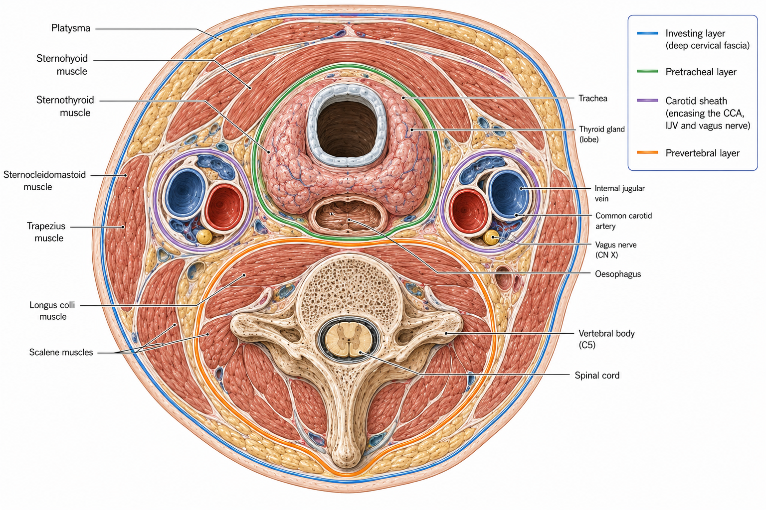
| Layer | Encloses | Clinical relevance |
|---|---|---|
| Investing (superficial) layer of deep cervical fascia | Surrounds the neck; splits to enclose trapezius and SCM, and the submandibular and parotid glands | The plane of the superficial nerves; abscesses tracking within it |
| Pretracheal | Thyroid, trachea, oesophagus; continues as the buccopharyngeal fascia | Infection can track into the superior mediastinum |
| Prevertebral | Prevertebral and postvertebral muscles and the vertebral column; prolongs laterally as the axillary sheath around the brachial plexus | The basis of interscalene and supraclavicular brachial plexus blocks. A prevertebral abscess (classically tuberculous) points into the retropharyngeal space |
| Carotid sheath | Common/internal carotid artery, internal jugular vein, vagus nerve (artery medial, vein lateral, nerve posterior between them); deep cervical nodes | The dissection plane in neck surgery |
| Retropharyngeal space | Between buccopharyngeal and prevertebral fascia | The “danger space” — infection descends directly to the posterior mediastinum |
Common carotid — bifurcates at the upper border of the thyroid cartilage (C4) into:
The carotid sinus (at the bifurcation) is a baroreceptor supplied by the glossopharyngeal nerve; the carotid body is a chemoreceptor. Carotid sinus hypersensitivity means firm pressure over the bifurcation can cause bradycardia and syncope — a reason to avoid heavy anterolateral neck pressure in older patients.
Vertebral artery — the first branch of the subclavian, ascending through the foramina transversaria of C6 to C1, then turning sharply medially in the groove on the posterior arch of the atlas before entering the foramen magnum to form the basilar artery. It supplies the brainstem, cerebellum and posterior cerebrum.
The vertebral artery is most tortuous and most mechanically stressed in the atlanto-axial segment, where it is stretched by rotation and by rotation with extension. Cervical arterial dissection — vertebral or internal carotid — is rare but catastrophic, and it presents with neck pain and headache before any neurological sign, which is exactly why patients present to physiotherapists.
The IFOMPT framework has replaced pre-manipulative positional testing (which had poor validity) with a reasoning process across the whole examination:
The clinical rule: if the presentation is a new, unusual, severe headache or neck pain — particularly with any of the above features — it is a medical presentation until proved otherwise, and end-range rotation techniques and manipulation are contraindicated.
Mechanism: the trigeminocervical nucleus. The spinal nucleus of the trigeminal nerve extends caudally into the upper cervical cord and converges with the afferents from C1, C2 and C3. Nociceptive input from upper cervical structures — the atlanto-occipital and atlanto-axial joints, the C2–C3 facet joint, the suboccipital muscles, the upper cervical discs and dura — is therefore perceived as head pain in trigeminal territory.
Features: unilateral, side-consistent headache, starting in the neck and radiating fronto-orbitally; provoked by neck movement or sustained posture; reduced cervical range (especially the flexion-rotation test, which specifically assesses C1–C2 and is the single most useful test); tenderness of the upper cervical segments.
Management has good trial evidence (Jull et al.): manual therapy plus low-load craniocervical flexor and scapular retraining, sustained over 6 weeks, with effects maintained at 12 months.
A diagnosis of exclusion, made after vestibular and vascular causes are ruled out. The mechanism is disturbance of cervical proprioceptive input — the deep upper cervical muscles have among the highest muscle spindle densities in the body (Chapter 17), and their afferents converge with vestibular and visual input in the central integrator. Altered or asymmetrical cervical input therefore produces sensory conflict, experienced as unsteadiness or light-headedness.
Distinguishing features: dizziness described as unsteadiness or disorientation rather than true spinning, temporally related to neck pain and neck position, lasting minutes to hours, without hearing loss or true nystagmus. Managed with cervical treatment plus oculomotor and cervical joint position sense retraining, which has supportive evidence.
But rule out first: BPPV (positional, brief, rotatory, with a positive Dix–Hallpike), vestibular neuritis, Ménière’s, orthostatic hypotension, medication, and — critically — vertebrobasilar insufficiency or dissection.
| Type | Features | Management |
|---|---|---|
| Congenital muscular | SCM fibrosis, often with a palpable mass in the first weeks; head laterally flexed towards and rotated away from the affected side | Positioning, stretching, active rotation, tummy time, plagiocephaly management; screen for DDH, which co-occurs |
| Acquired / acute (wry neck) | Sudden painful restriction, often on waking; facet or discogenic | Reassurance, movement, manual therapy, analgesia |
| Spasmodic (cervical dystonia) | Adult-onset involuntary sustained contraction | Botulinum toxin plus physiotherapy in the post-injection window |
Compression of the neurovascular bundle at three possible sites:
| Site | Boundaries | Compressed |
|---|---|---|
| Interscalene (scalene) triangle | Anterior scalene, middle scalene, first rib | Brachial plexus and subclavian artery (the vein passes anterior to anterior scalene, so it escapes here) |
| Costoclavicular space | Clavicle, first rib, subclavius | All three — the commonest site for venous TOS |
| Subcoracoid / pectoralis minor space | Pectoralis minor, coracoid, ribs | Plexus and vessels in hyperabduction |
Types: neurogenic (~95%, but true neurogenic TOS with objective wasting is rare), venous (~3%, Paget–Schroetter effort thrombosis), arterial (~1%, but the most serious). A cervical rib or fibrous band is present in a minority.
Provocation tests (Adson’s, Roos/EAST, Wright’s, costoclavicular) all have poor specificity — they are positive in a substantial proportion of asymptomatic people — so diagnosis rests on the pattern, and on excluding cervical radiculopathy and peripheral entrapment. Management is postural, scalene and pectoralis minor loading and length work, nerve gliding, and breathing pattern retraining.
Worth noting here which cranial nerves a physiotherapist tests routinely and why: V (facial sensation, masticatory power), VII (facial expression, forehead rule), VIII (hearing and vestibular function), IX/X (swallow, gag, palate), XI (trapezius and SCM), XII (tongue). Together they form the bulbar screen that determines swallow safety and stroke localisation.
15 questions on this chapter. Tap one to see the answer and the reasoning.
Answer: (C) The posterior closes at 2–3 months.
Answer: (B) Fracture here causes extradural haematoma.
Answer: (C) V₃ passes through the foramen ovale.
Answer: (B) IX, X and XI use the jugular foramen.
Answer: (B) Rotation occurs in the lower compartment, giving the first 20–25 mm of opening.
Answer: (D) It also attaches to the disc.
Answer: (B) With opening usually limited to 25–30 mm and no click.
Answer: (B)
Answer: (C) Both are associated with synkinesis.
Answer: (C) Hence its vulnerability in lymph node biopsy.
Answer: (B)
Answer: (B) Which is why the interscalene triangle compresses plexus and artery but not vein.
Answer: (B)
Answer: (B) The trigeminocervical nucleus.
Answer: (B)
Everything on this page, in one screen
| Source | Why it earns its place |
|---|---|
| Standring S (ed.) — Gray’s Anatomy, 42nd edn | Definitive head and neck anatomy |
| Moore KL, Dalley AF, Agur AMR — Clinically Oriented Anatomy | The best clinical treatment of the triangles, fascia and foramina |
| Chaurasia BD — Human Anatomy, Vol 3: Head, Neck and Brain | The standard Indian regional text for this region |
| Jull G, Sterling M, Falla D, Treleaven J, O’Leary S — Whiplash, Headache and Neck Pain | The reference for cervicogenic headache, dizziness and deep flexor retraining |
| Jull G et al. — “A randomized controlled trial of exercise and manipulative therapy for cervicogenic headache”, Spine, 2002 | The trial evidence |
| Rushton A et al. — IFOMPT International Framework for Examination of the Cervical Region (2020 update) | The current cervical arterial screening standard |
| Sullivan FM et al. — “Early treatment with prednisolone or acyclovir in Bell’s palsy”, NEJM, 2007 | The steroid evidence |
| Ross B, Fradet G, Nedzelski J — Sunnybrook Facial Grading System | Facial palsy outcome measurement |
| Okeson JP — Management of Temporomandibular Disorders and Occlusion | The definitive TMD text |
| Armijo-Olivo S et al. — physical therapy for TMD systematic reviews, Phys Ther | The conservative management evidence |
Chapter 22 of 24 · Human Anatomy · Physiotherapist India Next: Chapter 23 — The Nervous System: brain, spinal cord, tracts and the cranial nerves.
Reviewed by the Physiotherapist India Team. · Human Anatomy contents