Evidence-based physiotherapy and rehabilitation resources
Editorial & review policy
Evidence-based physiotherapy and rehabilitation resources
Editorial & review policyHuman Anatomy · General anatomy
Skin is the organ you touch at every treatment and the one that records what went wrong. Development explains why a nerve root supplies both a strip of skin and a set of muscles, and why pain from a deep organ is felt somewhere else entirely.
Part 1 · General anatomy
Skin layers and repair, the germ layers, and why one nerve root has both a muscle and a skin patch
Skin is the organ you touch every time you treat a patient, the organ that fails first in the immobile, and the organ whose repair you either help or hinder. Embryology is what explains why the body is organised the way it is — why a nerve root supplies both a strip of skin and a group of muscles, why cardiac pain is felt in the left arm, why the ureter and the testis share a pain pathway, and why the thumb is lateral and the great toe medial.
They belong in one chapter because the answer to the chapter’s subtitle question — why one nerve root has both a muscle and a skin patch — comes from development, and is the single most clinically useful idea in either subject.
Figure 1 · What skin does
Skin is the largest organ: 1.5–2 m² in an adult, ~15% of body weight, thickness from 0.5 mm on the eyelid to 4–5 mm on the sole and palm.
Its functions: protection (mechanical, chemical, microbial, ultraviolet), thermoregulation, sensation, vitamin D synthesis, fluid and electrolyte conservation, immune surveillance, excretion, absorption (the basis of transdermal drug delivery and of many topical physiotherapy agents), and social and communicative function.
Skin is classified as thick (palms and soles: thick epidermis with a stratum lucidum, no hair follicles or sebaceous glands, abundant sweat glands, epidermal ridges producing fingerprints) or thin (everywhere else: thinner epidermis, hair follicles and sebaceous glands present).
Figure 2 · The structure of the skin
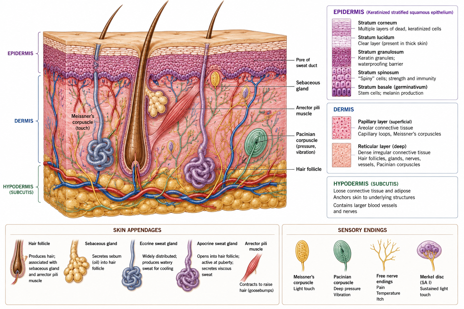
From deep to superficial:
| Layer | Features |
|---|---|
| Stratum basale (germinativum) | A single layer of columnar cells on the basement membrane; the mitotic layer. Contains melanocytes, Merkel cells and epidermal stem cells |
| Stratum spinosum | Several layers of polyhedral keratinocytes joined by desmosomes (the “prickles”); contains Langerhans cells |
| Stratum granulosum | 3–5 layers with keratohyalin granules (filaggrin) and lamellar bodies releasing lipid — the water barrier is formed here |
| Stratum lucidum | A clear layer, present only in thick skin |
| Stratum corneum | 15–30 layers of anucleate, flattened, keratin-filled corneocytes in a lipid matrix; continuously desquamated |
Epidermal turnover is approximately 28–40 days from basal cell to desquamation — accelerated dramatically in psoriasis (to ~4–7 days), which is why psoriatic plaques are scaly.
Non-keratinocyte cells:
| Cell | Origin | Function |
|---|---|---|
| Melanocyte | Neural crest | Produces melanin in melanosomes, transferred to keratinocytes to shield nuclear DNA from UV. Skin colour reflects melanin type and distribution, not melanocyte number, which is similar across populations |
| Langerhans cell | Bone marrow | Dendritic antigen-presenting cell; the afferent limb of contact hypersensitivity |
| Merkel cell | Neural crest / epidermal | Slowly adapting type I mechanoreceptor; light touch and texture discrimination |
| Layer | Structure |
|---|---|
| Papillary dermis | Loose connective tissue forming dermal papillae that interdigitate with epidermal ridges (increasing adhesion and surface area for diffusion); contains capillary loops and Meissner’s corpuscles |
| Reticular dermis | Dense irregular connective tissue: thick type I collagen bundles and elastic fibres; contains appendages, Pacinian and Ruffini corpuscles, vessels and nerves |
The predominant collagen bundle orientation in the reticular dermis creates lines of cleavage (Langer’s lines). Incisions parallel to them gape less and scar better; incisions across them gape and produce wider scars. Wound orientation relative to these lines partly predicts scar quality — a point of direct relevance in scar management.
Ageing skin shows epidermal thinning, flattening of the dermo-epidermal junction (reducing adhesion and increasing shear vulnerability — the reason elderly skin tears with adhesive tape removal), collagen and elastin loss, reduced vascularity, reduced sweat and sebaceous output, and slower healing.
| Appendage | Notes |
|---|---|
| Hair follicle | Epidermal downgrowth; cycles through anagen (growth), catagen, telogen (rest). Arrector pili is smooth muscle, sympathetically innervated |
| Sebaceous gland | Holocrine; opens into the follicle; sebum lubricates and is antimicrobial; androgen-sensitive (hence acne) |
| Eccrine sweat gland | Merocrine; over the whole body, densest on palms, soles and forehead; sympathetic cholinergic innervation (the exception to the adrenergic rule); the principal thermoregulatory effector; 2–4 million glands producing up to several litres per hour in extreme conditions |
| Apocrine sweat gland | Axilla, areola, perineum; opens into hair follicles; active from puberty; odour results from bacterial action on the secretion |
| Nail | Keratinised plate; grows ~3 mm/month (fingernails) from the matrix under the lunula |
Two horizontal plexuses — deep (at the dermo-hypodermal junction) and superficial (subpapillary) — connected by perforating vessels, with arteriovenous anastomoses (glomus bodies) in the digits, palms, soles, ears and nose.
Skin blood flow can vary from about 0.5% to 30% of cardiac output. Sympathetic vasoconstrictor tone controls it; heat causes vasodilatation and sweating, cold causes vasoconstriction and arteriovenous shunting to preserve core temperature.
Clinical implications for physiotherapy: all thermal and electrophysical agents act on and through this system. In any patient with impaired sensation, impaired circulation or impaired cognition, the protective withdrawal from a damaging thermal stimulus is absent — which is why thermal agents are contraindicated over areas of sensory loss, arterial insufficiency, or in a patient who cannot report sensation reliably. Burns from hot packs and paraffin in neuropathic feet remain a persistent and entirely avoidable source of harm.
| Receptor | Adaptation | Modality |
|---|---|---|
| Merkel disc | Slowly adapting type I | Sustained light touch, edges, texture; small receptive field |
| Meissner corpuscle | Rapidly adapting type I | Light touch, low-frequency flutter, slip detection; dense in fingertips |
| Pacinian corpuscle | Rapidly adapting type II | High-frequency vibration, deep pressure; large receptive field |
| Ruffini ending | Slowly adapting type II | Skin stretch, sustained pressure, joint position contribution |
| Free nerve endings | Non-adapting | Nociception (Aδ and C), temperature, crude touch, itch |
| Hair follicle receptors | Rapidly adapting | Hair movement |
Two-point discrimination — approximately 2–4 mm on the fingertip, 40–50 mm on the back — reflects receptor density and cortical representation, and is a standard clinical measure of peripheral nerve regeneration.
| Phase | Timing | Events | Physiotherapy implication |
|---|---|---|---|
| Haemostasis | Minutes | Vasoconstriction, platelet plug, fibrin clot; platelets release PDGF, TGF-β | — |
| Inflammation | ~0–5 days (up to 7) | Vasodilatation, increased permissibility; neutrophils (24–48 h) then macrophages (48–72 h, the key orchestrating cell); debridement | Protect; manage oedema; maintain adjacent joint range |
| Proliferation | ~3 days – 3 weeks | Fibroplasia (type III collagen), angiogenesis, granulation tissue, epithelialisation (from wound edges and appendage remnants), wound contraction by myofibroblasts | Begin controlled movement; protect the fragile new epithelium from shear |
| Remodelling (maturation) | 3 weeks – 1–2 years | Type III replaced by type I; fibres realign along stress lines; cross-links mature; vascularity and cellularity decline; the scar pales and softens | The phase physiotherapy influences most: scar mobilisation, sustained low-load stretch, pressure therapy, silicone, and progressive functional loading |
Tensile strength timeline (a frequently examined sequence): ~5% at 1 week, ~20% at 3 weeks, ~50–60% at 6 weeks, and a plateau at ~70–80% of the original at 6–12 months. Scar never regains normal strength, and it lacks appendages, elastic recovery and normal sensation.
Primary versus secondary intention: a clean approximated wound heals by primary intention — minimal granulation, minimal contraction, a fine scar. A wound with tissue loss or contamination heals by secondary intention — abundant granulation, marked wound contraction, prolonged epithelialisation and a broad scar. Secondary healing is where contracture, and therefore physiotherapy, matters most.
Factors impairing healing: age; malnutrition (protein, vitamin C, zinc); diabetes; smoking; corticosteroids and immunosuppression; ischaemia and venous insufficiency; infection; repeated mechanical shear and pressure; radiotherapy; and foreign material.
| Hypertrophic scar | Keloid | |
|---|---|---|
| Extent | Remains within the original wound boundary | Extends beyond the original wound |
| Onset | Within weeks | May be months later |
| Course | Often improves over 1–2 years | Rarely regresses; recurs after excision |
| Sites | Across flexor surfaces, joints, high-tension areas | Sternum, shoulder, ear lobe, jaw |
| Predisposition | Any; burns, prolonged healing | Genetic; more common in darker-skinned individuals |
| Management | Pressure garments, silicone, massage, stretch, sustained positioning, splinting | The same plus intralesional corticosteroid; excision alone is not advised |
Scar management principles for physiotherapy: sustained low-load stretch and positioning (exploiting creep — Chapter 7), pressure therapy (24 mmHg or more, worn 23 hours a day for months in burns), silicone gel or sheeting, massage once epithelialisation is complete, splinting in the anti-deformity position, and desensitisation. All of this works during the remodelling phase, which is the window that closes.
| Depth | Layers involved | Appearance | Sensation | Healing |
|---|---|---|---|---|
| Superficial (1st degree) | Epidermis only | Erythema, dry, no blisters | Painful | 3–7 days, no scar |
| Superficial partial (2nd, superficial dermal) | Epidermis + papillary dermis | Blisters, moist, pink, blanches | Very painful | 1–3 weeks, minimal scar |
| Deep partial (2nd, deep dermal) | Into reticular dermis | Blotchy red/white, less blanching, drier | Reduced sensation | 3–8 weeks; hypertrophic scarring likely; often grafted |
| Full thickness (3rd) | Entire dermis ± appendages | White, waxy, leathery, charred; does not blanch | Insensate | Will not heal without grafting; contracture guaranteed |
| Fourth degree | Into fascia, muscle, bone | Charred, exposed deep structures | Insensate | Surgical; often amputation |
Extent is estimated by the rule of nines in adults (head 9%, each arm 9%, each leg 18%, anterior trunk 18%, posterior trunk 18%, perineum 1%) — modified in children, whose head is proportionally larger; the Lund and Browder chart is more accurate.
Physiotherapy in burns is dominated by two aims: preventing contracture (positioning in the anti-deformity position — neck extension, shoulder abduction to ~90° with slight horizontal flexion, elbow extension and forearm supination, wrist slight extension with MCPs flexed to 70–90° and IPs extended, hips extended and abducted, knees extended, ankles neutral — which is the opposite of the comfortable flexed posture the patient adopts), and maintaining respiratory and functional capacity. Splinting, sustained stretch, early mobilisation, and later pressure therapy and scar management are the core interventions, delivered over months.
Mechanism. Sustained pressure exceeding capillary closing pressure (~32 mmHg) occludes perfusion; shear compounds it by distorting and occluding vessels obliquely; moisture macerates and reduces tissue tolerance; friction removes the stratum corneum. Muscle is more susceptible than skin, so damage is frequently deeper than it appears — the “iceberg” pattern.
Sites: sacrum, ischial tuberosities, greater trochanters, heels, lateral malleoli, occiput, scapulae, elbows; and, increasingly, device-related sites (oxygen tubing, catheters, casts).
Staging (NPIAP/EPUAP):
| Stage | Description |
|---|---|
| 1 | Intact skin with non-blanchable erythema |
| 2 | Partial-thickness loss with exposed dermis; a shallow open ulcer or intact/ruptured blister |
| 3 | Full-thickness skin loss; subcutaneous fat visible; no exposed bone, tendon or muscle |
| 4 | Full-thickness skin and tissue loss; exposed bone, tendon or muscle |
| Unstageable | Base obscured by slough or eschar |
| Deep tissue injury | Persistent non-blanchable deep red, maroon or purple discoloration, or a blood-filled blister |
Prevention is the physiotherapy contribution, and it is substantial: repositioning schedules, pressure-redistributing surfaces, teaching independent pressure relief (lift-offs or leans every 15–30 minutes in wheelchair users), wheelchair seating and cushion prescription, transfer technique that eliminates dragging, maintaining mobility and nutrition, and skin inspection education. A stage 4 pressure injury in a spinal cord injured patient can cost a year of rehabilitation progress.
Figure 3 · Neurulation and the neural crest
| Stage | Timing | Event |
|---|---|---|
| Fertilisation | Day 0 | Zygote formed in the ampulla of the uterine tube |
| Cleavage → morula | Days 1–4 | Successive divisions |
| Blastocyst | Days 5–6 | Inner cell mass (embryoblast) + trophoblast + blastocyst cavity |
| Implantation | Days 6–10 | In the uterine endometrium |
| Bilaminar disc | Week 2 | Epiblast and hypoblast; amniotic cavity and yolk sac form. “The week of twos” |
| Gastrulation | Week 3 | The primitive streak appears; epiblast cells migrate through it to form the three germ layers. “The week of threes” |
| Neurulation | Weeks 3–4 | Notochord induces the overlying ectoderm to form the neural plate → neural groove → neural tube; neural crest cells separate |
Gastrulation is the pivotal event: it converts a two-layered disc into a three-layered embryo with an established body plan and axes. Every tissue in the body traces to one of the three layers formed in that week.
Figure 4 · The three germ layers and what each becomes
| Germ layer | Derivatives |
|---|---|
| Ectoderm — surface | Epidermis, hair, nails, sweat and sebaceous glands, mammary glands; lens and cornea epithelium; enamel of teeth; anterior pituitary; inner ear epithelium; epithelium of the mouth and anal canal below the pectinate line |
| Ectoderm — neuroectoderm (neural tube) | Brain, spinal cord, retina and optic nerve, posterior pituitary, pineal gland, motor neurons, preganglionic autonomic neurons |
| Ectoderm — neural crest | An extraordinary list: dorsal root and autonomic ganglia, Schwann cells, adrenal medulla, melanocytes, enteric nervous system, meninges (pia and arachnoid), most of the skull and facial skeleton (via ectomesenchyme), odontoblasts, aorticopulmonary septum, parafollicular C cells |
| Mesoderm — paraxial (somites) | Sclerotome → vertebrae, ribs, part of the skull base; Myotome → skeletal muscle; Dermatome → dermis and hypodermis of the back |
| Mesoderm — intermediate | Kidneys, ureters, gonads, genital ducts |
| Mesoderm — lateral plate (somatic) | Limb bones, connective tissue and dermis of the limbs and body wall; parietal serous membranes |
| Mesoderm — lateral plate (splanchnic) | Heart, blood and lymphatic vessels, blood cells, spleen, smooth muscle and connective tissue of the gut, visceral serous membranes, suprarenal cortex |
| Endoderm | Epithelial lining of the gastrointestinal and respiratory tracts; liver, pancreas, gallbladder; thyroid, parathyroid, thymus; bladder and urethral epithelium; tympanic cavity and auditory tube; tonsils |
Neural crest cells delaminate from the crests of the neural folds and migrate widely. Because they produce structures as unrelated as the facial skeleton, the adrenal medulla and skin melanocytes, a neurocristopathy produces baffling multi-system combinations: Hirschsprung disease (absent enteric ganglia), neurofibromatosis, Waardenburg syndrome (pigmentary changes with deafness), DiGeorge syndrome (thymic, parathyroid and conotruncal cardiac defects), and CHARGE syndrome. If you ever wonder why a child with a cardiac outflow defect also has a facial anomaly and pigmentary change, the answer is neural crest.
Figure 5 · What a somite becomes
From day 20, paraxial mesoderm on each side of the notochord segments into somites, forming craniocaudally at roughly three pairs per day, reaching 42–44 pairs (4 occipital, 8 cervical, 12 thoracic, 5 lumbar, 5 sacral, 8–10 coccygeal, several of which regress). Somite count is the standard measure of embryonic age in weeks 4–5.
Each somite differentiates into three parts:
| Part | Becomes |
|---|---|
| Sclerotome (ventromedial) | Vertebrae, ribs, and their ligaments; migrates around the notochord and neural tube |
| Myotome (dorsolateral) | Skeletal muscle — dividing into a dorsal epimere (innervated by the dorsal ramus, becoming the deep back extensors) and a ventral hypomere (innervated by the ventral ramus, becoming everything else) |
| Dermatome (dorsolateral, superficial) | Dermis of the back; limb and ventral body wall dermis derives from lateral plate mesoderm, though it retains segmental innervation |
Each somite is supplied by its own spinal nerve, and that relationship is retained for life however far the tissue migrates. This is the whole basis of segmental clinical anatomy.
| Term | Definition | Clinical use |
|---|---|---|
| Dermatome | The area of skin supplied by a single spinal nerve root | Sensory level in spinal cord injury; radiculopathy mapping; shingles distribution |
| Myotome | The muscle group supplied by a single spinal nerve root | Motor level testing; root versus peripheral nerve localisation |
| Sclerotome | The bone and deep tissue derived from one somite and supplied by that root | Explains deep, poorly localised, aching bone pain that does not follow a dermatome |
| Viscerotome | The viscera whose afferents enter at a given spinal level | The basis of referred visceral pain |
Dermatome maps do not agree with each other (Keegan and Garrett vs Foerster vs the modern evidence-based maps), because there is substantial overlap between adjacent dermatomes — usually enough that section of a single dorsal root produces little detectable sensory loss, and at least two adjacent roots must be lost for a definite area of anaesthesia. Use dermatomes as a probabilistic guide, not a map with fixed borders. The most reliable single points (C5 lateral elbow, C6 thumb, C7 middle finger, C8 little finger, T4 nipple, T10 umbilicus, L1 groin, L3 medial knee, L4 medial malleolus, L5 dorsum of foot / first web space, S1 lateral heel) are the ones worth memorising, and are the ASIA key sensory points.
Visceral afferents accompany sympathetic fibres back to the spinal cord and converge on the same second-order neurons as somatic afferents from the corresponding dermatome. The brain, which receives far more somatic than visceral input over a lifetime, misattributes the signal to the skin — the convergence–projection theory.
| Structure | Referred to | Segments | Developmental explanation |
|---|---|---|---|
| Heart | Central chest, left arm and medial forearm, jaw | T1–T5 | Cardiac afferents enter at the same levels as the medial arm dermatomes |
| Diaphragm (central part) | Tip of the shoulder | C3, C4, C5 via the phrenic nerve | The diaphragm develops in the cervical region from the septum transversum at C3–C5 and migrates caudally, dragging its nerve supply. This is why splenic rupture, subphrenic abscess, hepatic irritation or gas after laparoscopy all present as shoulder-tip pain |
| Diaphragm (peripheral part) | Lower chest wall and upper abdomen | T7–T11 (intercostal nerves) | Peripheral diaphragm acquires segmental innervation locally |
| Gallbladder | Right hypochondrium, inferior angle of the right scapula | T7–T9 | — |
| Ureter | Loin to groin, testis or labium | T11–L2 | The gonad develops on the posterior abdominal wall at T10 and descends, retaining its nerve supply |
| Testis / ovary | Periumbilical / loin | T10 | Same reason — descent from the T10 level |
| Appendix | Periumbilical initially, then right iliac fossa | T10 visceral → then somatic parietal peritoneum | The shift from vague central to localised right-sided pain is the classic diagnostic sequence |
| Hip joint | Knee | L2–L4 via the obturator nerve | Hilton’s law and shared segmental supply |
You will see patients presenting with musculoskeletal-sounding pain of visceral origin. Left shoulder or interscapular pain that is exertional, unrelated to position or movement, and accompanied by breathlessness or nausea is a cardiac presentation until proved otherwise. Right shoulder-tip pain with abdominal tenderness may be hepatobiliary or a ruptured viscus. Thoracic pain with a band-like distribution and no mechanical behaviour may be visceral or neoplastic.
The screening rule: musculoskeletal pain has a mechanical behaviour — it is reproduced and eased by specific positions and movements. Pain that is entirely unrelated to position, load or movement, or that is accompanied by systemic features, requires medical screening before it receives a mechanical diagnosis.
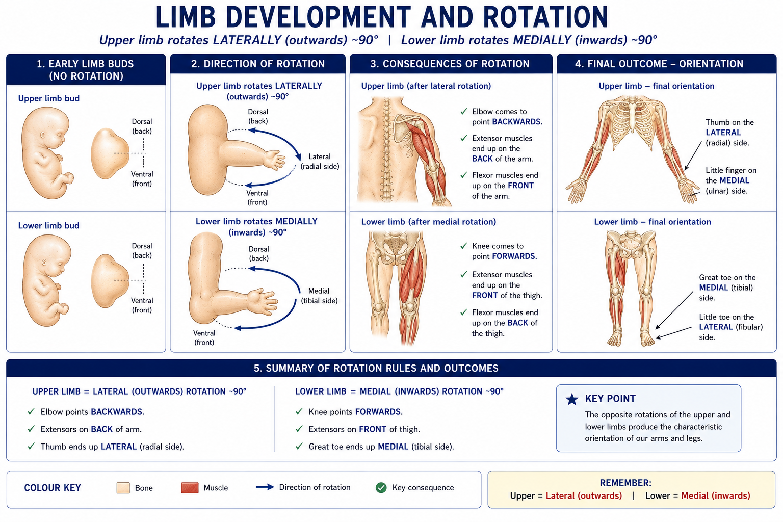
Figure 6 · Limb development and rotation
| Stage | Timing | Event |
|---|---|---|
| Limb bud appears | Week 4 (upper) and shortly after (lower) | Proliferation of lateral plate somatic mesoderm covered by ectoderm; myogenic cells migrate in from adjacent somites |
| Apical ectodermal ridge (AER) | Weeks 4–8 | A thickened ectodermal ridge at the bud apex; controls proximodistal outgrowth via FGFs. Removal truncates the limb |
| Zone of polarising activity (ZPA) | — | Posterior mesenchyme; secretes sonic hedgehog (SHH); establishes the anteroposterior (preaxial–postaxial, thumb–little finger) axis. Ectopic SHH → mirror-image duplication |
| Wnt7a / dorsal ectoderm | — | Establishes the dorsoventral axis |
| Hox genes | — | Specify segment identity along the limb |
| Digit separation | Weeks 6–8 | Interdigital apoptosis separates the digital rays. Failure → syndactyly |
| Rotation | Weeks 7–8 | Upper limb rotates laterally ~90°; lower limb rotates medially ~90° (Chapter 1) |
| Ossification | From week 8 | Endochondral, from primary centres |
Critical period: the limbs are most vulnerable to teratogens in weeks 4–8, which is exactly when thalidomide produced phocomelia and amelia.
| Condition | Developmental basis | Physiotherapy relevance |
|---|---|---|
| Spina bifida (occulta, meningocele, myelomeningocele) | Failure of caudal neuropore closure by day 28; folate-preventable | Level-dependent motor and sensory loss, neurogenic bladder and bowel, hydrocephalus, Chiari II; lifelong mobility, orthotic and skin-care management |
| Developmental dysplasia of the hip (DDH) | Abnormal acetabular development and instability, influenced by intrauterine packing and postnatal positioning | Ortolani and Barlow tests, Galeazzi sign, ultrasound screening; Pavlik harness; the swaddling posture matters — hips must be free to flex and abduct |
| Congenital talipes equinovarus (CTEV) | Multifactorial; abnormal muscle and connective tissue development | Ponseti serial casting followed by bracing; physiotherapy in bracing adherence, stretching and gait |
| Torticollis (congenital muscular) | Sternocleidomastoid fibrosis, often with a palpable mass | Positioning, stretching, active rotation, plagiocephaly management; screen for DDH, which co-occurs |
| Erb’s and Klumpke’s palsy | Traction on the brachial plexus at birth — upper roots (C5–6) or lower (C8–T1) | “Waiter’s tip” versus claw hand; positioning, range, sensory re-education, prevention of contracture and glenohumeral dysplasia |
| Arthrogryposis multiplex congenita | Fetal akinesia from any cause → multiple congenital contractures | Serial casting, splinting, strengthening, long-term function |
| Cerebral palsy | Non-progressive lesion of the developing brain (may be prenatal, perinatal or early postnatal) | The largest paediatric neurological caseload in physiotherapy |
| Syndactyly / polydactyly | Failed interdigital apoptosis / duplicated digital ray | Post-surgical hand therapy |
| Osteogenesis imperfecta; achondroplasia | Type I collagen defect; FGFR3 gain-of-function affecting the proliferative zone | Safe strengthening, mobility, fracture prevention (Chapter 2) |
15 questions on this chapter. Tap one to see the answer and the reasoning.
Answer: (C) It also houses melanocytes and Merkel cells.
Answer: (C) Which is why pigmentary and neurological anomalies co-occur in neurocristopathies.
Answer: (B) The classic exception to sympathetic adrenergic transmission.
Answer: (B) It arrives at 48–72 hours and coordinates debridement and the proliferative phase.
Answer: (C) Remodelling continues for a year or more, plateauing at 70–80% of original.
Answer: (B)
Answer: (C) It usually requires grafting and scars hypertrophically.
Answer: (A)
Answer: (C)
Answer: (B) All other skeletal muscle derives from the hypomere and is supplied by ventral rami.
Answer: (B)
Answer: (C) The ZPA (via SHH) sets the anteroposterior axis.
Answer: (B)
Answer: (B) The thalidomide window.
Answer: (C) The adrenal cortex, by contrast, is mesodermal — two different origins in one gland.
Everything on this page, in one screen
| Source | Why it earns its place |
|---|---|
| Standring S (ed.) — Gray’s Anatomy, 42nd edn | Reference account of skin and of development, integrated by region |
| Sadler TW — Langman’s Medical Embryology | The standard student embryology text; the clearest treatment of somites, germ layers and limb development |
| Moore KL, Persaud TVN, Torchia MG — The Developing Human: Clinically Oriented Embryology | Stronger on clinical correlation and congenital anomaly |
| Ross MH, Pawlina W — Histology: A Text and Atlas | Epidermal layers, appendages and cutaneous receptors |
| Sussman C, Bates-Jensen BM — Wound Care: A Collaborative Practice Manual for Health Professionals | The physiotherapy-facing reference for wound and scar management |
| EPUAP/NPIAP/PPPIA — Prevention and Treatment of Pressure Ulcers/Injuries: Clinical Practice Guideline (2019) | The current international staging and prevention standard |
| Procter F — “Rehabilitation of the burn patient”, Indian J Plast Surg, 2010 | Anti-deformity positioning and burn rehabilitation, in an Indian context |
| Lee Dellon A; and Lee MWL, McPhee RW, Stringer MD — “An evidence-based approach to human dermatomes”, Clin Anat, 2008 | The evidence on dermatome variability and overlap |
| ASIA — International Standards for Neurological Classification of Spinal Cord Injury | The key sensory and motor points you will actually use |
| Palastanga N, Field D, Soames R — Anatomy and Human Movement | The physiotherapy-facing synthesis |
Chapter 8 of 24 · Human Anatomy · Physiotherapist India End of Part 1 — General anatomy. Next: Chapter 9 — Bones of the Upper Limb, opening Part 2.
Reviewed by the Physiotherapist India Team. · Human Anatomy contents