Evidence-based physiotherapy and rehabilitation resources
Editorial & review policy
Evidence-based physiotherapy and rehabilitation resources
Editorial & review policyHuman Anatomy · The trunk
A ring of bone carrying the whole weight of the trunk into the legs, closed underneath by a sheet of muscle most people never think about until it fails. The pelvis comes first and in detail. Then the glands — because the bone you have just learnt is one of the things they spend a lifetime regulating.
Part 4 · The trunk
The pelvic floor, and the glands that regulate the body
The pelvic floor is the floor of the abdominal canister, the bottom of the load-transfer chain from spine to legs, and a muscle group that a third of adult women and a growing number of men have a clinical problem with. Pelvic health is now a recognised physiotherapy specialty precisely because these are muscles, and muscles are what we treat. Yet it remains one of the least confidently taught regions in the undergraduate curriculum — largely because of embarrassment rather than difficulty.
The endocrine glands appear in an anatomy course because they are anatomical structures with positions, relations and blood supplies. But for a physiotherapist their relevance is different and specific:
Figure 1 · The bony pelvis, male and female
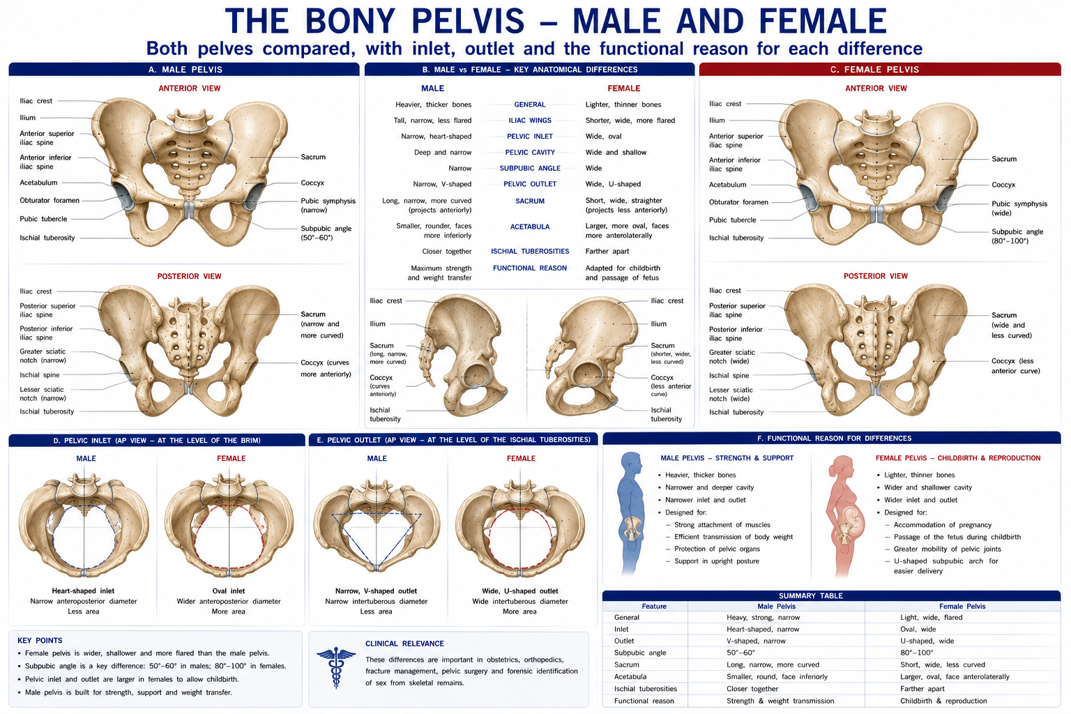
Figure 2 · Form closure and force closure
Figure 3 · Nutation and counternutation
Formed by the two hip bones, the sacrum and the coccyx, articulating at the two sacroiliac joints and the pubic symphysis.
The pelvic brim (inlet) divides it:
| Division | Boundaries | Contents |
|---|---|---|
| Greater (false) pelvis | Above the brim; iliac fossae, L5–S1 | Abdominal viscera |
| Lesser (true) pelvis | Below the brim, between the inlet and outlet | Bladder, rectum, reproductive organs |
The pelvic brim runs: sacral promontory → arcuate line of the ilium → pecten pubis → pubic crest → symphysis.
Pelvic inlet and outlet dimensions, and the male–female comparison, are covered in Chapter 13. The functional summary: the female pelvis is wider, shallower, with a rounder inlet, a wider subpubic angle (80–85° vs 50–60°) and everted ischial spines — adaptations for parturition that also give a longer abductor moment arm requirement and a larger Q angle.
| Feature | Detail |
|---|---|
| Type | An unusual composite joint: the anterior third is a synovial plane joint (with hyaline cartilage on the sacral side and fibrocartilage on the iliac side — a genuinely unique arrangement); the posterior two-thirds is a syndesmosis with the massive interosseous ligament |
| Surfaces | Auricular (ear-shaped), irregular and ridged — the ridges interlock, giving form closure |
| Ligaments | Interosseous sacroiliac (the strongest ligament in the body by cross-section), anterior and posterior sacroiliac, sacrotuberous, sacrospinous, iliolumbar |
| Movement | Very small: 2–4° of rotation and 1–2 mm of translation. Nutation = sacral base moves anteroinferiorly (the close-packed, stable position); counternutation = the reverse |
| Innervation | Posteriorly by the dorsal rami of L4–S3; anteriorly variable (L2–S2) — hence poorly localised referred pain |
Stability comes from two mechanisms:
Individual palpation and motion tests for the sacroiliac joint have poor reliability — inter-rater agreement for positional and motion palpation is consistently at or near chance level. What does perform acceptably is a cluster of pain-provocation tests (thigh thrust, distraction, compression, sacral thrust, Gaenslen’s): three or more positive out of five gives useful sensitivity and specificity against the reference standard of a diagnostic anaesthetic block.
Similarly, the notion of the SIJ being “out of place” and needing to be “put back” is not supported: the available movement is 2–4°, and no manual technique has been shown to produce a measurable, lasting positional change. Manual and exercise therapy for pelvic girdle pain works — but through load management, force closure and neurophysiological mechanisms, not through repositioning.
Pregnancy-related pelvic girdle pain is common (up to 20% of pregnancies) and responds to exercise, load management, and a pelvic belt where indicated — not to rest.
Figure 4 · The pelvic floor and the perineum
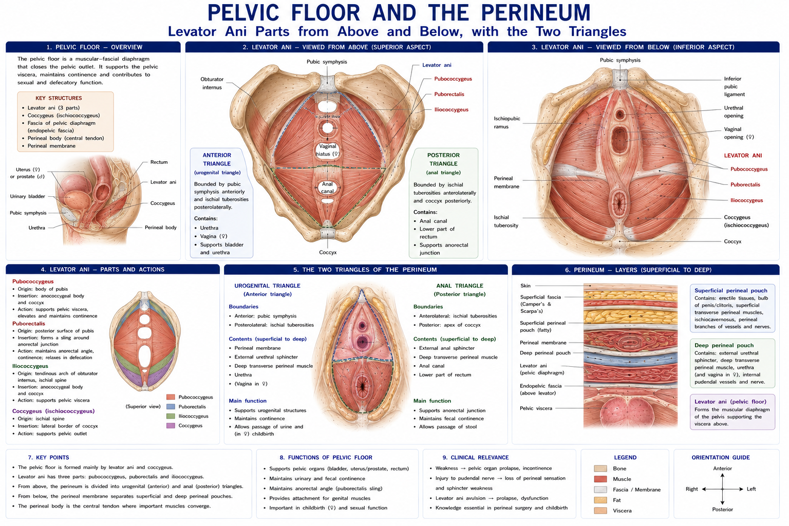
Figure 5 · The floor as the base of a canister
A funnel-shaped muscular sheet closing the pelvic outlet, supporting the viscera, maintaining continence and forming the floor of the abdominal canister.
| Muscle | Parts | Attachments | Nerve | Function |
|---|---|---|---|---|
| Levator ani | Pubococcygeus (with puborectalis and the pubovaginalis/puboprostaticus slips), iliococcygeus | From the pubis, the tendinous arch of the levator ani (over obturator internus fascia) and the ischial spine → the perineal body, anococcygeal ligament and coccyx | Nerve to levator ani (S3, S4) from above, with variable contribution from the pudendal nerve (S2–S4) below | Supports the pelvic viscera; maintains continence; puborectalis forms a U-shaped sling that pulls the anorectal junction forwards, creating the anorectal angle (~90° at rest) — the key mechanism of faecal continence |
| Coccygeus (ischiococcygeus) | — | Ischial spine → lateral coccyx and lower sacrum, on the sacrospinous ligament | S4, S5 | Supports; assists in counternutation |
The levator hiatus (urogenital hiatus) is the anterior midline gap through which the urethra, vagina (in females) and anal canal pass — necessarily a weak point, and the route of prolapse.
Diamond-shaped, divided by a line between the ischial tuberosities into:
| Triangle | Contents |
|---|---|
| Urogenital (anterior) | Deep perineal pouch: the external urethral sphincter, deep transverse perineal muscle, and (in the male) the bulbourethral glands, all within the perineal membrane. Superficial perineal pouch: ischiocavernosus, bulbospongiosus, superficial transverse perineal muscles, and the erectile tissues |
| Anal (posterior) | Anal canal, external anal sphincter, and the ischioanal (ischiorectal) fossae — fat-filled spaces permitting anal canal distension, and the site of ischioanal abscess. The pudendal canal (Alcock’s canal) in their lateral walls carries the pudendal nerve and internal pudendal vessels |
The perineal body — a fibromuscular node between the vagina/bulb of the penis and the anal canal, into which converge the bulbospongiosus, superficial and deep transverse perineal muscles, external anal sphincter, levator ani and the perineal membrane. It is the central anchor of the pelvic floor, and its disruption in obstetric tearing or episiotomy is a major cause of subsequent prolapse and incontinence.
“S2, 3, 4 keeps the pelvis off the floor.”
Leaves the pelvis through the greater sciatic foramen below piriformis, hooks around the ischial spine and sacrospinous ligament, and re-enters through the lesser sciatic foramen into the pudendal canal. Branches: inferior rectal, perineal (to the superficial pouch and the external urethral sphincter), and the dorsal nerve of the penis/clitoris.
Clinically:
The pelvic floor does five things: 1. Supports the pelvic viscera against gravity and intra-abdominal pressure. 2. Maintains urinary and faecal continence through sphincteric action and the anorectal angle. 3. Contributes to sexual function. 4. Forms the floor of the abdominal canister, co-contracting with the diaphragm and transversus abdominis and rising before an expected rise in IAP (a feedforward response, lost in many people with incontinence). 5. Contributes to lumbopelvic stability via force closure.
Continence mechanisms: urethral closure pressure exceeding bladder pressure, supported by intact urethral support (the “hammock” of the anterior vaginal wall and endopelvic fascia against which the urethra is compressed), the striated external urethral sphincter, the smooth internal sphincter, and the anatomical position of the bladder neck within the abdominal pressure zone.
| Condition | Mechanism |
|---|---|
| Stress urinary incontinence | Leakage on a rise in IAP (cough, laugh, lift, jump). Failure of urethral support or sphincter closure |
| Urgency urinary incontinence / OAB | Detrusor overactivity; a bladder problem more than a floor problem, though the floor is part of the treatment |
| Pelvic organ prolapse | Descent of the anterior wall (cystocele), posterior wall (rectocele), uterus or vault, through the levator hiatus. Graded by the POP-Q system |
| Faecal incontinence | Sphincter disruption (obstetric), pudendal neuropathy, or loss of the anorectal angle |
| Overactive (non-relaxing) pelvic floor | The mirror image: pelvic pain, dyspareunia, obstructed defecation, urinary hesitancy. Treated by downtraining, not by Kegels — a distinction that matters enormously and is frequently missed |
Supervised pelvic floor muscle training is first-line treatment for stress and mixed urinary incontinence in women, with Cochrane-level evidence: women are around five to eight times more likely to report cure than with no treatment, and it is recommended as first-line in NICE and international guidelines before surgery or medication.
It is also effective for pelvic organ prolapse symptoms (POPPY trial) and, in men, before and after radical prostatectomy, where it accelerates return of continence.
Key practice points: up to 30–50% of women cannot perform a correct contraction on verbal instruction alone — many bear down instead, which is actively harmful. Assessment of the contraction is therefore essential, and supervised programmes substantially outperform unsupervised leaflets. Training must be progressive and sustained (typically at least 3 months), and it must be integrated with breathing and load management rather than performed as isolated squeezes.
| Organ | Position and features | Clinical points |
|---|---|---|
| Bladder | Subperitoneal, behind the pubis; the trigone between the two ureteric orifices and the internal urethral orifice. When full it rises above the pubis and can be catheterised suprapubically without entering the peritoneum | Detrusor is parasympathetic (S2–S4, pelvic splanchnics) — contraction empties; the internal sphincter is sympathetic (L1–L2); the external sphincter is somatic (pudendal). Neurogenic bladder patterns after SCI: suprasacral lesions → reflex (spastic) bladder with detrusor–sphincter dyssynergia; sacral/cauda equina lesions → flaccid, areflexic bladder with retention and overflow |
| Urethra | Female ~4 cm, straight; male ~20 cm, with prostatic, membranous and spongy parts | The female urethra’s shortness explains the higher UTI rate; the male membranous urethra is the narrowest and most easily ruptured part |
| Rectum and anal canal | Rectum from S3 to the anorectal junction; anal canal ~4 cm. The pectinate (dentate) line divides endodermal from ectodermal origin | Above the line: visceral innervation, painless internal haemorrhoids, portal drainage. Below: somatic (inferior rectal nerve), painful external haemorrhoids and fissures, systemic drainage |
| Uterus | Normally anteverted and anteflexed, supported by the transverse cervical (cardinal), uterosacral and pubocervical ligaments and, principally, by the levator ani | The round ligament (through the inguinal canal) maintains anteversion but is not a major support. Retroversion is a normal variant in ~20% |
| Ovaries and uterine tubes | Ovary in the ovarian fossa, suspended by the suspensory ligament (carrying the ovarian vessels) | Ovarian pain refers to T10 (the level of gonadal development) — periumbilical and loin |
| Prostate | Below the bladder, surrounding the prostatic urethra; palpable on digital rectal examination | Benign hypertrophy affects the transitional zone (obstruction); carcinoma arises in the peripheral zone (palpable, and prone to metastasise to the vertebral column via the valveless internal vertebral venous plexus of Batson — a classic cause of back pain in an older man that must not be treated as mechanical) |
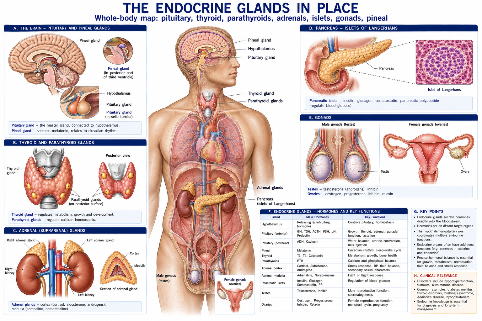
Figure 6 · The endocrine glands in place
| Gland | Position | Principal hormones | Physiotherapy relevance |
|---|---|---|---|
| Hypothalamus | Floor of the third ventricle | Releasing and inhibiting hormones; ADH and oxytocin (produced here, stored in the posterior pituitary) | Thermoregulation, autonomic control, circadian rhythm |
| Pituitary (hypophysis) | In the sella turcica of the sphenoid, below the optic chiasma | Anterior: GH, TSH, ACTH, FSH, LH, prolactin. Posterior: ADH, oxytocin | A tumour compresses the chiasma → bitemporal hemianopia. Acromegaly (GH excess in adults) → carpal tunnel syndrome, arthropathy, kyphosis, sleep apnoea, hypertension |
| Thyroid | Anterior neck, at C5–T1, in the pretracheal fascia; moves on swallowing | T3, T4 (metabolic rate); calcitonin from parafollicular C cells (neural crest) | Hypothyroidism → proximal myopathy, fatigue, carpal tunnel, adhesive capsulitis, myalgia, slowed reflex relaxation. Hyperthyroidism → proximal myopathy, osteoporosis, tremor, exercise intolerance, atrial fibrillation |
| Parathyroids | Four, on the posterior thyroid | PTH — raises serum calcium | Hyperparathyroidism → bone resorption, osteitis fibrosa cystica, fragility, proximal weakness, “stones, bones, abdominal groans and psychic moans” |
| Suprarenal (adrenal) | On the upper renal poles, retroperitoneal | Cortex (mesoderm): GFR = Glomerulosa/Aldosterone, Fasciculata/Cortisol, Reticularis/Androgens. Medulla (neural crest): adrenaline, noradrenaline | Cushing’s syndrome / exogenous corticosteroids → osteoporosis, proximal myopathy, avascular necrosis, tendon rupture, thin skin, poor healing, hyperglycaemia. Addison’s → fatigue, weakness, postural hypotension — exercise caution |
| Pancreas (islets of Langerhans) | Retroperitoneal, along the transpyloric plane | Insulin (β), glucagon (α), somatostatin (δ) | Diabetes mellitus — see below |
| Pineal | Posterior third ventricle | Melatonin | Circadian rhythm; sleep, which matters more in pain management than is usually taught |
| Thymus | Superior and anterior mediastinum; involutes after puberty | Thymosin; T-cell maturation | Myasthenia gravis association |
| Gonads | Testes (scrotum), ovaries (pelvis) | Testosterone; oestrogen and progesterone | Oestrogen deficiency → osteoporosis (Chapter 2); RED-S in athletes. Testosterone deficiency → sarcopenia |
This is the section that earns the glands their place in a physiotherapy anatomy course.
India has one of the largest diabetic populations in the world, and a physiotherapist there will see its musculoskeletal consequences daily.
| Manifestation | Mechanism |
|---|---|
| Adhesive capsulitis | Up to a five-fold increased risk; more resistant to treatment and more often bilateral |
| Limited joint mobility syndrome (diabetic cheiroarthropathy) | Non-enzymatic glycation of collagen creating irreversible cross-links (Chapter 7) → stiff, waxy skin and joint contractures. The “prayer sign” — inability to appose the palms fully |
| Dupuytren’s disease, trigger finger, carpal tunnel syndrome | All markedly more common |
| Tendinopathy | Stiffer, more glycated, less adaptable tendon; slower response to loading |
| Peripheral neuropathy | Distal symmetrical sensory loss → the diabetic foot (Chapter 16); no thermal modalities over insensate skin |
| Autonomic neuropathy | Blunted heart rate response to exercise, orthostatic hypotension, impaired thermoregulation and gastroparesis — exercise prescription must use RPE rather than heart rate, and hypoglycaemia risk rises |
| Impaired healing | Microvascular disease and glycation — every tissue timeline in this book is longer |
| Charcot neuroarthropathy | Chapter 16 — the emergency not to miss |
Exercise remains a cornerstone of diabetes management, improving glycaemic control, insulin sensitivity and cardiovascular risk. The practical cautions are hypoglycaemia (particularly with insulin or sulfonylureas), foot inspection, autonomic and retinal screening, and avoiding heavy Valsalva work in proliferative retinopathy.
Long-term corticosteroid use is one of the most common causes of secondary musculoskeletal pathology a physiotherapist will encounter:
Fluoroquinolone antibiotics deserve mention in the same breath: they carry a recognised risk of tendinopathy and rupture, particularly of the Achilles, greatly amplified by concurrent corticosteroids and by age over 60. A patient with sudden Achilles pain on a recent course of ciprofloxacin or levofloxacin needs the drug reviewed, not a loading programme.
Both under- and over-activity cause proximal myopathy, which is a common cause of “deconditioning” that does not respond to training. Hypothyroidism additionally causes carpal tunnel syndrome, adhesive capsulitis, myalgia and slow-relaxing reflexes; hyperthyroidism causes osteoporosis, tremor and exercise intolerance.
The screening implication: a patient with bilateral, symmetrical, proximal weakness and fatigue that does not improve with graded exercise should prompt consideration of a metabolic or endocrine cause and appropriate medical referral — not more exercise.
Bone is an endocrine target organ (Chapter 2). The commonest causes of secondary osteoporosis in a physiotherapy caseload are corticosteroid therapy, hypogonadism (including RED-S in athletes and post-menopausal oestrogen loss), hyperthyroidism, hyperparathyroidism and malabsorption. Recognising them changes both the exercise prescription and the referral decision.
15 questions on this chapter. Tap one to see the answer and the reasoning.
Answer: (B)
Answer: (B)
Answer: (C)
Answer: (C)
Answer: (C) “S2, 3, 4 keeps the pelvis off the floor.”
Answer: (B) Hooking around the ischial spine, where it can be blocked.
Answer: (B)
Answer: (B)
Answer: (C) Which is why assessment of the contraction is essential.
Answer: (B)
Answer: (B) Sacral and cauda equina lesions give the flaccid pattern.
Answer: (B)
Answer: (B) Up to a five-fold increased risk, and more resistant to treatment.
Answer: (B)
Answer: (B) Along with proximal myopathy and fragility fracture, it is a hallmark steroid complication.
Everything on this page, in one screen
| Source | Why it earns its place |
|---|---|
| Standring S (ed.) — Gray’s Anatomy, 42nd edn | Definitive pelvic and endocrine anatomy |
| Moore KL, Dalley AF, Agur AMR — Clinically Oriented Anatomy | The best clinical account of the pelvic floor and perineum |
| Bø K, Berghmans B, Mørkved S, Van Kampen M — Evidence-Based Physical Therapy for the Pelvic Floor | The reference text for pelvic health physiotherapy |
| Dumoulin C, Cacciari LP, Hay-Smith EJC — “Pelvic floor muscle training versus no treatment for urinary incontinence in women”, Cochrane, 2018 | The first-line evidence |
| Hagen S et al. — “Individualised pelvic floor muscle training in women with pelvic organ prolapse (POPPY)”, Lancet, 2014 | The prolapse evidence |
| Laslett M et al. — “Diagnosis of sacroiliac joint pain: validity of individual provocation tests and composites”, Aust J Physiother, 2005 | The provocation test cluster |
| Vleeming A et al. — “European guidelines for the diagnosis and treatment of pelvic girdle pain”, Eur Spine J, 2008 | Form and force closure, and clinical management |
| Goodman CC, Fuller KS — Pathology: Implications for the Physical Therapist | The definitive account of endocrine disease as it presents in physiotherapy |
| Goodman CC, Snyder TEK — Differential Diagnosis for Physical Therapists | Screening for systemic and endocrine causes |
| Chaurasia BD — Human Anatomy, Vol 2 | Indian syllabus-matched descriptive account |
Chapter 21 of 24 · Human Anatomy · Physiotherapist India End of Part 4 — The trunk. Next: Chapter 22 — Skull, Face and Neck, opening Part 5.
Reviewed by the Physiotherapist India Team. · Human Anatomy contents