Evidence-based physiotherapy and rehabilitation resources
Editorial & review policy
Evidence-based physiotherapy and rehabilitation resources
Editorial & review policyHuman Anatomy · General anatomy
Muscle is the tissue you actually treat. Everything a physiotherapist prescribes -- load, range, speed, repetition -- is an instruction to muscle, and it answers in ways that follow directly from how it is built, from the whole belly down to the sarcomere.
Part 1 · General anatomy
From whole muscle to sarcomere, fibre types, and the motor unit
Muscle is the only tissue you can instruct directly. You cannot tell a ligament to shorten or a disc to rehydrate, but you can tell a muscle to contract, and by choosing how often, how hard, how fast and through what range, you can change its size, its fibre-type expression, its neural drive, its length, its stiffness and its metabolic capacity. Every exercise prescription is an intervention on the material in this chapter.
By the end you should be able to:
Figure 1 · The three types of muscle
| Skeletal | Cardiac | Smooth | |
|---|---|---|---|
| Striated | Yes | Yes | No |
| Control | Voluntary (somatic) | Involuntary (autonomic + intrinsic) | Involuntary (autonomic, hormonal, local) |
| Cell shape | Long cylindrical syncytial fibres, up to 30 cm | Branched cells, one or two central nuclei | Fusiform, single central nucleus |
| Nuclei | Multiple, peripheral | Central | Central |
| Cell junctions | None | Intercalated discs with gap junctions and desmosomes | Gap junctions |
| T-tubules | At the A–I junction; triads (1 T-tubule + 2 terminal cisternae) | At the Z line; dyads | Absent (caveolae instead) |
| Calcium source | Sarcoplasmic reticulum, via DHPR–ryanodine receptor mechanical coupling | Extracellular Ca²⁺ triggering calcium-induced calcium release | Mainly extracellular |
| Calcium receptor | Troponin C | Troponin C | Calmodulin → myosin light chain kinase |
| Regeneration | Limited, via satellite cells | Essentially none | Good (smooth muscle cells retain mitotic capacity) |
| Fatigue | Yes | No | No |
The rest of this chapter concerns skeletal muscle, of which there are over 600 named muscles making up roughly 40–45% of body mass in a healthy adult.
Figure 2 · Levels of skeletal muscle structure
A muscle is a composite of contractile cells and a continuous connective tissue framework. The framework is not packing material: it transmits force, stores elastic energy, carries the vessels and nerves, and determines the muscle’s passive properties.
| Layer | Investing | Content and function |
|---|---|---|
| Epimysium | The whole muscle | Dense irregular collagen; continuous with deep fascia and with tendon; resists over-stretch |
| Perimysium | A fascicle (a bundle of fibres) | Carries the main intramuscular vessels and nerves; the plane a surgeon separates along; the visible “grain” of meat |
| Endomysium | Each individual muscle fibre | Fine reticular network; carries capillaries and the site of lateral force transmission between adjacent fibres (myofascial force transmission) |
| Basal lamina + sarcolemma | The fibre membrane | Satellite cells lie between basal lamina and sarcolemma — the muscle stem cell reservoir |
All three layers converge distally and continue into the tendon (or aponeurosis, its flattened form), which inserts into bone through a fibrous or fibrocartilaginous enthesis, anchored by Sharpey’s fibres.
Clinical relevance. The muscle–tendon junction is the commonest site of strain injury, because it is the stiffness transition where strain concentrates. The classic examples — the long head of biceps femoris, the medial head of gastrocnemius, rectus femoris — are all biarticular, superficial, with a high proportion of type II fibres and a long intramuscular tendon. Those four features together define the muscles you should expect to strain.
Figure 3 · Fascicular arrangements
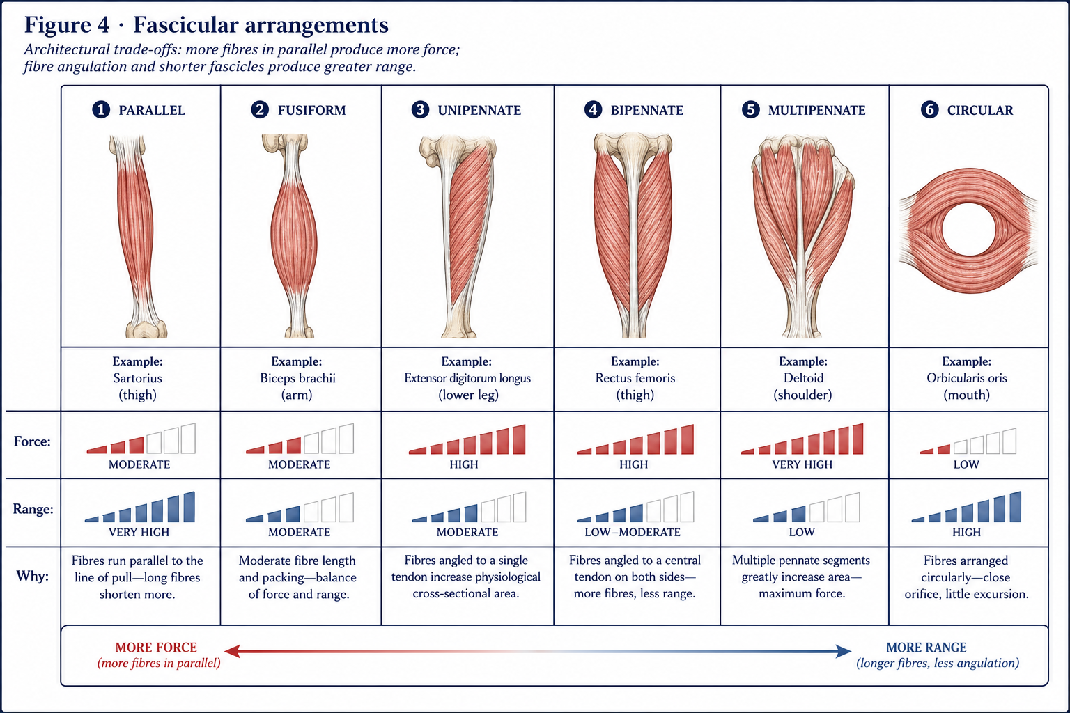
The single most useful architectural concept is physiological cross-sectional area (PCSA) — the cross-sectional area measured perpendicular to the fibres, not to the muscle. Force is proportional to PCSA (roughly 20–35 N/cm² of specific tension). Excursion and velocity are proportional to fibre length. A muscle of fixed volume must trade one against the other.
| Architecture | Description | Consequence | Examples |
|---|---|---|---|
| Parallel / strap | Fibres run the length of the muscle | Long fibres → large excursion and high velocity, low force | Sartorius, sternocleidomastoid, rectus abdominis (with tendinous intersections) |
| Fusiform | Spindle-shaped, parallel fibres with a central belly | Intermediate | Biceps brachii, brachialis |
| Unipennate | Fibres attach at an angle to one side of a tendon | Short fibres, large PCSA → high force, small excursion | Flexor pollicis longus, extensor digitorum longus, semimembranosus |
| Bipennate | Fibres on both sides of a central tendon | Higher force still | Rectus femoris, dorsal interossei, tibialis anterior |
| Multipennate | Multiple tendinous septa | Highest force, least excursion | Deltoid (middle fibres), subscapularis |
| Circular / sphincteric | Concentric fibres around an orifice | Closure | Orbicularis oris and oculi, external anal sphincter |
| Triangular / convergent | Broad origin converging on a narrow insertion | Versatile line of pull depending on which fibres act | Pectoralis major, trapezius, gluteus maximus |
| Digastric / multi-bellied | Two or more bellies with an intervening tendon | Change of direction of pull | Digastric, omohyoid |
Pennation angle costs force: only the component along the tendon (cos θ) is transmitted, so at 30° pennation about 87% of fibre force reaches the tendon. The gain in PCSA from packing more, shorter fibres into the same volume far outweighs this loss. Pennation angle increases with hypertrophy, which is why ultrasound-measured pennation is used as a training-adaptation marker.
Worked comparison. Sartorius and gluteus maximus have comparable volumes, but sartorius has very long parallel fibres and a small PCSA (large excursion, small force), while gluteus maximus has short fibres and a huge PCSA (small excursion, enormous force). The architecture is a direct statement of function: sartorius positions the limb through range; gluteus maximus extends the hip against body weight.
A single multinucleated cell, 10–100 µm in diameter, formed by fusion of myoblasts. Key components:
The contractile unit, ~2.0–2.5 µm at resting length, running Z line to Z line.
| Structure | Composition | Behaviour during contraction |
|---|---|---|
| Z line (Z disc) | α-actinin; anchors thin filaments; desmin links adjacent myofibrils at the Z line, keeping them in register | Z lines approach each other |
| I band | Thin filaments only; light, isotropic; bisected by the Z line | Shortens |
| A band | The full length of the thick filaments, including overlap; dark, anisotropic | Unchanged |
| H zone | The part of the A band with thick filaments only | Shortens / disappears |
| M line | Centre of the H zone; myomesin and creatine kinase; anchors thick filaments | Unchanged |
The single most examined fact: during contraction, the A band length does not change; the I band and H zone shorten. Filaments slide, they do not shorten — the sliding filament theory (Huxley and Huxley, 1954).
| Protein | Role |
|---|---|
| Myosin II | Thick filament. Two heavy chains with globular heads (cross-bridges) carrying actin-binding and ATPase sites, plus light chains. ~300 molecules per thick filament, arranged tail-to-tail so that the two halves pull toward the M line |
| Actin (F-actin) | Thin filament: two helical strands of G-actin monomers, each with a myosin-binding site |
| Tropomyosin | A fibrous protein lying in the actin groove, blocking the myosin-binding sites at rest |
| Troponin complex | TnT binds tropomyosin, TnI inhibits, TnC binds calcium — the calcium switch |
| Titin | The largest protein in the body; spans Z line to M line; provides passive elasticity, prevents over-stretch, keeps the thick filament centred; a major contributor to passive muscle stiffness and to residual force enhancement |
| Nebulin | Runs along the thin filament; a “molecular ruler” setting its length |
| Dystrophin | Links the actin cytoskeleton through the dystrophin–glycoprotein complex to the basal lamina, transmitting force laterally and protecting the sarcolemma. Its absence causes Duchenne muscular dystrophy; its reduction, Becker |
| Desmin | Intermediate filament linking myofibrils at the Z line to each other and to the sarcolemma |
Learn this as a sequence; it is asked in every examination and it explains several drugs and diseases.
| Step | Event |
|---|---|
| 1. Attachment | The energised myosin head (carrying ADP + Pi) binds the exposed actin site |
| 2. Power stroke | Pi is released; the head pivots ~45°, pulling the thin filament toward the M line; ADP is released |
| 3. Rigor state | Myosin remains tightly bound to actin — the state that persists in rigor mortis when ATP is exhausted |
| 4. Detachment | A new ATP binds the myosin head, reducing its affinity for actin; the head detaches |
| 5. Re-cocking | Myosin ATPase hydrolyses ATP to ADP + Pi; the head returns to the high-energy 90° position, ready to reattach |
ATP is therefore required for both contraction and relaxation. Two clinical facts follow immediately: rigor mortis (no ATP → no detachment) and the fact that a muscle in energy crisis becomes stiff, not floppy.
| Condition | Lesion | Consequence |
|---|---|---|
| Myasthenia gravis | Antibodies against postsynaptic ACh receptors (or MuSK) | Fatigable weakness, worse with activity and through the day; ptosis, diplopia, bulbar and proximal weakness; improved by anticholinesterases. Exercise prescription must be short, frequent and sub-fatiguing |
| Lambert–Eaton myasthenic syndrome | Antibodies against presynaptic voltage-gated Ca²⁺ channels; paraneoplastic (small-cell lung cancer) | Weakness that improves briefly with sustained activity; reduced reflexes that potentiate |
| Botulinum toxin | Cleaves SNARE proteins, blocking ACh release | Flaccid paralysis; therapeutically used for spasticity, with effect over ~2–4 weeks and lasting 3–4 months — the window in which stretching, casting and motor training must be delivered |
| Curare / non-depolarising blockers | Competitive ACh receptor antagonists | Surgical paralysis |
| Organophosphate poisoning | Acetylcholinesterase inhibition | Persistent depolarisation, fasciculation, then block |
| Malignant hyperthermia | Ryanodine receptor mutation | Uncontrolled SR calcium release under volatile anaesthetics; treated with dantrolene |
Active tension depends on the degree of overlap between thick and thin filaments:
Passive tension comes from the connective tissue framework and, principally, from titin. It is negligible at short lengths and rises steeply — exponentially — beyond resting length.
Total tension = active + passive. This is why a muscle can generate high total force at long lengths despite low active force, and why eccentric work at long muscle lengths (Nordic curls, Copenhagen adductor exercise) is such a potent stimulus: it loads both the contractile and the elastic elements, and it increases fascicle length by adding sarcomeres in series — an adaptation that shifts the whole curve rightward and is protective against strain injury.
Hill’s classic relationship:
The eccentric plateau is explained by forcible detachment of cross-bridges under strain plus the contribution of titin, which stiffens on calcium binding. It has three clinical consequences:
An eccentric contraction immediately followed by a concentric one produces more force than the concentric contraction alone, through: elastic energy storage in tendon and titin, stretch reflex facilitation, and the time available to build active state. It is the basis of walking, running, jumping and every plyometric exercise. The amplification is lost if the coupling time between phases exceeds roughly a second.
Figure 4 · The motor unit
A motor unit = one alpha motor neuron + all the muscle fibres it innervates. It is the smallest functional unit of voluntary movement; every fibre in a unit is of the same type and contracts together, all-or-none.
| Muscle | Approximate fibres per motor unit | Implication |
|---|---|---|
| Extraocular muscles | ~5–10 | Extremely fine control |
| Intrinsic muscles of the hand, larynx | ~100 | Fine control |
| Biceps brachii | ~750 | Moderate |
| Gastrocnemius, quadriceps | ~1,000–2,000 | Gross, powerful control |
Low innervation ratio = fine control. High innervation ratio = gross power. The motor unit territory is scattered, not clustered: the fibres of a unit are distributed across the muscle cross-section, interdigitating with other units, which produces smooth force rather than lumpy local contraction.
The nervous system increases force by two mechanisms operating together:
Most muscles complete recruitment at ~50–85% of maximum voluntary contraction, above which additional force comes from rate coding alone; the hand muscles complete recruitment much earlier.
Type II fibres are recruited only at high force or high velocity demands. It follows that:
The first 4–6 weeks of a strength programme produce large strength gains with minimal hypertrophy. These are neural: increased motor unit recruitment, increased firing rate, reduced antagonist co-contraction, improved intermuscular coordination, and reduced neural inhibition. This is directly clinically useful — it explains why an early post-operative patient gains strength quickly, and why cross-education (training the uninjured limb produces measurable strength gain in the immobilised contralateral limb, typically 5–15%) is a legitimate intervention when one side cannot be loaded.
Figure 5 · Skeletal muscle fibre types
| Type I (slow oxidative) | Type IIa (fast oxidative-glycolytic) | Type IIx (fast glycolytic) | |
|---|---|---|---|
| Contraction speed | Slow | Fast | Fastest |
| Myosin ATPase activity | Low | High | Highest |
| Fatigue resistance | High | Moderate | Low |
| Force per fibre | Low | High | Highest |
| Diameter | Small | Intermediate | Large |
| Mitochondria | Many | Many | Few |
| Capillary density | High | Intermediate | Low |
| Myoglobin | High (red) | Intermediate | Low (white) |
| Glycogen | Low–moderate | High | High |
| Principal energy system | Oxidative | Oxidative + glycolytic | Anaerobic glycolytic |
| Motor neuron | Small, low threshold | Large | Largest |
| Typical roles | Posture, endurance | Sustained power | Sprint, jump |
| Muscles rich in them | Soleus, erector spinae, deep neck flexors | Mixed muscles | Gastrocnemius, triceps, orbicularis oculi |
Human muscle is mixed, in proportions that vary by muscle, by individual and to a limited extent with training. Fibre-type conversion within the type II family (IIx ⇄ IIa) is readily achieved with training; I ⇄ II conversion is limited and requires extreme stimuli such as prolonged electrical stimulation or spinal cord injury.
Disuse and ageing. Both cause preferential type II atrophy. Sarcopenia involves loss of motor units (particularly the large, fast ones), denervation of type II fibres with partial reinnervation by slow motor neurons, fibre-type grouping, and a fall in muscle quality. The consequence is that older adults lose power faster than strength, and strength faster than mass — which is why power, not just mass, should be the training target.
| Receptor | Location | Stimulus | Afferent | Effect |
|---|---|---|---|---|
| Muscle spindle | In parallel with extrafusal fibres, encapsulated, containing intrafusal fibres (nuclear bag and nuclear chain) | Muscle length and rate of change of length | Ia (primary, rate-sensitive) and II (secondary, static) | Monosynaptic excitation of the homonymous alpha motor neuron (stretch reflex), reciprocal inhibition of antagonists |
| Golgi tendon organ | In series, at the musculotendinous junction | Tension (highly sensitive to active contraction) | Ib | Di/polysynaptic inhibition of the homonymous muscle (autogenic inhibition), excitation of antagonists |
| Free nerve endings | Throughout | Pain, metabolites, pressure | III (Aδ) and IV (C) | Nociception; the metaboreflex driving the cardiovascular response to exercise |
Gamma motor neurons innervate the contractile poles of intrafusal fibres. Their function is alpha–gamma co-activation: as the muscle shortens under alpha drive, gamma drive keeps the spindle taut so it continues to signal. Without this the spindle would fall slack and go silent during contraction.
Blood supply enters at the neurovascular hilum and forms an extensive capillary bed running longitudinally in the endomysium, with 3–5 capillaries per type I fibre. Muscle blood flow rises 20-fold or more during exercise. Because intramuscular pressure occludes flow during contraction, sustained isometric contraction above roughly 15–20% MVC progressively restricts its own perfusion — which is why isometric holds fatigue disproportionately and why they raise blood pressure through the pressor reflex.
Energy systems, in order of onset:
| System | Fuel | Duration of dominance | Power |
|---|---|---|---|
| Phosphagen (ATP–PCr) | Stored ATP, phosphocreatine | ~0–10 s | Highest |
| Anaerobic glycolysis | Muscle glycogen → lactate | ~10 s – 2 min | High |
| Oxidative phosphorylation | Glycogen, glucose, fatty acids, some protein | >2 min, indefinitely | Lowest, but greatest capacity |
Fatigue is not lactic acid. Contemporary accounts distinguish:
Mechanical tension is the principal driver, acting through mechanotransduction → mTOR pathway → increased myofibrillar protein synthesis, with metabolic stress and muscle damage as secondary contributors. Adaptation includes increased myofibril number and size, increased pennation angle, and satellite cell donation of new myonuclei to maintain the myonuclear domain.
Myonuclear permanence (“muscle memory”): nuclei acquired during hypertrophy appear to be retained through subsequent atrophy, which provides a cellular explanation for the rapidity with which previously trained muscle regains size. This is a directly usable message for a patient facing a long immobilisation.
Driven by the ubiquitin–proteasome system (atrogin-1/MAFbx and MuRF1) and autophagy. Rates are sobering: quadriceps cross-sectional area falls measurably within a week of immobilisation, and disuse atrophy is greatest when the muscle is immobilised in a shortened position — which also causes loss of sarcomeres in series and true contracture. Immobilising in a lengthened position preserves both length and mass far better, and this should guide splinting and positioning decisions.
Delayed-onset muscle soreness follows unaccustomed, predominantly eccentric work: onset 6–12 h, peak 24–72 h, resolution by 5–7 days. Mechanism: sarcomere strain and popping, Z-line streaming, membrane and excitation–contraction coupling disruption, calcium influx and a secondary inflammatory response sensitising group III/IV afferents. Creatine kinase rises and peaks later; strength deficits and reduced range accompany it.
The repeated-bout effect is the striking protective adaptation: a single bout of eccentric work confers substantial protection against damage from a repeat bout for weeks. It is why load should be introduced gradually and why an early sub-maximal eccentric exposure is a legitimate way to prepare an athlete.
DOMS is not a marker of a good session, nor an injury. Its clinical value is as a warning that load was progressed too fast.
Satellite cells (Pax7-positive), quiescent between basal lamina and sarcolemma, activate after injury, proliferate as myoblasts, and either fuse with damaged fibres, fuse into new myotubes, or return to quiescence to replenish the pool. Regeneration is effective when the basal lamina scaffold survives; where it is destroyed (complete tear, deep laceration, severe contusion), fibrous scar forms instead, with reduced compliance and a predisposition to re-injury.
Muscle strain grading and the practical implication:
| Grade | Description | Typical timeline |
|---|---|---|
| I | Few fibres, minimal strength loss, full or near-full range | Days–2 weeks |
| II | Partial tear, definite strength and range loss, palpable defect possible | 3–8 weeks |
| III | Complete rupture, palpable gap, marked weakness, retraction | Months; often surgical |
Rehabilitation principles: brief protection, then early controlled loading (which orients regenerating fibres and limits scar), progressive eccentric work at long muscle lengths, restoration of fascicle length, and criteria-based rather than time-based return to sport. Prolonged immobilisation produces a larger, denser, more disorganised scar.
15 questions on this chapter. Tap one to see the answer and the reasoning.
Answer: (C) The A band equals the thick filament length, which is constant.
Answer: (B) Calmodulin is the smooth-muscle pathway.
Answer: (C) Absence of ATP produces the rigor state.
Answer: (B) The Golgi tendon organ is the in-series tension receptor.
Answer: (B)
Answer: (C) Eccentric force plateaus at ~1.2–1.8 × maximum isometric.
Answer: (C) Fibre length determines excursion and velocity.
Answer: (C)
Answer: (C) Titin spans Z line to M line and behaves as a molecular spring.
Answer: (B) Dystrophin links the cytoskeleton to the basal lamina, protecting the sarcolemma during contraction.
Answer: (B) Improvement with activity suggests Lambert–Eaton.
Answer: (B) Shortened immobilisation causes both greater atrophy and loss of sarcomeres in series.
Answer: (B) It follows unaccustomed eccentric work and is protected against by the repeated-bout effect.
Answer: (B) Loss of the scaffold results in fibrous scarring.
Answer: (B) It is a neural phenomenon, useful when one limb cannot be loaded.
Everything on this page, in one screen
| Source | Why it earns its place |
|---|---|
| MacIntosh BR, Gardiner PF, McComas AJ — Skeletal Muscle: Form and Function | The definitive single text on everything in this chapter |
| Lieber RL — Skeletal Muscle Structure, Function and Plasticity | The reference on architecture, PCSA and adaptation, written for rehabilitation |
| Standring S (ed.) — Gray’s Anatomy, 42nd edn | Descriptive and histological reference |
| Hall JE — Guyton and Hall Textbook of Medical Physiology | The clearest sequential account of excitation–contraction coupling |
| Huxley AF, Niedergerke R; Huxley HE, Hanson J — Nature, 1954 | The two original sliding-filament papers |
| Henneman E, Somjen G, Carpenter DO — J Neurophysiol, 1965 | The size principle |
| Enoka RM, Duchateau J — “Rate coding and the control of muscle force”, Cold Spring Harb Perspect Med, 2017 | Modern account of force gradation |
| Schoenfeld BJ — Science and Development of Muscle Hypertrophy | Evidence-based translation into training prescription |
| Järvinen TAH et al. — “Muscle injuries: biology and treatment”, Am J Sports Med, 2005 | The reference account of strain injury and regeneration |
| Palastanga N, Field D, Soames R — Anatomy and Human Movement | The physiotherapy-facing synthesis |
Chapter 5 of 24 · Human Anatomy · Physiotherapist India Next: Chapter 6 — How Muscles Work Together: agonists and fixators, contraction types, levers, and the two insufficiencies.
Reviewed by the Physiotherapist India Team. · Human Anatomy contents