Evidence-based physiotherapy and rehabilitation resources
Editorial & review policy
Evidence-based physiotherapy and rehabilitation resources
Editorial & review policyHuman Anatomy · Lower limb
The knee carries more load than any other joint and has almost no bony stability to do it with. A large round femur sits on a nearly flat tibia, and everything holding that arrangement together is soft tissue. It is the most commonly injured joint in the body, and this chapter explains why.
Part 3 · The lower limb
The knee joint, menisci and cruciates, and the compartments of the leg
The knee has to do two incompatible things. It must permit a large sagittal range — around 140° — so that we can squat, sit, climb and run. And it must be stable under loads of several times body weight, in a joint whose articular surfaces are two convex femoral condyles sitting on a nearly flat tibial plateau.
There is almost no bony congruence. Every bit of stability is bought from ligaments, menisci and muscle. That is the whole explanation for the knee’s clinical profile: the highest ligament injury burden of any joint, the highest meniscal injury rate, and one of the highest rates of symptomatic osteoarthritis.
Figure 1 · The knee joint
The joint is classified as a modified hinge (bicondylar) synovial joint with two degrees of freedom: flexion–extension, and axial rotation (available only in flexion).
The capsule is deficient anteriorly, where the quadriceps tendon, patella and patellar ligament replace it, and is reinforced by the medial and lateral patellar retinacula.
The synovial membrane is the most extensive in the body and has an unusual arrangement: it invaginates from behind to cover the cruciates, so the cruciate ligaments are intracapsular but extrasynovial.
| Bursa | Location | Clinical |
|---|---|---|
| Suprapatellar | Between quadriceps and femur; communicates with the joint | Distends with any effusion — hence the “milking” or sweep test, and the patellar tap |
| Prepatellar | Subcutaneous over the patella | “Housemaid’s knee” — from kneeling upright |
| Superficial infrapatellar | Over the patellar ligament | “Clergyman’s knee” — from kneeling upright with the trunk erect |
| Deep infrapatellar | Between patellar ligament and tibia | Involved in Osgood–Schlatter and patellar tendinopathy |
| Pes anserine | Beneath the conjoint tendon, medial tibia | A common cause of medial knee pain mimicking meniscal or MCL pathology |
| Semimembranosus / gastrocnemius (popliteal) | Posteromedial | Distension = Baker’s (popliteal) cyst, usually secondary to intra-articular pathology; rupture mimics DVT |
The infrapatellar (Hoffa’s) fat pad deserves separate mention: it is one of the most densely innervated structures in the knee, a genuine and under-recognised source of anterior knee pain, and it is impinged in extension when swollen or when the knee hyperextends.
| Movement | Range | Notes |
|---|---|---|
| Flexion | 0–135–140° (150° passive) | Limited by soft-tissue apposition |
| Extension | 0° (5–10° hyperextension common) | Limited by the posterior capsule, cruciates and collaterals |
| Internal rotation of the tibia | ~10° | Available only in flexion |
| External rotation of the tibia | ~30–45° | Available only in flexion; greatest at ~90° |
Close-packed position: full extension with lateral rotation of the tibia. Resting position: ~25° flexion. Capsular pattern: flexion grossly more limited than extension (e.g. a knee losing 90° of flexion may lose only 10° of extension).
In the last ~20–30° of extension, the tibia rotates laterally ~10° on the femur (or, in weight bearing, the femur rotates medially on a fixed tibia). This is an automatic, obligatory, passive movement — not under voluntary control.
Why it happens — three contributing mechanisms: 1. The medial femoral condyle’s articular surface is longer than the lateral. As extension proceeds, the lateral compartment runs out of surface first, and continued extension forces the tibia to spin laterally around the medial condyle. 2. The anterior cruciate ligament tightens in extension, drawing the tibia into lateral rotation. 3. The lateral pull of the quadriceps contributes.
What it achieves: in full extension the joint is close-packed, the collaterals and cruciates are taut, the menisci are maximally congruent, and the knee is locked — allowing prolonged standing with minimal quadriceps activity, which is metabolically essential.
Unlocking requires popliteus, which internally rotates the tibia (or externally rotates the femur in weight bearing) at the start of flexion. Popliteus is a small muscle with a disproportionately important job, and it also retracts the lateral meniscus and acts as a posterolateral stabiliser.
Clinically: a knee that cannot achieve full extension cannot lock, so the quadriceps must work continuously in standing — which is fatiguing, painful and one reason that restoring full extension is the first priority after knee injury or surgery, ahead of flexion. A flexion contracture of even 10° imposes a substantial and continuous quadriceps demand.
Two semilunar fibrocartilaginous wedges, triangular in cross-section, thick peripherally and thin centrally, covering ~60–70% of the tibial plateau surface.
| Medial meniscus | Lateral meniscus | |
|---|---|---|
| Shape | C-shaped, larger radius | Almost circular (O-shaped), smaller |
| Coverage of plateau | ~50% | ~70% |
| Peripheral attachment | Firmly attached to the capsule and to the deep MCL | Attached to the capsule except at the popliteal hiatus, where popliteus passes |
| Mobility | Less mobile (~5 mm) | More mobile (~11 mm) |
| Injury rate | Higher — precisely because it is tethered | Lower |
| Additional attachments | — | Meniscofemoral ligaments (of Humphrey, anterior to PCL; of Wrisberg, posterior to PCL); popliteus |
Collagen fibres are predominantly circumferential, converting compressive load into hoop stress resisted at the anterior and posterior root attachments, with radial “tie” fibres preventing longitudinal splitting.
The root tear. If a meniscal root is avulsed, hoop stress can no longer be contained, the meniscus extrudes peripherally, and the joint behaves as though the meniscus were absent. A root tear is biomechanically equivalent to a total meniscectomy — which is why root repair is now pursued aggressively rather than treated as a degenerative finding.
Vascular zones (Arnoczky and Warren):
| Zone | Vascularity | Healing |
|---|---|---|
| Red–red (peripheral third) | Perimeniscal capillary plexus | Heals — repairable |
| Red–white (middle third) | Marginal | Variable |
| White–white (inner third) | Avascular, nourished by diffusion | Does not heal |
Vascularity declines with age: the whole meniscus is vascular at birth, and only the peripheral 10–30% remains so in adulthood.
Longitudinal, bucket-handle (a displaced longitudinal tear producing a springy block to extension — a locked knee), radial, horizontal cleavage (typically degenerative), flap, complex, and root tears.
A crucial distinction in management:
This is one of the clearest examples in musculoskeletal medicine of imaging findings driving unnecessary surgery, and it is worth being able to explain to patients who arrive with an MRI report and an expectation.
Figure 2 · What holds the knee together
Named for their tibial attachments, and crossing each other like the limbs of an X.
| Anterior cruciate ligament (ACL) | Posterior cruciate ligament (PCL) | |
|---|---|---|
| From | Anterior intercondylar area of the tibia | Posterior intercondylar area of the tibia |
| To | Posteromedial aspect of the lateral femoral condyle | Anterolateral aspect of the medial femoral condyle |
| Bundles | Anteromedial (taut in flexion) and posterolateral (taut in extension) | Anterolateral (taut in flexion) and posteromedial (taut in extension) |
| Primary function | Resists anterior translation of the tibia on the femur (~85% of the restraining force); also resists internal rotation and hyperextension | Resists posterior translation of the tibia (~95%); the primary restraint |
| Strength | Weaker; ~2,160 N | Stronger, roughly twice the ACL |
| Taut | In extension | In flexion |
| Blood supply | Middle genicular artery; poor, and bathed in synovial fluid | Middle genicular; better |
| Injury mechanism | Non-contact deceleration, pivoting, or landing with the knee near extension in valgus and internal rotation (~70% non-contact) | Dashboard injury; fall on the flexed knee with the foot plantarflexed |
ACL rupture produces an immediate haemarthrosis within 2 hours (the ligament is vascular and intracapsular), a “pop”, and giving way on pivoting. The Lachman test (30° flexion) is the most accurate single test (sensitivity ~85%, specificity ~94%); the pivot shift is the most specific (~98%) but hardest to perform in an acute knee; the anterior drawer at 90° is the least accurate.
Why the ACL does not heal: intra-articular position, synovial fluid preventing fibrin clot formation, and poor vascularity (Chapter 7). Hence reconstruction rather than repair — though non-operative management with structured rehabilitation is a legitimate option for many patients, and the decision is now framed around activity demands rather than automatic surgery.
Female ACL injury risk is 2–8 times that of males in comparable sports, attributed to a combination of anatomical (wider pelvis, greater Q angle, narrower intercondylar notch, steeper tibial slope), hormonal, and — most importantly, because it is modifiable — neuromuscular factors (dynamic valgus on landing, quadriceps-dominant strategies, reduced hamstring co-activation). Neuromuscular training programmes reduce ACL injury incidence by roughly 50%, and by more in females, making prevention one of the highest-value interventions in sports physiotherapy.
| Ligament | Attachments | Function | Notes |
|---|---|---|---|
| Medial (tibial) collateral ligament (MCL) | Medial epicondyle → medial tibia, ~7–10 cm below the joint line. Superficial and deep layers; the deep layer attaches to the medial meniscus | Primary restraint to valgus; secondary restraint to external rotation and anterior translation | Broad, flat, extracapsular (superficial layer), and well vascularised — so it heals well without surgery. Its meniscal attachment is why MCL and medial meniscal injuries occur together |
| Lateral (fibular) collateral ligament (LCL) | Lateral epicondyle → head of fibula | Primary restraint to varus | Cord-like, extracapsular, and NOT attached to the lateral meniscus — popliteus tendon intervenes |
| Posteromedial corner | Posterior oblique ligament, semimembranosus expansions, oblique popliteal ligament | Rotatory stability medially | |
| Posterolateral corner (PLC) | LCL, popliteus tendon, popliteofibular ligament, arcuate ligament, lateral capsule | Resists varus, external rotation and posterior translation | PLC injury missed alongside a PCL or ACL reconstruction is the commonest cause of graft failure — hence the dial test at 30° and 90° |
The “unhappy triad” (O’Donoghue) — ACL + MCL + medial meniscus — from a valgus, externally rotating force on a planted foot. Modern imaging shows the lateral meniscus is in fact more often torn in acute ACL injuries, so the classical triad is taught as history rather than as current fact.
| Test | Structure | Note |
|---|---|---|
| Lachman (20–30° flexion) | ACL | The most accurate single test |
| Anterior drawer (90°) | ACL | Less sensitive; hamstring guarding confounds it |
| Pivot shift | ACL (rotatory instability) | Most specific; difficult acutely |
| Posterior drawer / posterior sag / quadriceps active test | PCL | The sag sign at 90° is the most reliable |
| Valgus stress at 0° and 30° | MCL (30°); MCL + cruciates + capsule (0°) | Laxity at 0° implies a major multi-ligament injury |
| Varus stress at 0° and 30° | LCL and PLC | Same principle |
| Dial test (30° and 90°) | PLC (30° only) vs PLC + PCL (both) | |
| McMurray, Thessaly, joint line tenderness, Apley | Meniscus | Individually modest; a cluster performs better than any single test, and joint line tenderness is the most sensitive |
Ottawa knee rules determine the need for radiography after acute knee injury: age ≥55, isolated patellar tenderness, fibular head tenderness, inability to flex to 90°, or inability to weight-bear four steps both immediately and in the department.
A diamond-shaped space behind the knee.
| Boundary | Structure |
|---|---|
| Superolateral | Biceps femoris |
| Superomedial | Semimembranosus and semitendinosus |
| Inferolateral | Lateral head of gastrocnemius (+ plantaris) |
| Inferomedial | Medial head of gastrocnemius |
| Floor | Popliteal surface of the femur, posterior knee capsule with the oblique popliteal ligament, and popliteus |
| Roof | Popliteal fascia (dense, and the reason a popliteal aneurysm or abscess causes early pressure symptoms), pierced by the small saphenous vein |
Contents, from superficial to deep — the order matters and is examinable:
Plus the small saphenous vein, popliteal lymph nodes, and the posterior femoral cutaneous nerve.
Clinical: because the artery is the deepest structure and lies against the capsule, it is vulnerable in supracondylar femoral fractures and knee dislocation — popliteal artery injury complicates up to a third of knee dislocations, and a knee dislocation is therefore a limb-threatening emergency requiring vascular assessment (ankle–brachial index and often CT angiography) regardless of palpable pulses. The popliteal artery is also the second commonest site of peripheral aneurysm.
Popliteal (Baker’s) cyst: distension of the semimembranosus–gastrocnemius bursa, usually secondary to an intra-articular problem in adults (meniscal tear, osteoarthritis, inflammatory arthritis). Rupture produces acute calf pain and swelling that closely mimics DVT — and it is the physiotherapist who often sees it first.
Figure 3 · Compartments of the leg
Divided by the interosseous membrane, the anterior and posterior intermuscular septa, and the transverse intermuscular septum.
| Compartment | Muscles | Nerve | Artery | Action |
|---|---|---|---|---|
| Anterior | Tibialis anterior, extensor hallucis longus, extensor digitorum longus, fibularis tertius | Deep fibular (L4, L5, S1) | Anterior tibial | Dorsiflexion and toe extension; inversion (TA) |
| Lateral | Fibularis longus, fibularis brevis | Superficial fibular (L5, S1, S2) | Branches of fibular artery (no artery of its own) | Eversion; weak plantarflexion; fibularis longus supports the transverse and lateral arches |
| Superficial posterior | Gastrocnemius, soleus, plantaris | Tibial (S1, S2) | Posterior tibial (sural branches) | Powerful plantarflexion; gastrocnemius also flexes the knee |
| Deep posterior | Tibialis posterior, flexor digitorum longus, flexor hallucis longus, popliteus | Tibial (L4–S3) | Posterior tibial and fibular | Plantarflexion, inversion (TP), toe flexion; popliteus unlocks the knee |
| Muscle | Origin | Insertion | Functional note |
|---|---|---|---|
| Tibialis anterior | Lateral tibial condyle and upper shaft, interosseous membrane | Medial cuneiform and base of 1st metatarsal | The strongest dorsiflexor; eccentrically controls foot lowering after heel strike — its failure produces foot slap |
| Extensor hallucis longus | Middle fibula, interosseous membrane | Base of distal phalanx of hallux | Tested to assess L5 |
| Gastrocnemius | Femoral condyles (above the knee) | Calcaneal tendon | Biarticular — tested with the knee extended; a fast, powerful, type-II-rich propulsive muscle |
| Soleus | Soleal line of tibia, posterior fibula | Calcaneal tendon | Monoarticular, type-I-rich, postural; the “peripheral heart” whose contraction pumps venous blood back to the trunk. Tested with the knee flexed |
| Tibialis posterior | Interosseous membrane, adjacent tibia and fibula | Navicular tuberosity, with slips to all tarsals except the talus and to metatarsals 2–4 | The principal dynamic support of the medial longitudinal arch; its dysfunction is the commonest cause of acquired adult flatfoot |
| Fibularis longus | Head and upper fibula | Crosses the sole to the medial cuneiform and base of 1st metatarsal | With tibialis posterior forms the “stirrup” supporting the arches; plantarflexes the first ray at push-off |
| Popliteus | Lateral femoral condyle (intracapsular), running to the posterior tibia | Posterior tibia above the soleal line | Unlocks the knee; retracts the lateral meniscus; posterolateral stabiliser |
The calcaneal (Achilles) tendon — the thickest and strongest tendon in the body, formed from gastrocnemius and soleus, spiralling ~90° as it descends (which contributes to the stress concentration in its watershed zone, 2–6 cm above the insertion). It withstands loads of 6–12 × body weight in running.
The plantaris — small, with a very long tendon; absent in 7–20% of people; commonly harvested for graft; its rupture is one differential for acute calf pain.
Figure 4 · Cross-section of the leg and the popliteal fossa
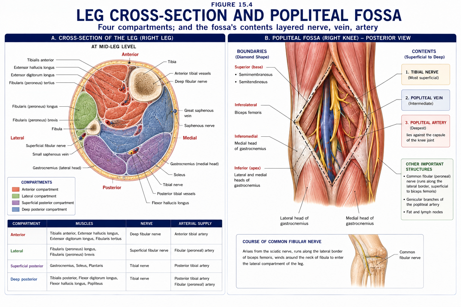
Passes vertically through the popliteal fossa, deep to the soleal arch (a potential entrapment point) into the deep posterior compartment, and runs with the posterior tibial vessels to pass behind the medial malleolus in the tarsal tunnel, dividing into medial and lateral plantar nerves.
Runs along the medial border of biceps femoris, crosses the lateral head of gastrocnemius, and winds subcutaneously around the neck of the fibula — where it is the most commonly injured nerve in the lower limb. It divides within fibularis longus into:
Common fibular nerve palsy produces:
Causes: fibular neck fracture, tight plaster or bandage, prolonged squatting or leg crossing, positioning during surgery or in the unconscious patient, ganglion, and weight loss (loss of protective fat). Every physiotherapist positioning or splinting a limb must know this.
The popliteal artery divides at the lower border of popliteus into:
Venous return depends on the calf muscle pump acting on the deep veins within their fascial envelope, with valves directing flow proximally and from superficial to deep via perforators. Immobility, valve incompetence and hypercoagulability form Virchow’s triad.
Presentation: unilateral calf pain, swelling (measure calf circumference 10 cm below the tibial tuberosity — a difference >3 cm is significant), warmth, erythema, and tenderness along the deep venous system. Homans’ sign is unreliable and should not be used to rule DVT in or out.
Use the Wells score and refer for D-dimer and ultrasound rather than relying on clinical signs. Differentials include ruptured Baker’s cyst, calf muscle tear, cellulitis and superficial thrombophlebitis.
If DVT is suspected, do not massage, do not perform vigorous passive movement, and do not apply compression until it has been excluded or treated — the risk is pulmonary embolism. Once anticoagulation is established, early mobilisation is safe and is recommended.
The commonest knee complaint in adolescents and young adults, and in runners.
Pain at the inferior pole of the patella, in jumping and change-of-direction athletes; a load-related tendinopathy following the continuum model (Chapter 7). Managed with isometrics for pain, then progressive heavy slow resistance or eccentric decline squats, plus jump-landing mechanics and load management. Deep knee flexion under load compresses the tendon against the patella — so, as with other insertional tendinopathies, deep painful loading early is counterproductive.
Lateral knee pain, classically in runners and cyclists, at around 30° of knee flexion where the ITB is closest to the lateral femoral epicondyle.
Exercise-induced pain along the posteromedial tibial border, over a length of ≥5 cm, in runners and military recruits.
| Midportion tendinopathy | Insertional tendinopathy | Rupture | |
|---|---|---|---|
| Site | 2–6 cm above insertion (the watershed zone) | At the calcaneal insertion | 2–6 cm above insertion |
| Aggravated by | Load | Load and compression in dorsiflexion | — |
| Management | Eccentric (Alfredson) or heavy slow resistance loading | Same, but avoid dorsiflexion beyond neutral early; heel raise | Non-operative functional bracing or surgical repair; comparable outcomes with modern accelerated protocols |
| Presentation of rupture | — | — | Sudden “kick in the back of the leg”, audible pop, inability to heel-raise on one leg, palpable gap, positive Simmonds/Thompson test (no plantarflexion on calf squeeze) |
The Thompson (Simmonds’ calf squeeze) test is the single most useful bedside test and is often the only one needed. Around 20–25% of Achilles ruptures are missed at first presentation, usually because some plantarflexion is preserved by the deep posterior compartment muscles and the examiner is reassured by it.
15 questions on this chapter. Tap one to see the answer and the reasoning.
Answer: (B)
Answer: (B)
Answer: (B) Unlocked by popliteus, which internally rotates the tibia.
Answer: (B)
Answer: (B)
Answer: (B) Hoop stress can no longer be contained and the meniscus extrudes.
Answer: (B) The pivot shift is the most specific but hardest to perform acutely.
Answer: (B)
Answer: (D) Hence its vulnerability in knee dislocation.
Answer: (C) A limb-threatening emergency requiring vascular assessment regardless of pulses.
Answer: (B)
Answer: (B) L5 radiculopathy also weakens inversion and hip abduction.
Answer: (B)
Answer: (C) Around a quarter of Achilles ruptures are missed at first presentation.
Answer: (B)
Everything on this page, in one screen
| Source | Why it earns its place |
|---|---|
| Standring S (ed.) — Gray’s Anatomy, 42nd edn | Definitive regional anatomy |
| Neumann DA — Kinesiology of the Musculoskeletal System | Knee arthrokinematics, screw-home and joint forces |
| Palastanga N, Field D, Soames R — Anatomy and Human Movement | Functional anatomy of knee and leg |
| Magee DJ — Orthopedic Physical Assessment | Knee special tests and their accuracy |
| Arnoczky SP, Warren RF — Am J Sports Med, 1982 | The meniscal vascular zones |
| Thorlund JB et al. — “Arthroscopic surgery for degenerative knee: systematic review and meta-analysis”, BMJ, 2015; Sihvonen R et al. (FIDELITY), NEJM, 2013 | The evidence against arthroscopy for degenerative tears |
| Collins NJ et al. — “2018 Consensus statement on exercise therapy and physical interventions for patellofemoral pain”, Br J Sports Med | Current PFP management |
| Fairclough J et al. — “The functional anatomy of the iliotibial band during flexion and extension of the knee”, J Anat, 2006 | The compression rather than friction model |
| Webster KE, Hewett TE — ACL injury prevention programme meta-analyses, J Orthop Res | Prevention effect sizes |
| Chaurasia BD — Human Anatomy, Vol 2 | Indian syllabus-matched descriptive account |
Chapter 15 of 24 · Human Anatomy · Physiotherapist India Next: Chapter 16 — Ankle and Foot: the ankle mortise, its ligaments, and the arches of the foot.
Reviewed by the Physiotherapist India Team. · Human Anatomy contents