Evidence-based physiotherapy and rehabilitation resources
Editorial & review policy
Evidence-based physiotherapy and rehabilitation resources
Editorial & review policyHuman Anatomy · General anatomy
Every joint in the body is a settlement between two things that cannot both be maximised: how far it moves and how well it holds. Learn where a joint sits on that scale and most of what you need to know about it follows.
Part 1 · General anatomy
How joints are classified, what makes them stable, and what stops them moving
Figure 1 · The stability and mobility trade-off
Every joint in the body is a solution to the same engineering problem, and every joint solves it differently:
Mobility and stability are inversely related. A joint that moves freely is easy to displace; a joint that is hard to displace does not move much.
The shoulder chose mobility and pays for it with dislocation. The hip chose stability and pays for it with a restricted range that the lumbar spine must compensate for. The sacroiliac joint chose stability almost entirely. The knee, uniquely, was asked for both — a large sagittal range with weight-bearing stability in a joint with almost no bony congruence — and the price is the highest ligament and meniscal injury burden in the body.
When you assess any joint, you are asking where on that spectrum it sits, and whether disease or injury has moved it.
By the end of this chapter you should be able to:
Figure 2 · Classification of joints by structure
Two classifications run in parallel. Learn both, and learn how they map onto each other.
| Class | Uniting material | Joint cavity? | Subtypes |
|---|---|---|---|
| Fibrous | Dense fibrous connective tissue | No | Suture, syndesmosis, gomphosis |
| Cartilaginous | Cartilage | No | Primary (synchondrosis, hyaline); Secondary (symphysis, fibrocartilage) |
| Synovial | A fluid-filled cavity enclosed by a capsule | Yes | Plane, hinge, pivot, condyloid, saddle, ball-and-socket |
| Class | Movement | Typically corresponds to |
|---|---|---|
| Synarthrosis | Immovable | Sutures, gomphoses, synchondroses |
| Amphiarthrosis | Slightly movable | Syndesmoses, symphyses |
| Diarthrosis | Freely movable | All synovial joints |
The mapping is not perfect — the interosseous membrane of the forearm is a fibrous syndesmosis but permits substantial movement, and the first sternocostal joint is a synchondrosis that behaves as a synarthrosis. State the structural class first and the functional consequence second.
| Type | Structure | Examples | Clinical note |
|---|---|---|---|
| Suture | Thin layer of fibrous tissue between interlocking margins of skull bones. Types: plane (internasal), serrate (sagittal), squamous (temporoparietal), denticulate (lambdoid), schindylesis (vomer in the sphenoidal rostrum) | Cranial vault sutures | Permit moulding at birth and vault growth in infancy; fontanelles are wide membranous sutural regions. Sutures ossify with age (synostosis), and premature fusion is craniosynostosis. Sutural mobility is the subject of persistent but poorly supported claims in some manual traditions |
| Syndesmosis | Bones united by an interosseous ligament or membrane, permitting some movement | Inferior tibiofibular joint; interosseous membranes of forearm and leg | The “high ankle sprain” is a distal tibiofibular syndesmotic injury: longer recovery than a lateral ligament sprain, and instability requires fixation. The forearm interosseous membrane transmits load from radius to ulna and is torn in the Essex-Lopresti injury |
| Gomphosis | Peg-in-socket: tooth root in alveolus, held by the periodontal ligament | Tooth–alveolar joint | The only true gomphosis. The periodontal ligament is proprioceptive and load-adaptive |
| Type | Uniting cartilage | Examples | Notes |
|---|---|---|---|
| Primary (synchondrosis) | Hyaline | Epiphyseal plate; first sternocostal joint; spheno-occipital synchondrosis; costochondral junctions | Usually temporary, being replaced by bone at maturity. Immovable while present. The epiphyseal plate is a joint by definition and the weak link of the growing skeleton |
| Secondary (symphysis) | Fibrocartilage, with a thin hyaline layer on each articular surface | Intervertebral (interbody) joints; pubic symphysis; manubriosternal joint; mandibular symphysis (fuses in infancy) | Permanent, midline, slightly movable, strong. Collectively the intervertebral symphyses account for the majority of spinal movement despite each contributing little |
Clinical note — the pubic symphysis in pregnancy. Relaxin and progesterone increase symphyseal and sacroiliac laxity, allowing physiological widening (normally up to ~10 mm; beyond this suggests symphyseal diastasis). Pregnancy-related pelvic girdle pain is best managed with load-management, pelvic-floor and gluteal training and, where indicated, a pelvic belt — not with rest.
Figure 3 · The structure of a synovial joint
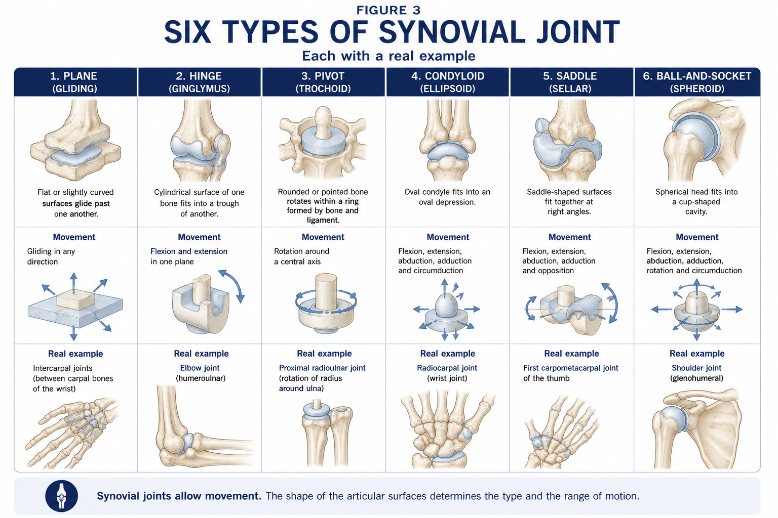
Figure 4 · The six types of synovial joint
Every synovial joint has all five:
| Feature | Function | Examples |
|---|---|---|
| Articular disc | Divides the cavity, improves congruence, distributes load, permits two different movements in one joint | Temporomandibular (rotation below the disc, translation above it), sternoclavicular, acromioclavicular, distal radio-ulnar (TFCC) |
| Meniscus | Incomplete disc; congruence, load distribution, shock absorption, secondary stability | Knee |
| Labrum | Deepens a shallow socket; forms a suction seal | Glenoid, acetabulum |
| Fat pad | Fills changing dead space; cushions; densely innervated | Infrapatellar (Hoffa’s), the pulvinar of the acetabulum, the elbow’s coronoid and olecranon pads |
| Bursa | Reduces friction between structures | Subacromial–subdeltoid, prepatellar, trochanteric, retrocalcaneal |
| Synovial sheath | A bursa wrapped around a tendon | Flexor tendons of the hand, tendons at the ankle |
| Synovial fold / plica | Embryological septal remnant | Mediopatellar plica (a recognised cause of anterior knee pain) |
| Tendon within the joint | — | Long head of biceps (intracapsular, extrasynovial); popliteus |
The synovium is a specialised connective tissue lining, 1–3 cells deep, with no basement membrane and no tight junctions — it is a discontinuous layer, not an epithelium, which is why it is freely permeable to solutes.
| Cell | Nature | Function |
|---|---|---|
| Type A synoviocyte | Macrophage-derived | Phagocytosis of debris and worn matrix; antigen presentation; the effector cell of synovitis |
| Type B synoviocyte | Fibroblast-derived | Secretes hyaluronan and lubricin (PRG4) into the fluid; produces matrix |
Beneath lies the subintima — areolar, adipose or fibrous — which carries a rich capillary network, lymphatics and nerve endings. Synovium is one of the most vascular tissues in the body relative to its mass, which is why it is a target in systemic inflammatory disease and why haemarthrosis follows even minor synovial injury.
Synovial fluid is a dialysate of plasma with added hyaluronan and lubricin.
| Property | Normal value / character |
|---|---|
| Volume (knee) | ~0.5–4 mL |
| Appearance | Clear, pale yellow, viscous |
| Cell count | < 200 cells/mm³, mostly mononuclear |
| Key solutes | Hyaluronan (viscosity), lubricin/PRG4 (boundary lubrication), glucose ≈ plasma, protein ~⅓ of plasma |
| Behaviour | Non-Newtonian and thixotropic: viscosity falls as shear rate rises |
The thixotropic property has a direct clinical meaning: fluid is thick and resistant at rest, and thins as movement speeds up. That is the physiological basis of morning stiffness and of the “warming up” of an arthritic joint, and part of the rationale for gentle early-range movement before loaded activity.
Functions of synovial fluid: lubrication, nutrition of articular cartilage and menisci, shock absorption, and waste removal.
| Normal | Osteoarthritis | Inflammatory (RA) | Septic | |
|---|---|---|---|---|
| Appearance | Clear | Clear/straw | Turbid, yellow | Purulent |
| WBC/mm³ | <200 | <2,000 | 2,000–50,000 | >50,000, mostly neutrophils |
| Viscosity | High | High | Low | Low |
| Glucose | Normal | Normal | Low-normal | Markedly low |
A hot, exquisitely painful joint with fever and a refusal to move is a septic arthritis until proved otherwise — a surgical emergency, not a physiotherapy referral. Do not mobilise; escalate.
| Type | Surfaces | DoF | Movements | Examples |
|---|---|---|---|---|
| Plane (gliding) | Flat or slightly curved | Non-axial (gliding) | Translation, small rotation | Intercarpal, intertarsal, acromioclavicular, zygapophysial (facet) joints, sternoclavicular (functionally saddle) |
| Hinge (ginglymus) | Convex cylinder in a concave trough | 1 | Flexion–extension | Humero-ulnar, interphalangeal, talocrural (approximately) |
| Pivot (trochoid) | Rounded peg within an osseoligamentous ring | 1 | Rotation about a longitudinal axis | Median atlanto-axial, proximal and distal radio-ulnar |
| Condyloid (ellipsoid) | Oval convex into oval concave | 2 | Flexion–extension, abduction–adduction, and circumduction; no true axial rotation | Radiocarpal, metacarpophalangeal 2–5, atlanto-occipital |
| Saddle (sellar) | Each surface concave in one direction, convex in the other | 2 (functionally 3, permitting conjunct rotation) | Flexion–extension, abduction–adduction, opposition | First carpometacarpal; sternoclavicular; calcaneocuboid |
| Ball-and-socket (spheroidal) | Sphere in a cup | 3 | Flexion–extension, abduction–adduction, rotation, circumduction | Glenohumeral, hip (a cotyloid variant, deeper) |
Two joints resist classification and are worth naming: the knee is a modified hinge (bicondylar) with obligatory axial rotation, and the temporomandibular joint is a modified hinge with both rotation and translation, divided by a disc.
Figure 5 · What stops a joint moving further
Stability is never a single structure. Rank the contributors for the joint in front of you.
| Factor | Mechanism | Where it dominates |
|---|---|---|
| Bony congruence and depth of socket | Geometry physically blocks displacement | Hip (deep acetabulum + labrum encloses >50% of the head); ankle mortise in dorsiflexion; elbow in full extension |
| Ligaments and capsule | Passive tension at end range; low energy cost; but they are static and cannot respond to unexpected load | Knee collaterals and cruciates; ankle lateral complex; interosseous sacroiliac ligaments |
| Muscles and tendons | Active, adjustable, and the only structure that can pre-tension in anticipation of load. Includes the dynamic compression of concavity–compression | Shoulder (rotator cuff), scapulothoracic articulation, lumbar spine (the local stabilisers), and the medial longitudinal arch of the foot |
| Negative intra-articular pressure | Intact capsule + fluid film generates suction; atmospheric pressure holds surfaces together | Hip (releasing suction is what makes hip arthroscopic distraction difficult); glenohumeral joint |
| Adhesion–cohesion of the fluid film | Surface tension between wetted surfaces | All synovial joints, minor contribution |
| Labra, menisci and discs | Deepen and seal the socket; increase congruence | Shoulder, hip, knee |
| Fascia and retinacula | Constrain and redirect | Iliotibial band; extensor retinaculum |
| Neuromuscular control (proprioception) | Reflex and feedforward muscle activation | Every joint — and the component most amenable to rehabilitation |
The glenoid fossa accommodates only about a third of the humeral head, and its passive ligaments are lax through most of the mid-range. Stability there is produced by concavity–compression: the rotator cuff compresses the humeral head into the labrum-deepened glenoid concavity, generating a stabilising force proportional to the compressive load. It follows that (a) shoulder stability is dynamic and trainable, (b) cuff weakness or inhibition produces instability without any structural lesion, and (c) rehabilitation of the unstable shoulder is largely a matter of restoring cuff and scapular control. This is a rare instance where the anatomy directly prescribes the treatment.
Generalised joint hypermobility is screened with the Beighton score (0–9): passive fifth MCP extension >90° (1 each side), thumb to forearm (1 each side), elbow hyperextension >10° (1 each side), knee hyperextension >10° (1 each side), and forward flexion with palms flat on the floor (1). A score of ≥5/9 in adults (≥6 in children, ≥4 over 50) suggests generalised hypermobility; the diagnosis of hypermobile Ehlers–Danlos syndrome or hypermobility spectrum disorder requires the full 2017 criteria, including a five-part questionnaire, systemic features and exclusion of other heritable connective tissue disorders. Clinically these patients need proprioceptive and strength training through range, load management and pacing, and they are typically made worse by the end-range stretching they instinctively seek.
The nerve supplying a joint also supplies the muscles that move the joint and the skin overlying their insertions.
Examples: the hip is supplied by branches of the femoral, obturator and sciatic nerves — which is why hip pathology refers to the knee via the obturator nerve, and why a child with a limp and knee pain must have the hip examined. The shoulder is supplied by the suprascapular, axillary and lateral pectoral nerves, hence pain referral over the deltoid.
Blood supply comes from periarticular anastomoses (the genicular anastomosis at the knee, the cruciate and trochanteric anastomoses at the hip, the anastomoses around the elbow), whose redundancy allows collateral flow when a main vessel is occluded or when the joint is held in an extreme position.
Four types, classified by Wyke, and the reason proprioceptive rehabilitation exists.
| Type | Receptor | Location | Threshold / adaptation | Function |
|---|---|---|---|---|
| I | Ruffini | Fibrous capsule, superficial layers | Low threshold, slowly adapting | Static joint position; intra-articular pressure; postural and tonic reflex control |
| II | Pacinian (and Golgi–Mazzoni) | Deep capsule, fat pads | Low threshold, rapidly adapting | Acceleration and deceleration — detects the onset of movement; phasic reflex control |
| III | Golgi–Mazzoni / Golgi ligament endings | Ligaments, menisci | High threshold, very slowly adapting | End-range tension; inhibitory reflex protection (the ligamento-muscular reflex) |
| IV | Free nerve endings | Capsule, ligaments, fat pads, adventitia of vessels, periosteum; not in articular cartilage or the inner meniscus | High threshold, non-adapting | Nociception; tonic reflex effects on muscle tone |
Why this matters clinically. Ligament rupture destroys type III receptors and part of the afferent supply, producing a measurable proprioceptive deficit and altered reflex muscle timing that persists after the ligament is reconstructed. This is the anatomical justification for balance and perturbation training as a mandatory component of ligament rehabilitation, and for the evidence that such training reduces recurrent ankle sprain and ACL injury rates.
Arthrogenic muscle inhibition is the same story in reverse: joint effusion, pain and abnormal afferent input reflexively inhibit motor drive — most famously quadriceps inhibition after knee injury or effusion, which can be produced experimentally by injecting as little as 20–30 mL of saline into a healthy knee. Effusion control is therefore not cosmetic; it is a prerequisite for strength gain.
These four concepts are the practical vocabulary of manual assessment.
| Concept | Definition | Clinical use |
|---|---|---|
| Close-packed position | The position of maximum congruence: articular surfaces are maximally contacted, capsule and ligaments maximally taut, joint surfaces cannot be separated, and the joint is effectively locked | The most stable position — but also the position of greatest compressive load, so it is where fractures occur and where an inflamed joint hurts most. Avoid it when mobilising or when testing an irritable joint |
| Loose-packed (open-packed) position | Any position other than close-packed; the capsule is relatively lax and some accessory movement is available | — |
| Resting position | The specific loose-packed position of maximum capsular laxity and greatest joint volume | The position the patient with an effusion adopts spontaneously (because it minimises intra-articular pressure), and the position in which joint mobilisation and distraction are performed |
| Capsular pattern | The characteristic proportional pattern of passive range limitation seen when the whole capsule is involved (arthritis, capsulitis) | Distinguishes capsular from non-capsular restriction (which suggests a loose body, internal derangement, ligament adhesion or extra-articular block) |
| Joint | Close-packed | Resting position | Capsular pattern |
|---|---|---|---|
| Glenohumeral | Full abduction + lateral rotation | ~55° abduction, ~30° horizontal adduction | Lateral rotation > abduction > medial rotation |
| Elbow (humero-ulnar) | Full extension + supination | ~70° flexion, ~10° supination | Flexion > extension |
| Radiocarpal | Full extension + radial deviation | Neutral with slight ulnar deviation | Flexion ≈ extension, equally limited |
| Hip | Full extension + medial rotation + abduction | ~30° flexion, ~30° abduction, slight lateral rotation | Medial rotation > flexion > abduction (variable in the literature) |
| Knee (tibiofemoral) | Full extension + lateral rotation of tibia | ~25° flexion | Flexion > extension (grossly disproportionate) |
| Talocrural | Full dorsiflexion (the wider anterior talus wedges into the mortise) | ~10° plantarflexion, mid-inversion/eversion | Plantarflexion > dorsiflexion |
The ankle example is worth dwelling on. The talar trochlea is wider anteriorly than posteriorly. In dorsiflexion the wide part engages the mortise, the syndesmosis splays slightly, and the joint is close-packed and stable. In plantarflexion the narrow part sits in the mortise, the joint is loose, and it is vulnerable — which is precisely why the classic inversion sprain occurs in plantarflexion and inversion, and why the anterior talofibular ligament is the first structure to fail.
Chapter 1 introduced roll, glide and spin and the convex–concave rule. Applied to treatment:
Two honest caveats. First, the convex–concave rule is a heuristic, and in-vivo imaging at the shoulder shows translations that do not always follow it; treat it as a starting hypothesis tested against the patient’s response. Second, the mechanism of manual therapy is now understood to be substantially neurophysiological — descending inhibition, temporary changes in mechanosensitivity, and non-specific contextual effects — rather than a durable structural change in capsular length. That does not make it useless; it makes it an adjunct that should be paired with active loading, not a treatment in itself.
| Condition | Nature | Key features |
|---|---|---|
| Osteoarthritis | Whole-joint degenerative disease (Chapter 3) | Activity-related pain, short-lived (<30 min) morning stiffness, crepitus, bony enlargement, capsular pattern. First-line care is exercise, education and weight management |
| Rheumatoid arthritis | Autoimmune synovitis; pannus erodes cartilage and bone | Symmetrical small-joint polyarthritis, prolonged (>1 h) morning stiffness, MCP/PIP involvement sparing the DIP, ulnar deviation, boutonnière and swan-neck deformities, atlanto-axial instability (screen before any cervical technique or intubation) |
| Spondyloarthropathies (ankylosing spondylitis, psoriatic, reactive, IBD-related) | Inflammatory, enthesis-centred, HLA-B27-associated | Inflammatory back pain (young onset, insidious, worse with rest, better with exercise, night pain), sacroiliitis, enthesitis, dactylitis. Exercise is disease-modifying, not merely symptomatic |
| Crystal arthropathy | Monosodium urate (gout) or calcium pyrophosphate (pseudogout) | Acute monoarthritis, exquisitely painful; first MTP in gout; knee and wrist in CPPD |
| Septic arthritis | Bacterial infection | Hot, swollen, exquisitely painful joint with fever, held rigidly still. Emergency — do not mobilise, escalate immediately |
| Haemarthrosis | Bleeding into the joint | Rapid (<2 h) tense effusion after knee trauma implies ACL rupture, osteochondral fracture, or patellar dislocation in ~75% of cases. In haemophilia, recurrent haemarthrosis causes synovial hypertrophy and joint destruction |
| Adhesive capsulitis (frozen shoulder) | Capsular fibrosis with contracture of the rotator interval and coracohumeral ligament | Global restriction in the capsular pattern, passive range as limited as active — the defining feature. Freezing / frozen / thawing phases; associated with diabetes and thyroid disease |
| Subluxation / dislocation | Partial / complete loss of surface contact | Direction and mechanism predict the structure injured: anterior glenohumeral → Bankart and Hill–Sachs lesions; posterior hip → sciatic nerve and AVN risk |
| Type | Tissue | Example |
|---|---|---|
| Arthrogenic | Capsule, intra-articular adhesion | Post-immobilisation stiffness; adhesive capsulitis |
| Myogenic | Muscle shortening or fibrosis | Gastrocnemius contracture after prolonged plantarflexed positioning |
| Neurogenic | Spasticity, tone, denervation imbalance | Equinus after stroke or cerebral palsy |
| Dermatogenic | Skin and scar | Post-burn contracture |
| Fasciogenic | Fascia | Dupuytren’s contracture; plantar fascia |
| Pathological / bony | Ankylosis, heterotopic ossification | Post-traumatic elbow HO; fibrous or bony ankylosis after septic arthritis |
The distinction is not academic: capsular contracture responds to mobilisation and prolonged low-load stretch; myogenic contracture to positioning and active lengthening; neurogenic contracture to tone management alongside both; and bony ankylosis to neither.
Within days to weeks of immobilisation a joint shows:
15 questions on this chapter. Tap one to see the answer and the reasoning.
Answer: (B) Hyaline unites a synchondrosis (primary cartilaginous joint), which is usually temporary.
Answer: (C) It also spares menisci and discs.
Answer: (B) Type A cells are macrophage-derived phagocytes.
Answer: (C) Its saddle geometry permits opposition.
Answer: (B) The wider anterior talus wedges into the mortise — which is why plantarflexion is the vulnerable position for inversion sprain.
Answer: (B)
Answer: (B) Hence hip pathology presenting as knee pain via the obturator nerve.
Answer: (B) Type I are slowly adapting Ruffini endings; type IV are free nerve endings.
Answer: (C) The capsuloligamentous restraints are lax in mid-range.
Answer: (B) It is the position an effused joint adopts and the position used for mobilisation.
Answer: (B) Approximately three-quarters of acute traumatic haemarthroses involve ACL rupture, osteochondral fracture or patellar dislocation.
Answer: (B) Transverse ligament and odontoid erosion can produce cord compromise.
Answer: (B) It can be produced experimentally by injecting saline into a healthy knee, which is why effusion control precedes strengthening.
Answer: (B) The score screens for hypermobility; the diagnosis requires the full 2017 criteria.
Answer: (B) Immobilisation weakens the insertion disproportionately — a key argument for early controlled motion after ligament injury.
Everything on this page, in one screen
| Source | Why it earns its place |
|---|---|
| Standring S (ed.) — Gray’s Anatomy, 42nd edn | Reference account of joint structure, synovium and innervation |
| Levangie PK, Norkin CC — Joint Structure and Function | The best single treatment of joint classification, stability and close-packed positions for physiotherapists |
| Neumann DA — Kinesiology of the Musculoskeletal System | Arthrokinematics, concavity–compression and joint-specific mechanics |
| Kaltenborn FM — Manual Mobilization of the Joints: The Extremities | Treatment planes, traction and glide grades, resting positions |
| Maitland GD, Hengeveld E, Banks K — Maitland’s Peripheral Manipulation | Grades of oscillatory movement and clinical reasoning |
| Cyriax J — Textbook of Orthopaedic Medicine | The origin of the capsular pattern and end-feel concepts |
| Wyke B — “The neurology of joints”, Ann R Coll Surg Engl, 1967 | The original classification of articular mechanoreceptors |
| Rice DA, McNair PJ — “Quadriceps arthrogenic muscle inhibition”, Semin Arthritis Rheum, 2010 | The evidence base for treating effusion before strength |
| Malfait F et al. — “The 2017 international classification of the Ehlers–Danlos syndromes”, Am J Med Genet C | Current diagnostic criteria for hypermobility disorders |
| Akeson WH, Amiel D, Woo SL — “Immobility effects on synovial joints: the pathomechanics of joint contracture”, Biorheology, 1980 | The classic experimental work behind the immobilisation box above |
| Palastanga N, Field D, Soames R — Anatomy and Human Movement | The physiotherapy-facing synthesis |
Chapter 4 of 24 · Human Anatomy · Physiotherapist India Next: Chapter 5 — Muscle: from whole muscle to sarcomere, fibre types, and the motor unit.
Reviewed by the Physiotherapist India Team. · Human Anatomy contents