Evidence-based physiotherapy and rehabilitation resources
Editorial & review policy
Evidence-based physiotherapy and rehabilitation resources
Editorial & review policyHuman Anatomy · General anatomy
Anatomy has its own language, and every later chapter assumes you speak it. This one teaches the position everything is described from, the planes and axes movement happens in, the terms that locate one structure against another, and the vocabulary of bone surfaces you will use at every joint you examine.
Part 1 · General anatomy
Anatomical terminology, position, planes and axes, kinematic conventions, and the vocabulary of bone surfaces
Anatomy is the only subject in your course that every other subject quotes. Kinesiology quotes it, electrotherapy quotes it when it tells you where a motor point lies, orthopaedics quotes it in every operative note, neurology quotes it in every localisation, and your own clinical reasoning quotes it every time you decide that a patient’s lateral elbow pain is tendinous rather than radicular.
But before any of that, anatomy has to be written down and spoken. A structure that cannot be described unambiguously cannot be handed from one clinician to another. The whole of this chapter is therefore about one problem and its solution:
The problem. The human body moves. Any description that depends on how the body happens to be arranged at the moment of writing becomes false the moment the body moves.
The solution. Agree on a fixed reference posture, a fixed set of reference planes and axes, and a fixed vocabulary — and describe everything from those, regardless of what the patient is actually doing.
Everything in this chapter follows from that single move. Master it and you will never again write a sentence in a clinical record that a colleague can misread.
By the end of this chapter you should be able to:
Anatomy (Greek ana-, up; tome, cutting) is the science of the structure of the body and of the spatial relationships between its parts. Physiology is the science of its function. The two are inseparable in practice — structure constrains function and function remodels structure — and the hybrid discipline you actually practise is best called functional anatomy or kinesiology.
| Branch | Scope | Where it becomes clinical for you |
|---|---|---|
| Gross (macroscopic) anatomy | Structures visible to the unaided eye. Taught either regionally (everything in the forearm together) or systemically (the whole skeletal system, then the whole muscular system). | The core of this subject. Regional teaching mirrors clinical presentation; systemic teaching mirrors pathology that follows a tissue type. |
| Histology (microscopic anatomy) | Tissue and cell architecture. | Explains healing timelines: why bone remodels and hyaline cartilage does not, why tendon is slow, why muscle regenerates only within limits. |
| Cytology | Cell structure. | Underpins motor-unit physiology and mitochondrial adaptation to endurance training. |
| Embryology (developmental anatomy) | Formation from zygote to birth, and postnatal growth. | Explains dermatome/myotome patterns, referred pain, congenital deformity (CTEV, DDH, spina bifida), and limb-bud rotation. |
| Surface anatomy (living anatomy) | What can be seen, palpated and inferred on a living subject. | Arguably the single most important branch for physiotherapy: your assessment is surface anatomy performed in real time. |
| Radiological / sectional anatomy | The body as rendered by radiograph, ultrasound, CT, MRI. | Reading the report — and increasingly, reading the images — that arrive with your patient. |
| Neuroanatomy | The nervous system in structural detail. | Lesion localisation, which is the entire basis of neurological physiotherapy. |
| Comparative anatomy | Structure across species. | Explains why the human pelvis, foot arch and lumbar lordosis carry the costs they do. |
| Applied / clinical anatomy | Anatomy interpreted through injury, disease and intervention. | Everything above, made useful. |
Both schemes are legitimate and you need both.
This subject uses the standard compromise: general anatomy systemically first (bone, cartilage, joint, muscle, connective tissue, skin), then the regions.
Until the late nineteenth century, anatomical naming was chaotic: a single structure might carry a dozen names in different European schools. Successive international committees produced standardised lists — Basle Nomina Anatomica (BNA, 1895), Nomina Anatomica (NA, from 1955), and since 1998 Terminologia Anatomica (TA), produced by the Federative International Programme for Anatomical Terminology (FIPAT) under the International Federation of Associations of Anatomists. A second edition (TA2) was released in 2019, alongside companion terminologies for histology (Terminologia Histologica) and embryology (Terminologia Embryologica).
TA has two working rules that are worth internalising:
The reason is not pedantry. A descriptive term teaches you something (the flexor digitorum profundus is the deep flexor of the fingers — you have learnt its compartment, its action and its depth from the name alone). An eponym teaches you nothing except a piece of nineteenth-century European history, and often the wrong history: many eponymous structures were described earlier by someone else, and a proportion of eponyms honour individuals whose conduct the profession would not now wish to commemorate.
But eponyms survive, and you must know them, because clinical practice is not governed by TA. You will meet, and must be fluent in, terms such as the circle of Willis, the ligament of Treitz, the canal of Guyon, the space of Poirier, Gerdy’s tubercle, Lister’s tubercle, the arcade of Frohse, and the anatomical snuffbox. The rule for your own writing: use the TA term, and give the eponym once in parentheses if the eponym is what the referring surgeon will use.
Anatomical vocabulary is a compositional system built from a few hundred Latin and Greek elements. Learning the elements converts thousands of terms from memorisation into reading.
| Element | Meaning | Worked example |
|---|---|---|
| supra- / infra- | above / below | supraspinatus — the muscle above the scapular spine |
| epi- / hypo- | upon / under | epicondyle — the eminence upon the condyle |
| ab- / ad- | away from / towards | abductor, adductor |
| pre- / retro- | in front of / behind | retropharyngeal space |
| inter- / intra- | between / within | interosseous membrane; intracapsular ligament |
| -osus / -osa | full of, rough | linea aspera — the rough line |
| brevis / longus / magnus | short / long / large | peroneus longus and brevis |
| teres | round, cylindrical | pronator teres |
| serratus | saw-toothed | serratus anterior, from its digitations |
| gastro- | belly | gastrocnemius — the belly of the leg |
| -glossus / -hyoid | tongue / hyoid | genioglossus, mylohyoid |
A muscle name in the standard form encodes up to five facts: shape (deltoid, trapezius, rhomboid), size (maximus, minimus), fibre direction (rectus, obliquus, transversus), location (brachii, femoris, abdominis), attachments (sternocleidomastoid, coracobrachialis), number of heads (biceps, triceps, quadriceps) and action (flexor, extensor, supinator, levator). Flexor pollicis longus is therefore not a name to memorise but a sentence to read: the long flexor of the thumb.
Wrong-site surgery remains a “never event” in most health systems, and ambiguous anatomical documentation is one of its recurrent contributors. Laterality (left / right), level (L4–L5 versus L5–S1) and digit naming (the thumb, index, middle, ring and little finger — never “the first finger”, which is ambiguous) must be written in unambiguous form every time. In the foot, the digits are conventionally numbered one to five from the great toe (hallux) laterally; the fifth is the little toe.
The anatomical position is the agreed reference posture from which every positional and movement term is defined:
At rest, standing normally, your forearms sit in mid-pronation: the palms face the thighs and the radius crosses obliquely over the ulna. In the anatomical position the forearm is supinated, so radius and ulna lie parallel and uncrossed, with the radius lateral and the ulna medial.
Every downstream statement about the forearm depends on this. The radius is the lateral bone and the ulna is the medial bone — in the anatomical position. Radial deviation of the wrist is abduction — in the anatomical position. The lateral cutaneous nerve of the forearm supplies the lateral forearm — in the anatomical position. Describe from the patient’s incidental posture instead and every one of these becomes wrong.
Some kinesiology texts also define the fundamental position: identical to the anatomical position except that the forearms are in mid-pronation with the palms facing the body. It is the more natural standing posture and is occasionally used as the starting position for describing forearm rotation itself (where “zero” is mid-position, not full supination). Where a text uses it, it will say so; unless told otherwise, assume the anatomical position.
This is the single piece of embryology that explains the most gross anatomy, and it is worth learning now rather than in the development chapter.
The limb buds appear in the fifth week as outgrowths of lateral plate mesoderm covered by ectoderm, with the upper bud a little ahead of the lower. Initially both buds project laterally with a preaxial (cranial) border — the future thumb / great toe side — and a postaxial (caudal) border. The palms and soles both face medially, and the future elbows and knees both point laterally.
During the seventh to eighth weeks the limbs rotate at their roots, and they rotate in opposite directions:
What this explains, immediately:
Learn this once and a large amount of otherwise arbitrary anatomy becomes derivable.
Figure 1 · The three planes of the body
A plane is a two-dimensional surface passing through the body. Three mutually perpendicular cardinal planes are defined, each passing through the body’s centre of mass in the anatomical position.
| Plane | Orientation | Divides the body into | Alternative names and notes |
|---|---|---|---|
| Sagittal | Vertical, running anteroposteriorly | Left and right portions | The plane passing exactly through the midline is the median (midsagittal) plane. Any parallel plane off the midline is paramedian or simply sagittal. From Latin sagitta, an arrow — the plane an arrow would travel in if fired at the back. |
| Coronal | Vertical, running side-to-side | Anterior and posterior portions | Also frontal. From corona, a crown — the plane a crown occupies as it passes ear to ear. |
| Transverse | Horizontal | Superior and inferior portions | Also axial or horizontal. This is the plane in which most CT and MRI slices are acquired. |
| Oblique | At an angle to all three | — | The plane of most real human movement. |
Three points that separate a competent answer from a superficial one:
A conventional anteroposterior (AP) radiograph or an axial CT/MR slice is displayed as though you are standing at the patient’s feet, looking towards the head. Consequently the patient’s left appears on the right of the image. Getting this wrong is a classic and dangerous error. Coronal images are viewed from the front (again, patient’s left on the viewer’s right); sagittal images are conventionally viewed from the patient’s left side.
Figure 2 · Planes, axes and the movements that pair with them
A plane tells you the surface along which a movement travels. An axis is the line about which the segment rotates — the pin of the hinge. The axis is always perpendicular to the plane of movement.
| Axis | Runs | Perpendicular to | Movements produced |
|---|---|---|---|
| Frontal (coronal, mediolateral) | Side to side, horizontally | Sagittal plane | Flexion, extension, dorsiflexion, plantarflexion |
| Sagittal (anteroposterior) | Front to back, horizontally | Coronal plane | Abduction, adduction, lateral flexion, radial/ulnar deviation, inversion, eversion |
| Vertical (longitudinal) | Head to foot | Transverse plane | Medial and lateral rotation, pronation and supination, horizontal abduction/adduction |
Learn the three pairings as sentences, not as a table:
Said this way, you can derive any movement name from first principles instead of recalling a list — which is what an examiner is actually testing when they ask about a movement you have not been taught.
Degrees of freedom (DoF) is the number of independent axes about which a joint can rotate (and, in full rigid-body mechanics, the number of independent translations it permits). It is a property of the joint, not of the plane.
| DoF | Joint type | Examples |
|---|---|---|
| 1 | Uniaxial — hinge (ginglymus) or pivot (trochoid) | Humero-ulnar joint, interphalangeal joints; atlanto-axial, proximal radio-ulnar |
| 2 | Biaxial — condyloid (ellipsoid) or saddle (sellar) | Radiocarpal, metacarpophalangeal 2–5; first carpometacarpal, sternoclavicular |
| 3 | Triaxial — ball and socket (spheroidal) | Glenohumeral, hip, subtalar (functionally triplanar about one oblique axis) |
Rigorously, every synovial joint also permits small translations (accessory glides), so a “three degrees of freedom” joint has six in full mechanical description: three rotations and three translations. Manual therapy is largely the exploitation of those translational degrees of freedom, which the patient cannot perform voluntarily.
This distinction — developed for clinical use in the Nordic manual-therapy tradition (Kaltenborn, Mennell, Maitland) — is fundamental and is examined constantly.
| Arthrokinematic motion | Definition | Analogy |
|---|---|---|
| Roll | New points on one surface meet new points on the other | A tyre rolling on tarmac |
| Glide (slide) | The same point on one surface meets new points on the other | A tyre skidding on ice |
| Spin | Rotation about a fixed mechanical axis; the same point stays in contact | A spinning top |
In a healthy joint, roll and glide are combined. Pure rolling would drive the moving surface off the edge of the stationary one; the accompanying glide keeps the surfaces centred. Loss of the glide component — the usual consequence of capsular fibrosis — produces the characteristic clinical picture of restricted, painfully compressed end-range movement despite intact muscle power.
The convex–concave rule (Kaltenborn) predicts the direction of glide, and therefore the direction in which a mobilisation should be applied:
(The rule is a clinically useful approximation rather than an exact law; in vivo imaging shows more complex translation at several joints, notably the shoulder. Treat it as a first hypothesis to be tested against the patient’s response, not as an inviolable principle. This is discussed further in Chapter 4, Joints.)
Textbook axes are drawn as fixed pins. Real joints have no such thing.
The instantaneous axis of rotation (IAR) is the axis about which a segment is rotating at one instant of a movement. Because articular surfaces are non-congruent and ligaments tighten progressively, the IAR migrates through the range. At the knee, the flexion–extension axis traces a J-shaped path (the classic evolute), moving posteriorly with flexion — which is why a single-axis knee brace hinge can never track the knee exactly, and why the moment arms of the quadriceps and hamstrings change through the range.
The IAR is the two-dimensional simplification. In three dimensions the equivalent is the helical (screw) axis: any displacement of a rigid body from one position to another can be described as a rotation about, plus a translation along, a single unique axis. Three-dimensional joint kinematics research reports finite helical axes for exactly this reason.
Euler and Cardan angles. To describe a 3D joint orientation with three familiar clinical angles, the rotation is decomposed into three sequential rotations about defined axes. The result is sequence-dependent:
Codman’s paradox. Start with the arm at the side, palm facing medially. Flex the shoulder to 90°, then abduct horizontally through 90° to the coronal plane, then adduct back down to the side. The arm has returned to the start position, but the palm now faces laterally: the humerus has been rotated approximately 180° about its long axis without any rotation ever being performed. This is not a trick — it is a demonstration that finite rotations in three dimensions are non-commutative, and it is the clearest reason why clinical shoulder rotation values must be defined with respect to a stated sequence and starting position.
Figure 3 · Terms of position and direction
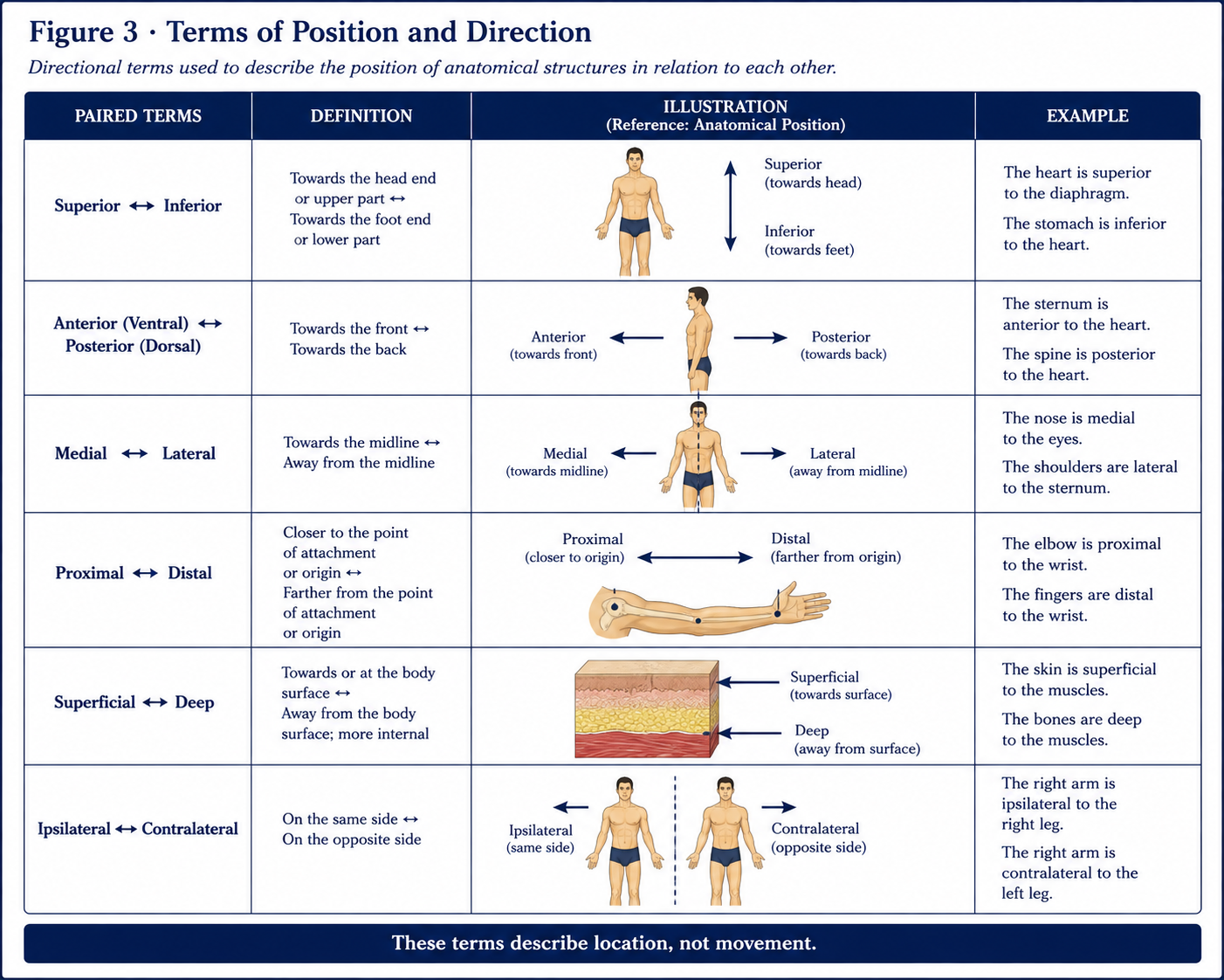
These terms come in opposing pairs and describe location, not movement.
| Pair | Meaning | Example |
|---|---|---|
| Superior / inferior | Nearer the head / nearer the feet | The shoulder is superior to the elbow |
| Cranial / caudal | Towards the head / towards the tail. Preferred in embryology and for the trunk | The caudal end of the neural tube |
| Anterior / posterior | Nearer the front / nearer the back | The sternum is anterior to the heart |
| Ventral / dorsal | Belly side / back side. Standard in embryology and neuroanatomy | The dorsal root carries afferents |
| Rostral / caudal | Towards the nose / towards the tail. Used within the brain, where “anterior” becomes ambiguous because the neuraxis bends | The rostral midbrain |
| Medial / lateral | Nearer / further from the median plane | The ulna is medial to the radius |
| Intermediate | Between a medial and a lateral structure | The intermediate cuneiform |
| Median | Exactly in the midline | The median nerve; the median plane |
| Proximal / distal | Nearer / further from the trunk or point of origin | The knee is proximal to the ankle |
| Superficial / deep | Nearer / further from the surface | Skin is superficial to fascia |
| Internal / external | Nearer the inside / outside of a cavity or organ | The internal surface of the ribs |
| Ipsilateral / contralateral | Same side / opposite side | Corticospinal lesions cause contralateral weakness above the decussation |
| Bilateral / unilateral | Both sides / one side | Bilateral pitting oedema |
| Palmar (volar) / dorsal | Palm side / back of hand | Palmar aponeurosis |
| Plantar / dorsal | Sole / upper surface of foot | Plantar fascia |
| Radial / ulnar | Thumb side / little-finger side of the forearm and hand | Ulnar deviation |
| Tibial / fibular (peroneal) | Medial / lateral side of the leg | The fibular (peroneal) nerve |
| Preaxial / postaxial | The thumb/great-toe border / the little-finger/little-toe border of the limb bud | Preaxial polydactyly |
| Prone / supine | Lying face down / face up | Position the patient prone |
| Parietal / visceral | Relating to the wall of a cavity / to the organ within it | Parietal pleura |
| Luminal / mural / adventitial | Facing the lumen / within the wall / outermost | Mural thrombus |
Proximal/distal and superficial/deep are relational only: nothing is “distal” in itself, only distal to something. “Pain in the distal thigh” is loose writing; “pain in the anteromedial thigh, 5 cm proximal to the superior pole of the patella” can be reproduced by another clinician a month later. The same applies to superficial and deep — always name both terms of the comparison.
Proximal and distal belong to the limbs. On the trunk, head and neck, use superior/inferior. “Distal cervical spine” is not standard usage; “lower cervical spine” or “C6–C7” is.
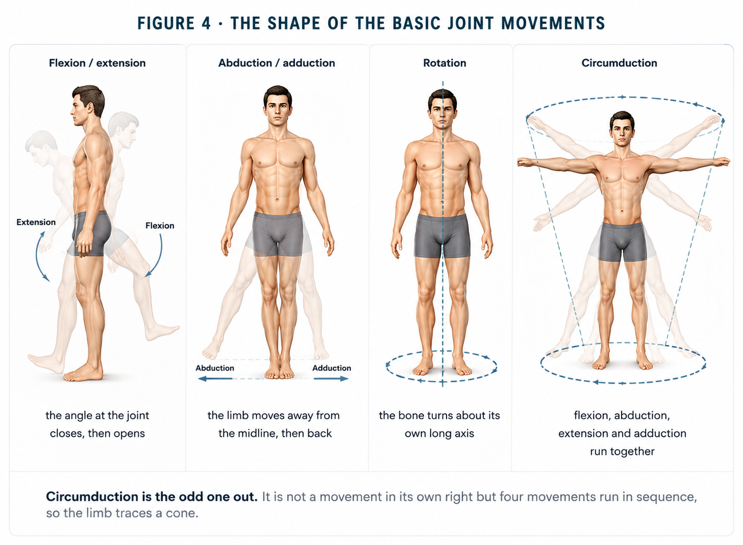
Figure 4 · The shape of the basic joint movements
| Movement | Definition | Plane and axis |
|---|---|---|
| Flexion | The angle between the segments decreases in the sagittal plane; the anterior surfaces approach (except at the knee and below, where posterior surfaces approach) | Sagittal / frontal axis |
| Extension | Return from flexion towards, or beyond, the anatomical position. Movement beyond neutral is hyperextension | Sagittal / frontal axis |
| Abduction | Movement away from the median plane (or, in the hand and foot, from the axial line of the limb) | Coronal / sagittal axis |
| Adduction | Movement towards the median plane | Coronal / sagittal axis |
| Medial (internal) rotation | The anterior surface of the limb turns towards the midline | Transverse / vertical axis |
| Lateral (external) rotation | The anterior surface turns away from the midline | Transverse / vertical axis |
| Circumduction | Sequential flexion, abduction, extension and adduction, so that the distal segment traces a cone. A combination, not an independent movement | All three |
| Horizontal abduction / adduction | Movement of the 90°-elevated limb posteriorly / anteriorly in the transverse plane. Also called horizontal extension / flexion | Transverse / vertical axis |
| Lateral flexion | Side-bending of the trunk or neck | Coronal / sagittal axis |
| Gliding / translation | Linear movement of one surface on another, without angular change | Not applicable (translation, not rotation) |
Note the axial-line convention. In the hand, abduction and adduction of the fingers are defined relative to the axis of the middle finger, not the midline of the body — so both index and ring finger move away from the middle finger in abduction. In the foot, the reference axis is the second toe.
| Region | Movement | Description |
|---|---|---|
| Forearm | Pronation | The radius crosses over the ulna; the palm turns to face posteriorly (or downwards, with the elbow flexed) |
| Supination | The bones lie parallel; the palm faces anteriorly (or upwards) — the position for carrying soup | |
| Wrist | Radial deviation (abduction) | The hand moves towards the thumb side |
| Ulnar deviation (adduction) | The hand moves towards the little-finger side | |
| Ankle (talocrural) | Dorsiflexion | The dorsum of the foot approaches the shin |
| Plantarflexion | The foot points downwards | |
| Subtalar / midfoot | Inversion / eversion | The sole turns to face medially / laterally (frontal-plane component) |
| Supination / pronation of the foot | Triplanar composites. Supination = inversion + adduction + plantarflexion. Pronation = eversion + abduction + dorsiflexion | |
| Scapula | Elevation / depression | Superior / inferior translation on the thoracic wall |
| Protraction (abduction) / retraction (adduction) | Movement away from / towards the vertebral column | |
| Upward / downward rotation | The glenoid turns to face superiorly / inferiorly — essential to full arm elevation | |
| Anterior / posterior tilt; internal / external rotation | Sagittal- and transverse-plane components measured in 3D shoulder analysis | |
| Clavicle | Elevation, depression, protraction, retraction, axial rotation | At the sternoclavicular joint; posterior long-axis rotation accompanies elevation |
| Thumb | Flexion / extension; abduction / adduction; opposition / reposition | Named relative to the plane of the palm, because the thumb ray is rotated ~90° |
| Mandible | Elevation, depression, protrusion, retrusion, lateral excursion | Combined rotation and translation at the temporomandibular joint |
| Spine | Flexion, extension, lateral flexion, axial rotation | With coupled motion: lateral flexion and rotation are mechanically linked, the coupling direction differing between the cervical and lumbar regions |
The first metacarpal is rotated approximately 90° about its long axis relative to the other four during development. Because thumb movements are named with reference to the palm rather than the body, the plane–axis pairings invert: thumb flexion and extension occur in the coronal plane of the body, and thumb abduction and adduction in the sagittal plane. Opposition is a composite of abduction, flexion and medial rotation at the saddle-shaped first carpometacarpal joint that brings the thumb pulp to meet the pulp of another digit. It is the movement that defines the human hand, and its loss (median nerve injury) costs approximately 40–50% of hand function in most impairment schedules.
A joint may have full accessory movement with restricted physiological movement (a muscular or neurological problem) or restricted accessory movement with restricted physiological movement (an articular problem). Distinguishing the two is the core purpose of a manual joint examination.
Movement terminology is incomplete without the terms for how muscle produces it. These are treated fully in Chapter 6, but note the vocabulary now:
| Contraction | Length change | Function | Example |
|---|---|---|---|
| Isometric | None | Stabilisation | Holding a load steady |
| Concentric | Shortening | Acceleration; muscle is the agonist doing positive work | Rising from a squat (quadriceps) |
| Eccentric | Lengthening under tension | Deceleration and shock absorption; negative work; highest force capacity; associated with delayed-onset muscle soreness | Descending stairs (quadriceps) |
| Isotonic / isokinetic | Constant load / constant angular velocity | Testing and training constructs, not naturally occurring states | Dynamometry |
Terminology becomes clinically usable only when attached to numbers.
The standard convention (AAOS; also the basis of the SFTR system) sets the anatomical position as 0° for every joint. Movement is recorded as degrees away from zero. A knee that lacks 10° of full extension and flexes to 120° is recorded as 10–120°, not as “−10 to 120”. A joint moving from zero is recorded as 0–120°. This removes the ambiguity of negative numbers, which different services interpret differently.
SFTR records movement plane by plane, with three numbers per plane in a fixed order: movement away from the body / starting position / movement towards the body.
A shoulder recorded as S: 50–0–170, F: 180–0–40, R (F90): 90–0–70 is fully described in one line, and unambiguously.
Every goniometric measurement requires four things to be stated: the patient’s position, the axis (fulcrum), the stationary arm alignment, and the moving arm alignment. Universal goniometry has good intra-rater reliability at most peripheral joints (ICC typically >0.90 at the knee and elbow) and lower inter-rater reliability, particularly at the shoulder, hip rotation and the subtalar joint. In practice this means: the same clinician should re-measure where possible, and a change should exceed the minimal detectable change (commonly quoted as roughly 5–10° at peripheral joints) before it is called real improvement.
The quality of resistance at the end of passive range (Cyriax) is diagnostic information the number alone does not carry.
| Normal end-feels | Produced by | Example |
|---|---|---|
| Bony (hard) | Bone-on-bone contact | Elbow extension |
| Firm (elastic) | Capsuloligamentous or muscular tension | Hip medial rotation; ankle dorsiflexion |
| Soft (tissue approximation) | Soft tissue compression | Knee flexion; elbow flexion |
Abnormal end-feels
Suggests
Empty
Pain stops the movement before tissue resistance is reached — consider acute inflammation, fracture, neoplasm; a red-flag finding
Springy block
An intra-articular displaced fragment — classically a bucket-handle meniscal tear
Muscle spasm
Sudden, guarded arrest — acute injury or instability
Boggy
Joint effusion
Capsular
Fibrosis; correlates with the joint’s characteristic capsular pattern of proportional restriction
Figure 5 · The surface features of a long bone
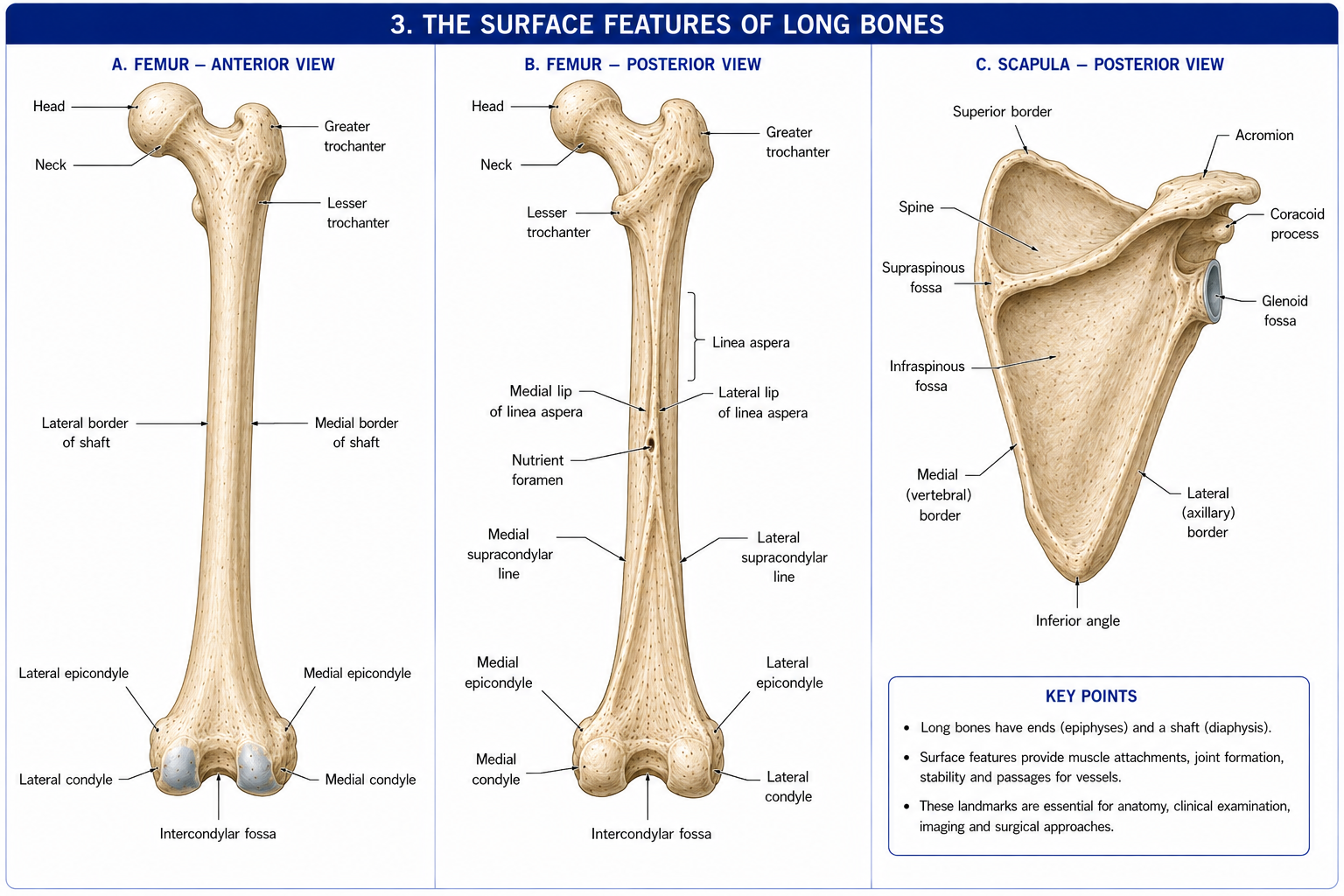
A dried bone is a record of the forces that acted on it. Every named feature is evidence of a soft-tissue event: something pulled there, something slid across, something passed through, or something articulated with it. Learn the features as clues, and the names come free.
| Term | Description | Example |
|---|---|---|
| Process | Any bony projection (generic) | Spinous process; coracoid process |
| Tuberosity | Large, rounded, roughened elevation | Ischial tuberosity; deltoid tuberosity |
| Tubercle | Small rounded elevation | Greater tubercle of the humerus |
| Trochanter | Very large blunt elevation — the word is reserved for the femur | Greater trochanter |
| Crest | Prominent ridge | Iliac crest |
| Line (linea) | Less prominent ridge, typically a sheet-muscle attachment | Linea aspera; soleal line |
| Spine | Sharp slender projection | Spine of the scapula; anterior superior iliac spine |
| Ramus | An arm or branch of a bone | Superior pubic ramus |
| Malleolus | Hammer-shaped process | Medial and lateral malleoli |
| Cornu | Horn | Greater cornu of the hyoid |
| Term | Description | Example |
|---|---|---|
| Head (caput) | Rounded articular expansion, usually on a narrowed neck | Head of the femur |
| Neck | Constriction supporting the head — a common fracture site | Surgical neck of the humerus |
| Condyle | Rounded articular knuckle | Femoral condyles |
| Epicondyle | Non-articular eminence above a condyle, for attachment | Medial epicondyle of the humerus |
| Facet | Small, flat, smooth articular surface | Zygapophysial (facet) joints; costal facets |
| Trochlea | Pulley-shaped articular surface | Trochlea of the humerus; trochlea of the talus |
| Capitulum | Small rounded articular head | Capitulum of the humerus |
| Term | Description | Example |
|---|---|---|
| Fossa | Shallow depression | Olecranon fossa; iliac fossa |
| Fovea | Small pit | Fovea capitis of the femoral head |
| Groove (sulcus) | Furrow carrying a tendon, vessel or nerve | Intertubercular (bicipital) groove; radial groove |
| Notch (incisura) | Indentation of a bone margin | Greater sciatic notch; trochlear notch |
| Foramen | Hole through bone | Foramen magnum; obturator foramen |
| Fissure | Narrow slit | Superior orbital fissure |
| Canal / meatus | Tunnel | Carpal canal; external acoustic meatus |
| Sinus | Air-filled cavity within bone | Frontal sinus; mastoid air cells |
| Antrum | A large sinus or cavity | Maxillary antrum |
| Division | Regions |
|---|---|
| Head and neck | Cranial, frontal, orbital, nasal, oral, buccal, zygomatic, auricular, occipital, temporal; cervical (anterior and posterior triangles), nuchal |
| Trunk, anterior | Sternal, pectoral, mammary, axillary, hypochondriac, epigastric, umbilical, lumbar (flank), inguinal, hypogastric (suprapubic), pubic |
| Trunk, posterior | Vertebral, scapular, interscapular, infrascapular, lumbar, sacral, gluteal, perineal |
| Upper limb | Deltoid, brachial (anterior/posterior), cubital (with the cubital fossa anteriorly), antebrachial, carpal, palmar (thenar, hypothenar), dorsum of hand, digital |
| Lower limb | Coxal (hip), femoral (thigh), patellar, popliteal, crural (leg), sural (calf), tarsal, plantar, dorsum of foot, digital |
In precise usage the arm is shoulder to elbow only, the forearm elbow to wrist, the thigh hip to knee, and the leg knee to ankle only. A patient reporting “leg pain” may mean anywhere from buttock to toes. Your record must name the region you examined, not repeat the patient’s word.
Two schemes coexist; know both.
Key surface planes worth knowing now: the transpyloric plane (L1, midway between jugular notch and pubic symphysis — crossing the pylorus, pancreatic neck, hila of the kidneys, origin of the superior mesenteric artery and the ninth costal cartilages), the subcostal plane (L3), the transtubercular plane (L5), and the supracristal plane (the highest points of the iliac crests, at L4 — the landmark for lumbar puncture).
| Cavity | Contents | Serous lining |
|---|---|---|
| Cranial | Brain, meninges | — (meninges rather than serosa) |
| Vertebral | Spinal cord, meninges, roots | — |
| Thoracic | Two pleural cavities and the mediastinum | Parietal and visceral pleura |
| — Pericardial (within the mediastinum) | Heart | Parietal and visceral pericardium |
| Abdominal | Digestive viscera, kidneys, suprarenals | Parietal and visceral peritoneum |
| Pelvic | Bladder, rectum, reproductive organs | Peritoneum superiorly; extraperitoneal below |
The universal serous pattern: a parietal layer lining the wall, a visceral layer covering the organ, continuous with each other and enclosing a potential space containing a thin film of lubricating fluid. Pathology in every one of these cavities is a variation on the same theme — fluid (effusion, haemothorax, ascites), air (pneumothorax), or fibrous adhesion of the two layers. For physiotherapy the pleural version is the operational one: pleural pain, restricted chest expansion and reduced diaphragmatic excursion.
Terminology is not decoration. Its purpose is that a description you write today can be reconstructed exactly by a clinician who was not present, months later, possibly in a medico-legal context.
Vague: > “Pain in the outer part of the arm near the top. Worse lifting the arm out to the side. Tender on pressing.”
Precise: > “Pain over the lateral aspect of the proximal right arm, referred distally to the deltoid insertion, non-dermatomal. Reproduced on active abduction in the coronal plane between 60° and 120°, with a painful arc; passive abduction full and less painful. Tender on palpation over the greater tubercle of the humerus at the supraspinatus footprint, with the arm in extension and internal rotation. Resisted abduction in the scapular plane at 30° reproduces pain without weakness. Cervical spine screening: full painless range, negative Spurling.”
The second entry names side, region, plane, arc, landmark, test position and the structures excluded. Another clinician can repeat that examination and compare findings. That is the entire purpose of the vocabulary — not to sound authoritative, but to be reproducible.
Where the record feeds a rehabilitation plan, this precision also maps directly onto the ICF framework: the anatomical finding is a body structure impairment, the restricted abduction a body function impairment, the inability to reach an overhead shelf an activity limitation, and the inability to return to painting work a participation restriction. Anatomy sits at the base of that chain; it does not replace the rest of it.
15 questions on this chapter. Tap one to see the answer and the reasoning.
Answer: (B) Palms face anteriorly; radius and ulna lie parallel and uncrossed. This is the basis for calling the ulna the medial bone.
Answer: (C) The movement travels along an anteroposterior surface, rotating about a mediolateral line.
Answer: (B) Only the plane dividing the body into equal halves is median (midsagittal).
Answer: (B) Medial rotation of ~90° brings the extensor surface anteriorly and the preaxial (great toe) border medially — the opposite of the upper limb, which rotates laterally.
Answer: (B) Kaltenborn’s convex–concave rule. Hence inferior glide of the humeral head during abduction.
Answer: (A) It matches the scapula’s resting orientation, minimises capsular twist, and aligns deltoid with supraspinatus.
Answer: (B) The subtalar axis is oblique, so its motions are triplanar.
Answer: (B) Order of rotation matters, which is why sequence must be stated in 3D kinematic reporting.
Answer: (C) Pain arrests the movement before any tissue resistance is met — a red flag requiring investigation.
Answer: (C) The term is reserved for the greater and lesser trochanters.
Answer: (B) The neutral-zero method avoids negative numbers by recording the limitation as the starting figure.
Answer: (B) In the foot the equivalent reference is the second toe.
Answer: (B) Because the neuraxis bends at the cephalic flexure, rostral/caudal are defined along the axis itself.
Answer: (B) Hence the adolescent apophysitis pattern — Osgood–Schlatter at the tibial tuberosity, Sever at the calcaneus.
Answer: (B) The convention is a view from the patient’s feet looking cranially.
Everything on this page, in one screen
| Source | Why it earns its place |
|---|---|
| Standring S (ed.) — Gray’s Anatomy: The Anatomical Basis of Clinical Practice, 42nd edn | The reference standard. Use it to settle disputes, not to learn from cover to cover. |
| Palastanga N, Field D, Soames R — Anatomy and Human Movement: Structure and Function | Written for physiotherapists; muscles taught by function, not only attachment. The single best purchase for this subject. |
| Neumann DA — Kinesiology of the Musculoskeletal System: Foundations for Rehabilitation | The definitive treatment of osteokinematics, arthrokinematics, axes and moment arms. Chapter 1 covers the material above at biomechanical depth. |
| Levangie PK, Norkin CC — Joint Structure and Function: A Comprehensive Analysis | Excellent on degrees of freedom, planes and axes, and clinical application. |
| Norkin CC, White DJ — Measurement of Joint Motion: A Guide to Goniometry | The reference for measurement technique, reliability data and the neutral-zero method. |
| Snell RS — Clinical Anatomy by Regions | Region by region with clinical meaning integrated rather than appended. |
| Chaurasia BD — Handbook of General Anatomy | The standard Indian text for general anatomy; closely matched to Indian university syllabi. |
| Moore KL, Dalley AF, Agur AMR — Clinically Oriented Anatomy | Strong on surface anatomy, cavities and the clinical blue boxes. |
| Drake RL, Vogl W, Mitchell AWM — Gray’s Atlas of Anatomy | An atlas, not a text. Keep it open beside whatever you are reading. |
| FIPAT — Terminologia Anatomica, 2nd edn (2019) | The authority on nomenclature; freely consultable online. |
| Grood ES, Suntay WJ (1983), J Biomech Eng | The original joint coordinate system paper — the basis of modern 3D knee kinematics. |
| Wu G et al. (2002, 2005), J Biomech | ISB recommendations on joint coordinate systems for the ankle, hip, spine, shoulder, elbow, wrist and hand. |
| Kaltenborn FM — Manual Mobilization of the Joints | The source of the convex–concave rule and the clinical grading of accessory movement. |
| Cyriax J — Textbook of Orthopaedic Medicine | The origin of end-feel classification and capsular patterns. |
Chapter 1 of 24 · Human Anatomy · Physiotherapist India Next: Chapter 2 — Bone: living tissue that rebuilds along the lines of force.
Reviewed by the Physiotherapist India Team. · Human Anatomy contents