Skip to content

Human Anatomy · General anatomy

How Muscles Work Together

A muscle learnt on its own is a list entry. The same muscle learnt as part of a movement explains why a patient compensates the way they do. This chapter is about what muscles do together -- which one moves, which one holds, and which one has to let go.

8Sections
5Figures
7Tables
15Questions

Part 1 · General anatomy

Agonists and fixators, contraction types, levers, moment arms, and the two insufficiencies

The problem with learning muscles one at a time

A muscle table tells you that biceps brachii flexes the elbow. It does not tell you that biceps is a weak elbow flexor when the forearm is pronated, that it is a powerful supinator, that it cannot flex the elbow effectively without the scapula and shoulder being stabilised, that it becomes actively insufficient when the shoulder is flexed and the elbow fully flexed, or that in a pull-up its “insertion” is fixed and its “origin” moves.

No muscle acts alone, and no muscle has a single action. This chapter is the grammar that turns a vocabulary of muscles into an account of movement.

Learning outcomes

  • Define and identify agonist, antagonist, synergist, fixator and neutraliser in a described movement.
  • Explain reciprocal inhibition and co-contraction, and when each is appropriate.
  • Distinguish isometric, concentric and eccentric contraction, and identify which is occurring in a given task.
  • Distinguish open- and closed-chain movement and explain the mechanical differences.
  • Define torque, moment arm and lever, classify levers, and calculate simple mechanical advantage.
  • Explain why most human levers are third-class and what that costs.
  • Explain active and passive insufficiency and demonstrate them at named joints.
  • Explain the tenodesis effect and its clinical use.
  • Explain force couples, with the scapula and pelvis as worked examples.
  • Explain the difference between local stabilisers and global mobilisers, and evaluate the concept critically.

The roles a muscle can play

Figure 1 · The four roles a muscle can play

The four roles a muscle can play Agonist, antagonist, synergist and fixator, each defined with a worked example. THE SAME MUSCLE PLAYS DIFFERENT ROLES IN DIFFERENT MOVEMENTS Agonist The prime mover. Produces the movement. EXAMPLE Biceps brachii, bending the elbow Antagonist Opposes it, and pays out under control. EXAMPLE Triceps, lengthening as the elbow bends Synergist Assists the agonist, or cancels a movement it would otherwise cause. EXAMPLE Wrist extensors, stopping the long finger flexors bending the wrist when you grip Fixator Holds a bone steady so the agonist has something firm to pull against. EXAMPLE Scapular stabilisers, holding the scapula while the arm lifts
These are jobs, not labels. The same muscle is an agonist in one movement and a fixator in the next, and the clinical problem is usually in the roles students skip.

Figure 2 · Origin, insertion and reversed action
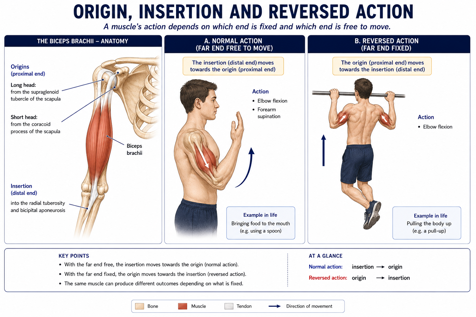
Biceps brachii with both heads shown, the long head from the supraglenoid tubercle and the short head from the coracoid process, insertion at the radial tuberosity, then the same muscle acting with the far end free and with the far end fixed.
A muscle always pulls; which end moves depends on which end is fixed. Normally the insertion travels towards the origin, as in lifting food to the mouth. Fix the hands on a bar and it reverses - the origin travels towards the insertion, and the body rises.

A muscle’s role is defined by the task, not by the muscle. The same muscle takes different roles in different movements.

RoleDefinitionExample
Agonist (prime mover)The muscle principally responsible for producing the movementBrachialis in elbow flexion
Assistant mover (secondary)Contributes, usually when the prime mover is disadvantaged or resistance is highBrachioradialis in rapid or loaded elbow flexion
AntagonistOpposes the agonist; usually relaxes (reciprocal inhibition) but may co-contract to control the movementTriceps during elbow flexion
SynergistWorks with the agonist to produce the desired movement more efficientlyLong finger flexors with wrist extensors
Neutraliser (a specific synergist)Cancels an unwanted secondary action of the agonistPronator teres cancelling the supination action of biceps during pure elbow flexion
Fixator (stabiliser)Contracts isometrically to steady the proximal segment so the agonist has a stable baseRotator cuff and scapular muscles during any hand activity; trunk muscles during any limb movement

Worked example — making a fist

The agonists are flexor digitorum superficialis and profundus. But those muscles cross the wrist, and if the wrist were free it would flex, shortening the flexors into active insufficiency and destroying grip. So the wrist extensors contract as synergists, holding the wrist in ~30° extension where the finger flexors are at their optimal length.

This is not theory. Denervate or inhibit the wrist extensors — as in a radial nerve palsy — and grip strength falls by roughly 70%, despite the finger flexors and their median/ulnar nerve supply being entirely intact. A patient with wrist drop is not primarily complaining of an inability to extend the wrist; they are complaining that they cannot hold anything. Splinting the wrist in extension restores most of the grip immediately, which is one of the most satisfying interventions in hand rehabilitation.

Reciprocal inhibition versus co-contraction

  • Reciprocal inhibition — Ia afferents from the contracting agonist’s spindles inhibit the antagonist’s motor neuron pool through an inhibitory interneuron. This is efficient and is the default for fast, unopposed movement.
  • Co-contraction — simultaneous activation of agonist and antagonist. It increases joint stiffness and stability at a metabolic cost and with a loss of speed. It is appropriate and normal in unstable, unfamiliar or high-precision tasks, in the early stage of motor learning, and around a joint whose passive restraints are compromised.

Excessive persistent co-contraction is, however, a common maladaptive pattern — after knee injury, in chronic low back pain, in anxiety-related bracing, and in the neurologically impaired patient. Distinguishing protective co-contraction (appropriate) from habitual co-contraction (a treatment target) is a core clinical judgement.

Types of muscle work

Figure 3 · The three kinds of contraction

The three kinds of muscle contraction Isometric, concentric and eccentric contraction shown by a schematic muscle of unchanged, shortened and lengthened length. Isometric no change Tension rises, length does not change. Holding a shopping bag still. Concentric shortens The muscle shortens while producing tension. Lifting the bag to the table. Eccentric lengthens The muscle lengthens while still producing tension. Lowering the bag slowly to the floor.
Eccentric work produces the most force and the most soreness. It is also most of what muscles do during an ordinary day.
TypeLength changeRoleTask examples
Isometric (static)None (at the whole-muscle level; the fascicles may shorten while the tendon lengthens)Stabilisation, holding, fixationHolding a shopping bag; postural muscle activity; rotator cuff during reaching
ConcentricShortensAcceleration; positive work; the muscle overcomes the loadStanding up (quadriceps); the upward phase of a curl
EccentricLengthens under tensionDeceleration, shock absorption, energy dissipation; negative work; the load overcomes the muscleSitting down; descending stairs; the landing phase of a jump; hamstrings in terminal swing

Three properties of eccentric work matter clinically:

  • Highest force capacity (~1.2–1.8 × isometric maximum), so it permits loading beyond concentric capacity.
  • Lowest metabolic cost for a given force, which is why eccentric-biased training is feasible in cardiac and respiratory populations.
  • Greatest mechanical stress, hence the most damage, the most DOMS, the strongest hypertrophic and tendon-remodelling stimulus, and the highest strain-injury risk.

Eccentric loading is the single best-evidenced intervention in tendinopathy (Alfredson’s protocol for the Achilles; heavy slow resistance as the modern alternative), and eccentric hamstring work at long muscle length (Nordic curl) has the strongest evidence of any single intervention for reducing hamstring strain incidence.

Naming the work, not the muscle

Ask “which muscle is working, and in which direction is gravity acting?” Walking downstairs, the knee flexes while the quadriceps works — eccentrically, controlling the descent. Students routinely name the hamstrings because the knee is flexing. The joint movement does not tell you which muscle is active; the direction of the resisting force does.

Open and closed kinetic chain

Open chainClosed chain
Distal segmentFree to moveFixed against resistance
Movement patternIsolated, at one joint; distal segment moves on proximalMulti-joint, predictable co-movement; proximal segment moves on distal
Muscle actionTypically single, concentric/eccentric at one jointCo-contraction; multiple muscles across multiple joints
Joint forcesMore shear, less compressionMore compression, less shear; greater joint stability
ExamplesSeated knee extension; biceps curlSquat, lunge, push-up, gait stance phase
ClinicalIsolates and quantifies a specific muscle; higher ACL strain in terminal knee extension (0–30°)Functional; safer for the reconstructed ACL; more proprioceptive input

Neither is superior. The modern position is that both are used, sequenced by tissue tolerance: closed chain early for safety and function, open chain added deliberately because it is the only reliable way to correct an isolated quadriceps deficit — a deficit that persists for years after ACL reconstruction if never specifically addressed.

Torque, moment arm and mechanical advantage

Figure 4 · The three classes of lever

The three classes of lever Three lever diagrams showing the arrangement of fulcrum, effort and load in first, second and third class levers. First class fulcrum effort load Effort — fulcrum — load Nodding the head. The skull balances on the atlas. Second class fulcrum effort load Fulcrum — load — effort Rising on to tiptoe. Rare in the body. Third class fulcrum effort load Fulcrum — effort — load Bending the elbow. Almost every muscle in the body.
Almost every joint in the body is a third class lever. The muscle sits close to the fulcrum and the load sits far from it, which costs force and buys speed.

Torque

Torque (moment) = Force × Moment arm

The moment arm is the perpendicular distance from the line of action of the force to the joint’s axis of rotation. Because it is perpendicular, it changes continuously through range as the joint angle changes.

This one equation explains a great deal of clinical reasoning:

  • A patient’s strength curve through range reflects the changing moment arm as much as the length–tension relationship.
  • Resistance is angle-dependent. A dumbbell held out at 90° of shoulder abduction imposes far more torque than the same dumbbell at 20°, because the external moment arm has lengthened, even though the load is unchanged.
  • Modifying the external moment arm is the simplest way to grade an exercise: bend the elbow during a straight-arm raise; hold a weight closer to the body; shorten a lever by holding at the knee rather than the ankle.
  • Surgical procedures manipulate internal moment arms deliberately — a tendon transfer changes the line of action; a high tibial osteotomy changes the ground reaction force’s moment arm at the knee.

Levers

A lever is a rigid bar (bone) rotating about a fulcrum (joint), acted on by an effort (muscle) and a resistance (load, including segment weight).

ClassArrangementCharacteristicAnatomical examples
First classFulcrum between effort and resistance (E–F–R)Balance; may favour force or speedAtlanto-occipital joint (neck extensors vs head weight); triceps extending the elbow; hip abductors in single-leg stance (a first-class arrangement about the hip)
Second classResistance between fulcrum and effort (F–R–E)Effort arm always longer → mechanical advantage >1; force at the cost of speedRising onto tiptoe (MTP joints as fulcrum, body weight as resistance, calf as effort); rare in the body
Third classEffort between fulcrum and resistance (F–E–R)Effort arm always shorter → mechanical advantage <1; requires more force, but gains speed and rangeThe great majority of body levers: biceps flexing the elbow; deltoid abducting the shoulder; hamstrings flexing the knee

Why the body chose the inefficient option. A third-class lever means the muscle must generate several times the external load — biceps typically produces around 6–8 times the weight held in the hand. In exchange, a small amount of muscle shortening produces a large, fast movement of the hand. Muscle is compact and can generate large force in a small space; long, fast limb movement cannot be generated any other way. The body trades force, which it has, for range and speed, which it needs.

The clinical corollary is that joint compressive loads are large. Hip joint reaction force in single-leg stance is approximately 2.5–3 × body weight, most of it generated by the abductors to counterbalance a body-weight moment arm roughly 2.5 times longer than their own. It follows that:

  • A stick held in the contralateral hand lengthens the counterbalancing moment arm enormously, reducing hip joint reaction force by up to 20–30% — far more than the small force applied through the stick would suggest.
  • A Trendelenburg gait (lateral trunk lean toward the stance side) is the patient’s own solution: it shifts the centre of mass over the hip, shortening the body-weight moment arm and reducing the abductor force required.
  • Losing 5 kg reduces peak knee joint load during walking by considerably more than 5 kg, because of the multiplier effect of the moment arms.

Active and passive insufficiency

Figure 5 · What stops a joint moving further

What stops a joint moving further Six panels naming the factors that limit joint range: bone contact, ligament, capsule, muscle, soft tissue apposition and pain. SIX THINGS CAN STOP A JOINT. KNOWING WHICH ONE CHANGES YOUR TREATMENT. Bone against bone The olecranon striking its fossa stops the elbow straightening further. Ligament tension The cruciates check the knee. Strong, and slow to recover if torn. Capsule tension Tightens at the end of range in every direction. Muscle tension A two-joint muscle limits one joint according to the other. Hamstrings limit hip flexion when the knee is straight. Soft tissue meeting The calf and thigh meet in full knee flexion. Pain and guarding Not anatomical, but it is what actually stops many patients.
Two of these six are muscle. If range changes when you move a neighbouring joint, a two-joint muscle is the limiter — not the joint you were testing.

These phenomena occur only in multi-joint (biarticular or polyarticular) muscles, and they are the reason multi-joint muscles cannot be understood one joint at a time.

Active insufficiencyPassive insufficiency
DefinitionA muscle cannot shorten further and cannot generate effective tension because it is already maximally shortened over all the joints it crossesA muscle cannot lengthen further, so it restricts movement at one joint when already stretched over another
Applies toThe agonistThe antagonist
MechanismExcessive filament overlap → few available cross-bridgesPassive tension limit of the muscle–tendon unit
DemonstrationFlex the wrist fully, then try to make a tight fist — grip is markedly weak because the long finger flexors are shortened across both wrist and fingersExtend the wrist fully, then try to fully flex the fingers — you cannot, because the long extensors are passively insufficient
Second exampleFlex the hip, then try to flex the knee fully — hamstring cramp and weakness, because they are shortened at both endsExtend the knee, then flex the hip (straight leg raise) — the hamstrings limit hip flexion
Third exampleExtend the hip, then try to extend the knee — rectus femoris is shortened at both endsFlex the knee (Ely’s test), then extend the hip — rectus femoris limits it

Clinical uses

  • Muscle testing. To test gastrocnemius, test plantarflexion with the knee extended; to test soleus in isolation, test it with the knee flexed (gastrocnemius is actively insufficient there). To differentiate flexor digitorum superficialis from profundus, block the adjacent digits’ profundus tendons.
  • Length testing. Every classic muscle length test is an application of passive insufficiency: straight leg raise (hamstrings), Thomas test (iliopsoas and, with the knee flexed, rectus femoris), Ober’s test (ITB and tensor fasciae latae), Ely’s test (rectus femoris), Silfverskiöld test (differentiating gastrocnemius from soleus contracture by testing ankle dorsiflexion with the knee extended and then flexed).
  • Stretch prescription. To stretch a biarticular muscle you must position both joints; hamstring stretching with the lumbar spine flexed loads the spine rather than the muscle.
  • Positioning. A patient positioned so that a biarticular muscle is shortened across both joints will develop contracture faster — hence the vigilance about hip and knee flexion positioning in the bed-bound patient, and about ankle plantarflexion in the patient with the foot unsupported.

The tenodesis effect

The passive-insufficiency phenomenon converted into function. Passively extending the wrist shortens the distance the long finger flexors must travel, causing the fingers to close passively into a functional grip; passively flexing the wrist opens the hand.

For a patient with a C6 spinal cord injury — who has active wrist extension (extensor carpi radialis longus, C6) but no active finger flexion (C8) — this is the entire basis of functional hand use. Two clinical implications follow, and they are critical:

  • Tenodesis grasp must be trained and is a primary rehabilitation goal after C6 tetraplegia.
  • The long finger flexors must be allowed to shorten slightly. Aggressive stretching of the finger flexors into full extension with the wrist extended destroys the tenodesis effect permanently and removes the patient’s grip. This is one of the few situations in physiotherapy where deliberate mild contracture is the correct goal, and it is a well-known cause of avoidable harm when a clinician unaware of it “corrects” the tightness.

Force couples

A force couple is two or more forces acting on the same segment in different directions, producing rotation.

Force couples are how the body produces rotation of a segment that has no fixed bony axis, and they are among the most useful concepts in musculoskeletal rehabilitation.

The scapular upward rotation couple

Full arm elevation requires roughly 60° of scapular upward rotation, contributed by three muscles pulling in three different directions:

  • Upper trapezius — elevates and upwardly rotates via the clavicle and acromion.
  • Lower trapezius — depresses the medial scapular spine, contributing an upward-rotation moment and stabilising against the upper fibres’ elevation.
  • Serratus anterior — protracts and upwardly rotates, and critically holds the medial border against the thorax.

Together they rotate the glenoid upwards; individually none of them can. This is why scapular dyskinesis is a coordination problem as much as a strength problem, why serratus anterior weakness produces medial border winging (long thoracic nerve, C5–7), and why trapezius palsy (spinal accessory nerve, often iatrogenic after posterior triangle lymph node surgery) produces lateral winging with a drooping shoulder and inability to elevate above shoulder height.

Other force couples worth knowing

CoupleMusclesFunction
Glenohumeral transverse coupleSubscapularis anteriorly vs infraspinatus and teres minor posteriorlyCompresses and centres the humeral head; balances deltoid’s superior translation force during abduction. Cuff dysfunction → superior migration
Deltoid–rotator cuff coronal coupleDeltoid (superior pull) vs inferior cuff (inferior/compressive pull)Produces rotation rather than translation of the head
Pelvic sagittal couplesAnterior tilt: hip flexors + erector spinae; posterior tilt: abdominals + hip extensorsControls lumbopelvic position
Trunk rotationExternal oblique on one side + internal oblique on the otherAxial rotation of the trunk
Foot arch supportTibialis posterior + fibularis longusThe “stirrup” supporting the medial longitudinal arch

Stabiliser and mobiliser systems — with a caveat

A widely taught model divides muscles into two systems:

Local (stabiliser) systemGlobal (mobiliser) system
PositionDeep, close to the joint axisSuperficial, distant from the axis
AttachmentsOften segmental, not crossing many jointsLong, multi-joint
Fibre typePredominantly type IHigher type II proportion
ActionLow-load tonic; controls segmental position and joint stiffness; often anticipatory (feedforward)Torque production; large ranges; power
ExamplesTransversus abdominis, multifidus, pelvic floor, diaphragm, rotator cuff, vastus medialis obliquus, deep neck flexorsRectus abdominis, external oblique, erector spinae (long fibres), latissimus dorsi, hamstrings, deltoid

The observation underpinning this — that transversus abdominis and multifidus normally activate before limb movement, and that this feedforward timing is delayed in people with low back pain, and that multifidus atrophies segmentally and does not spontaneously recover — is real and well replicated (Hodges, Richardson, Hides).

But the clinical inference drawn from it has not held up. Trials and meta-analyses consistently find that specific “core stability” or motor control training is no more effective than general exercise for chronic low back pain, and that the assumed mechanism (restoring transversus abdominis timing) does not explain the improvement observed. Contemporary practice therefore uses motor control training as one option among several, selected for particular patients (those with clear movement control impairment, or in the early painful phase where general loading is not tolerated), rather than as a universal prescription.

Teach the anatomy as anatomy and the model as a model. The deep muscles do behave differently from the superficial ones; the therapeutic conclusion once drawn from that fact was too strong.

Putting it together: analysing a movement

Use this sequence for any movement, in any examination or clinic:

  • Name the joint(s), and the movement occurring at each — with plane and axis (Chapter 1).
  • Identify the direction of the resistance, usually gravity plus any external load.
  • Decide which muscle group is working: the one on the side opposite to the direction the resistance is trying to move the segment.
  • Decide the type of contraction: shortening against resistance = concentric; lengthening under control = eccentric; no change = isometric.
  • Identify the fixators — what must be stabilised for this to work?
  • Identify the neutralisers — what unwanted actions must be cancelled?
  • Check for insufficiency — is any biarticular muscle shortened or lengthened across both joints?
  • Consider the lever and moment arm — how does the demand change through range?

Worked example — lowering slowly into a chair

  • Joints and movements: hip flexion, knee flexion, ankle dorsiflexion — all in the sagittal plane about frontal axes.
  • Resistance: gravity, acting to collapse the body downwards.
  • Muscles working: hip extensors (gluteus maximus, hamstrings), knee extensors (quadriceps), ankle plantarflexors (soleus) — the anti-gravity groups.
  • Contraction type: eccentric throughout; they are lengthening while resisting.
  • Fixators: trunk extensors and abdominals stabilising the spine; foot intrinsics and ankle everters/inverters stabilising the base.
  • Moment arm: the external moment arm at the knee increases as the knee flexes and the trunk moves back, so quadriceps demand rises through the descent — which is exactly why a patient with quadriceps weakness drops into the chair over the last 20–30°.
  • The rehabilitation conclusion writes itself: train eccentric quadriceps control through the outer range of knee flexion, and grade it by raising the seat height (shortening the range and the moment arm).

Where students consistently go wrong

  • Naming the muscle from the joint movement. The knee flexing downstairs does not mean the hamstrings are working. Look at gravity.
  • Assuming the origin is always fixed. In closed chain the insertion is fixed and the origin moves.
  • Ignoring fixators. A prime mover cannot function without a stabilised proximal base — this is the whole rationale for proximal-to-distal rehabilitation.
  • Forgetting insufficiency in muscle testing. Testing gastrocnemius with the knee flexed will underestimate it.
  • Calling every muscle-lengthening a stretch. Passive insufficiency is a normal mechanical limit, not tightness.
  • Stretching the finger flexors in a C6 tetraplegic patient. This destroys tenodesis grip.
  • Treating levers as trivia. Mechanical advantage explains gait aids, joint reaction forces, exercise grading and surgical decisions.
  • Treating a force couple’s components as independent muscles. Strengthening serratus without addressing lower trapezius rarely restores scapular rotation.
  • Prescribing core stability universally. The evidence does not support it as superior to general exercise.
  • Testing strength at only one joint angle. Torque varies with moment arm; a deficit may only appear in outer or inner range.

Check yourself

15 questions on this chapter. Tap one to see the answer and the reasoning.

Q1. A muscle that cancels an unwanted secondary action of the prime mover is a
  1. (A) fixator
  2. (B) neutraliser
  3. (C) antagonist
  4. (D) assistant mover

Answer: (B) Pronator teres neutralising biceps’ supination during pure elbow flexion.

Q2. Walking down stairs, the quadriceps works
  1. (A) concentrically
  2. (B) eccentrically
  3. (C) isometrically
  4. (D) not at all

Answer: (B) It lengthens under tension to control knee flexion against gravity.

Q3. Most levers in the human body are
  1. (A) first class
  2. (B) second class
  3. (C) third class
  4. (D) equally distributed

Answer: (C) Effort between fulcrum and resistance, favouring speed and range at the cost of force.

Q4. Rising onto tiptoe is an example of a
  1. (A) first-class lever
  2. (B) second-class lever
  3. (C) third-class lever
  4. (D) force couple

Answer: (B) The MTP joints are the fulcrum, body weight the resistance between fulcrum and the calf’s effort.

Q5. Grip strength falls markedly when the wrist is flexed because of
  1. (A) passive insufficiency of the flexors
  2. (B) active insufficiency of the long finger flexors
  3. (C) reciprocal inhibition
  4. (D) carpal tunnel compression

Answer: (B) They are shortened over both wrist and fingers, reducing cross-bridge availability.

Q6. Radial nerve palsy reduces grip strength principally because
  1. (A) the finger flexors are denervated
  2. (B) the wrist extensors cannot stabilise the wrist as synergists
  3. (C) sensation is lost
  4. (D) the thumb cannot oppose

Answer: (B) The flexors are intact; they simply have no stable wrist to work from.

Q7. Torque equals
  1. (A) force ÷ distance
  2. (B) force × moment arm
  3. (C) mass × acceleration
  4. (D) work ÷ time

Answer: (B) The moment arm is the perpendicular distance from the line of action to the axis.

Q8. A walking stick in the contralateral hand reduces hip joint reaction force mainly by
  1. (A) transferring body weight through the arm
  2. (B) providing a long counterbalancing moment arm, reducing required abductor force
  3. (C) reducing stride length
  4. (D) improving proprioception

Answer: (B) Which is why the stick is held on the opposite side to the affected hip.

Q9. Scapular upward rotation is produced by a force couple of
  1. (A) upper trapezius, lower trapezius and serratus anterior
  2. (B) rhomboids and levator scapulae
  3. (C) pectoralis minor and latissimus dorsi
  4. (D) deltoid and supraspinatus

Answer: (A)

Q10. Medial border winging of the scapula indicates weakness of
  1. (A) trapezius
  2. (B) rhomboids
  3. (C) serratus anterior
  4. (D) levator scapulae

Answer: (C) Long thoracic nerve, C5–C7.

Q11. The tenodesis effect in a C6 tetraplegic patient depends on
  1. (A) active finger flexion
  2. (B) passive tension in the long finger flexors during wrist extension
  3. (C) intact ulnar nerve function
  4. (D) intrinsic hand muscle activity

Answer: (B) Which is why over-stretching those flexors is harmful.

Q12. Compared with open-chain exercise, closed-chain exercise generally produces
  1. (A) more shear and less compression
  2. (B) more compression and less shear
  3. (C) less muscular co-contraction
  4. (D) isolated single-joint action

Answer: (B)

Q13. The Silfverskiöld test distinguishes
  1. (A) hamstring from gluteal tightness
  2. (B) gastrocnemius from soleus contracture
  3. (C) rectus femoris from iliopsoas tightness
  4. (D) ITB from TFL tightness

Answer: (B) By comparing ankle dorsiflexion with the knee extended and flexed.

Q14. Co-contraction of agonist and antagonist
  1. (A) is always pathological
  2. (B) increases joint stiffness and stability at a metabolic and speed cost
  3. (C) abolishes reciprocal inhibition permanently
  4. (D) occurs only in neurological disease

Answer: (B) It is appropriate in unstable or unfamiliar tasks and maladaptive when persistent.

Q15. Current evidence on specific core stability training for chronic low back pain shows it is
  1. (A) markedly superior to general exercise
  2. (B) broadly equivalent to general exercise
  3. (C) ineffective in all cases
  4. (D) contraindicated

Answer: (B) It remains a reasonable option for selected patients rather than a universal prescription.

Quick review

Everything on this page, in one screen

  • Roles are task-dependent: agonist, assistant mover, antagonist, synergist, neutraliser, fixator.
  • Reciprocal inhibition is the efficient default; co-contraction buys stiffness and stability at a cost, and is maladaptive when persistent.
  • Work types: isometric (hold), concentric (accelerate), eccentric (decelerate — highest force, lowest metabolic cost, most damage, best evidence in tendinopathy and hamstring injury prevention).
  • Ask what gravity is doing, not what the joint is doing, to identify the working muscle.
  • Open chain = isolated, more shear; closed chain = multi-joint, more compression, co-contraction. Use both, sequenced by tolerance.
  • Torque = Force × Moment arm. The moment arm changes through range, so demand and strength both vary with angle. Grade exercise by changing the external moment arm.
  • Levers: 1st = fulcrum in the middle (atlanto-occipital, triceps) · 2nd = resistance in the middle (tiptoe raise) · 3rd = effort in the middle (most of the body) — sacrificing force for speed and range, at the cost of high joint reaction forces.
  • Active insufficiency = agonist too short across both its joints. Passive insufficiency = antagonist too stretched across both. Both occur only in multi-joint muscles; both underpin muscle testing, length testing and stretching.
  • Tenodesis effect: wrist extension passively closes the fingers. Essential grip mechanism after C6 tetraplegia — never over-stretch those flexors.
  • Force couples produce rotation from non-parallel forces: scapular upward rotation (upper trapezius + lower trapezius + serratus anterior), glenohumeral transverse couple, pelvic tilt couples, trunk rotation obliques, foot stirrup.
  • Local vs global muscle systems is a useful anatomical description; specific core-stability training is not superior to general exercise for chronic low back pain.
  • Movement analysis sequence: joints and planes → resistance direction → working group → contraction type → fixators → neutralisers → insufficiency → lever and moment arm.

Further reading

SourceWhy it earns its place
Neumann DA — Kinesiology of the Musculoskeletal SystemThe best single treatment of torque, moment arms, force couples and joint reaction forces
Levangie PK, Norkin CC — Joint Structure and FunctionLevers, mechanical advantage and muscle roles, written for physiotherapists
Palastanga N, Field D, Soames R — Anatomy and Human MovementMuscles taught by function; the closest match to this chapter’s approach
Lieber RL — Skeletal Muscle Structure, Function and PlasticityArchitecture and its functional consequences
Kendall FP, McCreary EK, Provance PG — Muscles: Testing and Function with Posture and PainThe reference for muscle testing positions and length tests
Nordin M, Frankel VH — Basic Biomechanics of the Musculoskeletal SystemJoint reaction force calculations, including the classic hip and stick analysis
Alfredson H et al. — Am J Sports Med, 1998The original heavy eccentric Achilles protocol
van Dyk N, Behan FP, Whiteley R — “Including the Nordic hamstring exercise… meta-analysis”, Br J Sports Med, 2019The evidence for eccentric hamstring injury prevention
Hodges PW, Richardson CA — Spine, 1996The original feedforward transversus abdominis findings
Smith BE, Littlewood C, May S — “An update of stabilisation exercises for low back pain: a systematic review with meta-analysis”, BMC Musculoskelet Disord, 2014The evidence that tempers the clinical inference

Chapter 6 of 24 · Human Anatomy · Physiotherapist India Next: Chapter 7 — Connective Tissue, Ligaments and Fascia: one recipe behind tendon, ligament and fascia — and why compartments are dangerous.

Reviewed by the Physiotherapist India Team. · Human Anatomy contents
