Evidence-based physiotherapy and rehabilitation resources
Editorial & review policy
Evidence-based physiotherapy and rehabilitation resources
Editorial & review policyHuman Anatomy · The trunk
A muscular pump the size of a fist, slung in a tough bag in the middle of the chest. Its wiring and its blood supply are laid out so logically that you can predict what a blockage will damage. This chapter works from the outside in.
Part 4 · The trunk
Chambers, valves, the conducting system and the coronary supply
Cardiac rehabilitation is one of the best-evidenced interventions in the whole of physiotherapy — reducing cardiovascular mortality and hospital readmission in a way few of our treatments can claim. But you cannot prescribe exercise safely for a patient after myocardial infarction, valve surgery or heart failure without knowing which artery supplies which wall, why an inferior infarct causes bradycardia, what a sternotomy has actually divided, or why a patient in heart failure cannot lie flat.
Beyond cardiac rehabilitation, every patient you treat has a heart, and a proportion of the “musculoskeletal” chest, shoulder and interscapular pain that arrives in a physiotherapy clinic is not musculoskeletal at all. The anatomy in this chapter is what lets you tell the difference.
Figure 1 · The mediastinum, divided
The central compartment of the thorax, between the two pleural sacs, extending from the thoracic inlet to the diaphragm and from the sternum to the vertebral column.
Divided by the transverse thoracic plane — the plane of the sternal angle (T4–T5):
| Division | Contents |
|---|---|
| Superior mediastinum | Thymus (or its remnant), brachiocephalic veins and superior vena cava, arch of the aorta and its three branches (brachiocephalic trunk, left common carotid, left subclavian), trachea, oesophagus, thoracic duct, vagus and phrenic nerves, left recurrent laryngeal nerve, lymph nodes |
| Inferior — anterior | Thymic remnant, fat, lymph nodes, sternopericardial ligaments, internal thoracic vessels. A small space — but the site of retrosternal goitre, thymoma and lymphoma |
| Inferior — middle | The heart and pericardium, the roots of the great vessels, phrenic nerves, the main bronchi, and the pulmonary vessels |
| Inferior — posterior | Descending thoracic aorta, oesophagus and oesophageal plexus, thoracic duct, azygos and hemiazygos veins, sympathetic trunks and splanchnic nerves, lymph nodes |
The mediastinum is mobile in the living, which is why a tension pneumothorax can shift it and compress the great veins — an anatomical fact with an immediate clinical consequence.
Mediastinal shift: towards the lesion in collapse, fibrosis or pneumonectomy; away from the lesion in tension pneumothorax, large effusion or a large mass. Checking tracheal position is a two-second bedside test that detects it.
| Layer | Detail |
|---|---|
| Fibrous pericardium | Tough, inelastic outer sac. Fused below with the central tendon of the diaphragm and above with the adventitia of the great vessels; anchored to the sternum by the sternopericardial ligaments. Its inextensibility is why acute tamponade occurs with as little as 100–200 mL of rapidly accumulating fluid, while a slowly accumulating effusion may reach 1–2 litres |
| Parietal serous pericardium | Lines the fibrous layer |
| Visceral serous pericardium (epicardium) | Covers the heart; continuous with the parietal layer at the great vessel roots |
| Pericardial cavity | Potential space with ~15–50 mL of serous fluid |
Two sinuses, formed by the reflections of the serous layer:
Innervation: the fibrous and parietal serous pericardium are supplied by the phrenic nerve (C3–C5) — so pericarditis pain is referred to the shoulder tip and the neck, and is classically relieved by sitting forward and worsened by lying flat and by inspiration. The visceral layer is insensitive.
Cardiac tamponade — Beck’s triad of hypotension, distended neck veins and muffled heart sounds, with pulsus paradoxus (an inspiratory fall in systolic pressure >10 mmHg). An emergency requiring pericardiocentesis.
Figure 2 · The heart in the middle mediastinum
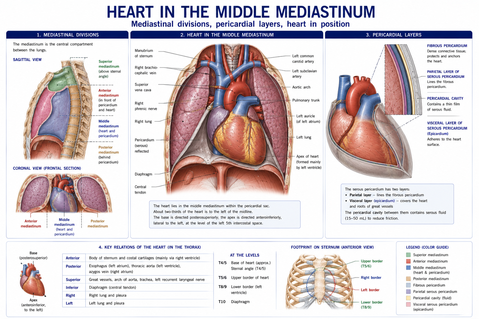
The heart lies obliquely, roughly two-thirds to the left of the midline, in the middle mediastinum. It is about the size of the owner’s closed fist and weighs 250–350 g.
| Surface / border | Formed by |
|---|---|
| Apex | Left ventricle — in the 5th intercostal space, midclavicular line, where the apex beat is palpated |
| Base (posterior surface) | Left atrium mainly, plus a little right atrium — receiving the pulmonary veins |
| Anterior (sternocostal) surface | Right ventricle mainly — hence its vulnerability in blunt sternal trauma |
| Inferior (diaphragmatic) surface | Left ventricle and part of the right ventricle |
| Right border | Right atrium |
| Left border | Left ventricle and the auricle |
| Inferior border | Right ventricle |
Surface projection (four corners, useful for the chest radiograph and for percussion):
Grooves: the coronary (atrioventricular) sulcus encircling the heart between atria and ventricles, and the anterior and posterior interventricular sulci — each carrying the corresponding arteries and veins.
Figure 3 · The chambers and valves of the heart
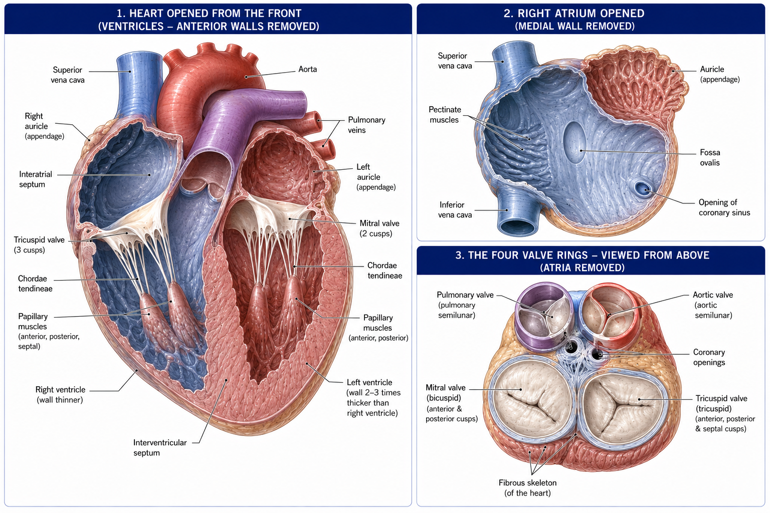
| Chamber | Wall | Key internal features |
|---|---|---|
| Right atrium | Thin | Crista terminalis dividing the smooth-walled posterior part (sinus venarum, from the sinus venosus) from the rough anterior part with pectinate muscles; the surface counterpart is the sulcus terminalis. Receives SVC, IVC (with its rudimentary valve) and the coronary sinus (with the valve of Thebesius). The fossa ovalis on the interatrial septum is the remnant of the foramen ovale — patent in ~25% of adults (patent foramen ovale, implicated in paradoxical embolism and cryptogenic stroke) |
| Right ventricle | ~3–5 mm | Trabeculae carneae; three papillary muscles with chordae tendineae to the tricuspid valve; the moderator band (septomarginal trabecula) carrying the right bundle branch to the anterior papillary muscle — ensuring it contracts before the valve is stressed. Outflow through the smooth conus arteriosus (infundibulum) to the pulmonary trunk |
| Left atrium | Thin | Mostly smooth; receives four pulmonary veins; pectinate muscle confined to the auricle — the left atrial appendage is the commonest site of thrombus formation in atrial fibrillation, and hence of cardioembolic stroke |
| Left ventricle | ~8–12 mm — three times the right | Conical; fine trabeculae; two large papillary muscles to the mitral valve; the aortic vestibule leading to the aortic valve. It must generate systemic pressure, hence the wall thickness |
Interventricular septum: a large muscular part and a small membranous part superiorly — the commonest site of ventricular septal defect.
Fibrous skeleton of the heart: four fibrous rings (anuli) around the valve orifices, with the right and left fibrous trigones. It anchors the valves, provides attachment for the myocardium, and — crucially — electrically insulates the atria from the ventricles, so the only normal conduction route is the atrioventricular bundle.
Figure 4 · Where a valve lies, and where it is heard
| Valve | Position | Cusps | Auscultation area |
|---|---|---|---|
| Tricuspid | Right AV orifice | 3 (anterior, posterior, septal) | Left 5th intercostal space at the lower sternal border |
| Pulmonary | Right ventricular outflow | 3 semilunar | Left 2nd intercostal space, parasternal |
| Mitral (bicuspid) | Left AV orifice | 2 (anterior, posterior) | Apex — 5th intercostal space, midclavicular line |
| Aortic | Left ventricular outflow | 3 semilunar (right, left, posterior) | Right 2nd intercostal space, parasternal |
The auscultation areas are NOT over the valves. They are the points to which the sound is best conducted downstream in the direction of blood flow. The valves themselves lie clustered close together behind the sternum, roughly along a line from the left 3rd costal cartilage to the right 6th. Students who try to listen “over the valve” hear little.
Mnemonic for the sequence Aortic (right 2nd), Pulmonary (left 2nd), Tricuspid (left 5th parasternal), Mitral (apex): All Physiotherapists Take Measurements.
Papillary muscles and chordae tendineae do not open or close the AV valves — pressure does that. They contract during systole to prevent the cusps from everting (prolapsing) into the atrium. Papillary muscle rupture after infarction therefore causes acute, severe mitral regurgitation and abrupt pulmonary oedema.
Semilunar valves have no chordae. Their cusps close passively in diastole as blood attempts to flow back; the coronary ostia lie in the aortic sinuses just above the right and left cusps, and coronary filling therefore occurs during diastole — an essential point (§19.7).
Figure 5 · The coronary arteries and the veins of the heart
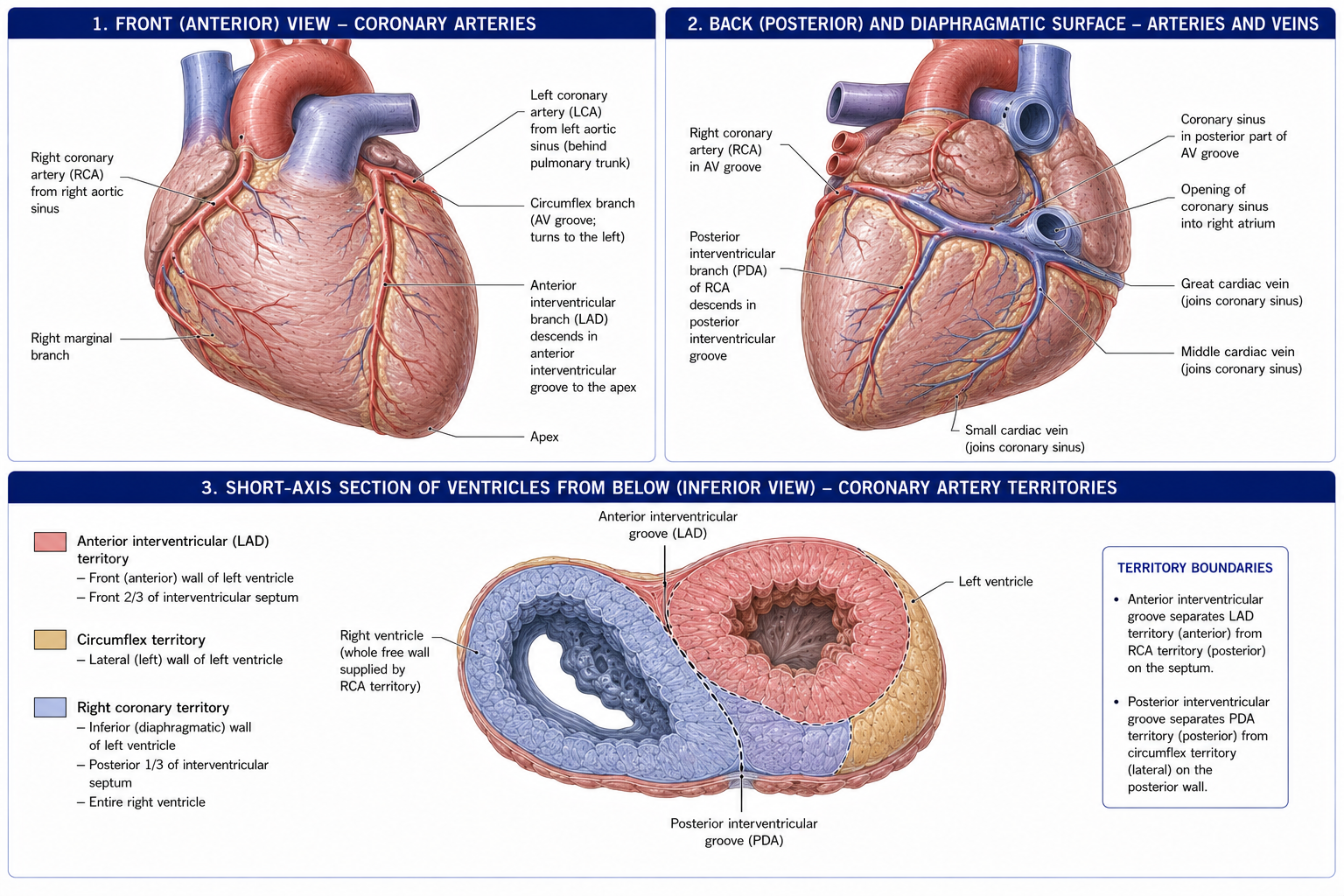
Both arise from the aortic sinuses immediately above the aortic valve.
| Artery | Branches | Supplies |
|---|---|---|
| Right coronary artery (RCA) | SA nodal branch (in ~60%), right marginal artery, AV nodal branch (in ~80–90%), posterior interventricular (posterior descending) artery in ~85% | Right atrium, right ventricle, SA and AV nodes, the inferior/diaphragmatic wall of the left ventricle, the posterior third of the interventricular septum |
| Left coronary artery (LCA) | Divides into the anterior interventricular (left anterior descending, LAD) and the circumflex | — |
| — LAD | Diagonal and septal branches | Anterior left ventricle, anterior two-thirds of the septum, apex — the largest territory. Occlusion is the “widow-maker” |
| — Circumflex | Left marginal artery; posterior interventricular artery in ~15% | Left atrium, lateral and posterior left ventricle; SA nodal branch in ~40% |
Dominance is defined by which artery gives the posterior interventricular artery: right dominant in ~85%, left dominant in ~8%, co-dominant in ~7%.
This table is the reason the chapter exists.
| Artery | Wall infarcted | ECG leads | Characteristic complications |
|---|---|---|---|
| LAD | Anterior / anteroseptal | V1–V4 | Largest infarcts; pump failure, cardiogenic shock, LV aneurysm, VSD; bundle branch blocks |
| Circumflex | Lateral | I, aVL, V5–V6 | Often electrically “silent”; mitral regurgitation |
| RCA | Inferior | II, III, aVF | Bradycardia and heart block (SA and AV nodal supply); right ventricular infarction (preload-dependent — nitrates and diuretics can cause catastrophic hypotension); vagally mediated nausea and vomiting |
Anastomoses between coronary branches exist but are functionally end-arteries in most people — collateral flow is inadequate to protect against acute occlusion, though it develops over time in chronic stenosis, which is one reason gradual disease can be less catastrophic than sudden thrombosis.
The left ventricular myocardium compresses its own intramural vessels during systole, so left coronary flow occurs almost entirely during diastole. Two consequences follow directly and are directly relevant to exercise prescription:
The rate–pressure product (heart rate × systolic BP) is the best simple index of myocardial oxygen demand, and the number that determines when a given patient develops angina. It reproduces reliably in the same individual, which is why an angina threshold is a usable prescription tool.
Figure 6 · The conducting system, step by step
| Structure | Location | Rate | Blood supply |
|---|---|---|---|
| Sinoatrial (SA) node | At the upper end of the crista terminalis, near the SVC opening in the right atrium | 60–100/min — the pacemaker | RCA ~60%, circumflex ~40% |
| Atrioventricular (AV) node | In the interatrial septum, in the triangle of Koch (bounded by the tendon of Todaro, the septal cusp of the tricuspid valve and the coronary sinus orifice) | 40–60/min | RCA ~80–90% |
| AV bundle (of His) | The only normal electrical connection between atria and ventricles, piercing the fibrous skeleton | — | Septal branches |
| Bundle branches | Right (running in the moderator band) and left (dividing into anterior and posterior fascicles) | — | LAD mainly |
| Purkinje fibres | Subendocardial network | 20–40/min | — |
The AV node delays conduction by ~0.1 s — the interval that allows atrial systole to complete ventricular filling before ventricular systole begins. It is the PR interval on the ECG.
Because the RCA supplies both nodes in most people, an inferior (RCA) infarct commonly produces bradycardia and AV block — usually transient and usually responsive to atropine, whereas block complicating an anterior (LAD) infarct implies extensive septal necrosis and carries a far worse prognosis. That difference is pure anatomy translated into prognosis.
ECG correlation:
| Wave / interval | Event |
|---|---|
| P wave | Atrial depolarisation |
| PR interval | AV nodal delay |
| QRS complex | Ventricular depolarisation (atrial repolarisation is buried within it) |
| ST segment | Plateau of ventricular action potential; elevation = transmural ischaemia |
| T wave | Ventricular repolarisation |
| Structure | Course and clinical points |
|---|---|
| Aorta | Ascending (from the left ventricle, giving the coronaries) → arch (at the level of the sternal angle, giving the brachiocephalic trunk, left common carotid, left subclavian) → descending thoracic aorta (T4 to T12, giving posterior intercostal, bronchial, oesophageal and phrenic branches). The ligamentum arteriosum (remnant of the ductus arteriosus) tethers the arch to the pulmonary trunk — and the aorta most often ruptures here in deceleration injury |
| Pulmonary trunk | Divides at the level of the sternal angle into right and left pulmonary arteries, carrying deoxygenated blood |
| Superior vena cava | Formed by the two brachiocephalic veins behind the right first costal cartilage; drains into the right atrium. SVC obstruction (usually bronchial carcinoma or lymphoma) causes facial and upper limb oedema, distended neck veins and headache |
| Azygos system | Azygos on the right, hemiazygos and accessory hemiazygos on the left; drains the posterior wall and arches over the right lung root into the SVC. The critical collateral route between SVC and IVC when either is obstructed |
| Thoracic duct | Begins at the cisterna chyli (L1–L2), enters through the aortic hiatus (T12), ascends to the right of the midline, crosses to the left at T5, and drains into the left venous angle. Drains the whole body except the right upper quadrant. Injury causes chylothorax |
| Oesophagus | From C6 to the cardia at T11. Three constrictions: at the cricopharyngeus (~15 cm from the incisors), where crossed by the aortic arch and left main bronchus (~25 cm), and at the diaphragmatic hiatus (~40 cm) — the sites where swallowed foreign bodies lodge and where corrosive strictures form |
| Trachea | C6 to the carina at the sternal angle (T4–T5). The right main bronchus is wider, shorter and more vertical — so inhaled foreign bodies and aspirated material preferentially enter the right lung, and specifically the posterior segment of the right upper lobe in the supine patient and the right lower lobe when upright. This determines where aspiration pneumonia occurs and where you position for postural drainage. |
| Phrenic nerves (C3–C5) | Pass anterior to the lung root, between the pericardium and mediastinal pleura. “Phrenic in front, vagus behind” |
| Vagus nerves (CN X) | Pass posterior to the lung root, forming the oesophageal plexus and continuing as the anterior (left) and posterior (right) vagal trunks |
| Left recurrent laryngeal nerve | Hooks under the arch of the aorta at the ligamentum arteriosum — vulnerable to aortic aneurysm, left atrial enlargement (mitral stenosis) and mediastinal tumour, producing hoarseness (Ortner’s syndrome). The right hooks around the subclavian artery |
| Sympathetic trunk and splanchnic nerves | On the vertebral bodies; the greater (T5–T9), lesser (T10–T11) and least (T12) splanchnic nerves pierce the crura to reach the abdomen |
| Phase | Duration | Events |
|---|---|---|
| Atrial systole | 0.1 s | Completes ventricular filling (~20–30% of total, the “atrial kick” — which is why atrial fibrillation reduces cardiac output most in patients with stiff ventricles) |
| Isovolumetric ventricular contraction | 0.05 s | All valves closed; pressure rises sharply |
| Ventricular ejection | 0.3 s | Semilunar valves open |
| Isovolumetric relaxation | 0.08 s | All valves closed; pressure falls |
| Ventricular filling | 0.4–0.5 s | AV valves open; rapid filling, diastasis, then atrial systole |
Diastole occupies about two-thirds of the cycle at rest — and this is the fraction that shrinks with tachycardia, compromising both ventricular filling and coronary perfusion.
| Sound | Cause | Notes |
|---|---|---|
| S1 (“lub”) | Closure of the mitral and tricuspid valves | Marks the start of systole |
| S2 (“dub”) | Closure of the aortic and pulmonary valves | Physiological splitting on inspiration (increased venous return delays pulmonary closure) |
| S3 | Rapid ventricular filling | Normal in children and young adults; in older adults suggests volume overload / heart failure — a “gallop” |
| S4 | Atrial contraction against a stiff ventricle | Always abnormal in adults; suggests hypertension, aortic stenosis, ischaemia |
| System | Origin | Effect |
|---|---|---|
| Sympathetic | T1–T5 via the cardiac plexus | ↑ rate (chronotropy), ↑ contractility (inotropy), ↑ conduction velocity, coronary vasodilatation |
| Parasympathetic | Vagus (CN X) | ↓ rate, ↓ AV conduction; little effect on ventricular contractility |
Cardiac pain afferents travel with the sympathetics, entering the cord at T1–T5 — which is exactly why cardiac pain refers to the central chest, the left arm and medial forearm (T1–T2 dermatomes), the neck and the jaw. That referral is a direct consequence of segmental convergence (Chapter 8), and it is the reason a physiotherapist must be able to recognise it.
The response to exercise:
Musculoskeletal pain has a mechanical behaviour: it is provoked and eased by specific positions, movements and palpation. Cardiac pain does not behave that way — it relates to exertion and emotion, not to position, and it is not reproduced by palpation or by movement testing.
Left shoulder, arm, jaw or interscapular pain that is exertional, unrelated to movement, accompanied by breathlessness, sweating or nausea, and not reproducible on examination is a cardiac presentation until proved otherwise. Refer immediately; do not treat, and do not arrange a follow-up.
| Lesion | Murmur | Physiotherapy implication |
|---|---|---|
| Aortic stenosis | Ejection systolic, radiating to the carotids | Severe symptomatic AS is a relative contraindication to vigorous and to heavy resistance exercise — fixed outflow obstruction means cardiac output cannot rise, risking syncope and sudden death |
| Mitral regurgitation | Pansystolic, radiating to the axilla | Volume overload; generally exercise-tolerant |
| Aortic regurgitation | Early diastolic, at the left sternal edge | Wide pulse pressure; reduced coronary perfusion pressure |
| Mitral stenosis | Mid-diastolic rumble with an opening snap | Left atrial enlargement → atrial fibrillation, thromboembolism, and hoarseness (Ortner’s). Still common in India as rheumatic heart disease |
Phase I (inpatient): early mobilisation, positioning, breathing exercises, education, and screening for sternal complications.
Sternal precautions after median sternotomy: the sternum is divided and wired; bony union takes 6–8 weeks, with full strength taking up to 3 months. Traditional advice restricted lifting to 5 kg and prohibited pushing, pulling and shoulder movement beyond 90° for 6–12 weeks. Contemporary practice (“Keep Your Move in the Tube”) has moved away from blanket restriction toward pain-guided, load-graded upper limb movement kept close to the trunk, because prolonged restriction produces stiffness, deconditioning and dependence without demonstrated reduction in sternal complications. Watch for sternal instability — clicking, a palpable rock, or a widening wound.
Phase II (outpatient): the core exercise programme, typically 6–12 weeks, prescribed by heart rate reserve or by rating of perceived exertion (11–14 on the Borg 6–20 scale), with resistance training added once aerobic conditioning is established.
Key prescription principles derived from this chapter:
The evidence: exercise-based cardiac rehabilitation reduces cardiovascular mortality and hospital admissions after myocardial infarction and in heart failure (Cochrane reviews, repeatedly). It is among the strongest evidence bases in the profession, and it is chronically under-referred.
15 questions on this chapter. Tap one to see the answer and the reasoning.
Answer: (B)
Answer: (C)
Answer: (B) Hence its vulnerability in blunt sternal trauma.
Answer: (B) To the anterior papillary muscle of the right ventricle.
Answer: (B)
Answer: (D)
Answer: (C) Usually an RCA territory infarct.
Answer: (B)
Answer: (B) Which is why tachycardia provokes ischaemia.
Answer: (C)
Answer: (B)
Answer: (B) Phrenic in front, vagus behind.
Answer: (B)
Answer: (C)
Answer: (B)
Everything on this page, in one screen
| Source | Why it earns its place |
|---|---|
| Standring S (ed.) — Gray’s Anatomy, 42nd edn | Definitive cardiac and mediastinal anatomy |
| Moore KL, Dalley AF, Agur AMR — Clinically Oriented Anatomy | The best clinical treatment of the mediastinum and cardiac surface anatomy |
| Hall JE — Guyton and Hall Textbook of Medical Physiology | The cardiac cycle, conduction and exercise response |
| Hough A — Physiotherapy in Respiratory and Cardiac Care | The physiotherapy-facing synthesis |
| American College of Sports Medicine — ACSM’s Guidelines for Exercise Testing and Prescription | Contraindications, termination criteria and prescription in cardiac disease |
| Dibben G et al. — “Exercise-based cardiac rehabilitation for coronary heart disease”, Cochrane Database Syst Rev, 2021 | The mortality and readmission evidence |
| Long L et al. — “Exercise-based cardiac rehabilitation for adults with heart failure”, Cochrane, 2019 | The heart failure evidence |
| Adams J et al. — “Sternal precautions and prone positioning… ‘Keep Your Move in the Tube’”, Proc (Bayl Univ Med Cent) | The contemporary sternal precaution approach |
| BACPR — Standards and Core Components for Cardiovascular Disease Prevention and Rehabilitation | Programme-level standards |
| Chaurasia BD — Human Anatomy, Vol 1: Upper Limb and Thorax | Indian syllabus-matched descriptive account |
Chapter 19 of 24 · Human Anatomy · Physiotherapist India Next: Chapter 20 — Abdomen: the abdominal wall, the peritoneum and the viscera.
Reviewed by the Physiotherapist India Team. · Human Anatomy contents