Evidence-based physiotherapy and rehabilitation resources
Editorial & review policy
Evidence-based physiotherapy and rehabilitation resources
Editorial & review policyHuman Anatomy · Lower limb
The hip is the opposite bargain to the shoulder. A deep socket, a strong capsule and heavy ligaments make it stable enough to carry you for a lifetime. What it asks in return is a set of muscles strong enough to hold the pelvis level every time you take a step.
Part 3 · The lower limb
The hip as the shoulder’s opposite, the abductor mechanism, and the femoral triangle
Set the hip beside the shoulder and every difference is a consequence of the same design decision.
| Shoulder | Hip | |
|---|---|---|
| Socket | Shallow glenoid, ⅓ of the head | Deep acetabulum, >½ of the head |
| Girdle attachment | One small joint, muscle-slung | Fused to the sacrum |
| Primary stabiliser | Muscle (concavity–compression) | Bone and capsule |
| Ligaments | Lax in mid-range | The strongest in the body; screw-home tightening in extension |
| Common failure | Dislocation | Osteoarthritis |
| Range | Greatest in the body | Large but constrained |
Both are triaxial ball-and-socket joints. One was optimised for reach, the other for load. Understanding the hip as the shoulder inverted makes both easier to remember.
The capsule runs from the acetabular margin to the intertrochanteric line anteriorly and to the medial two-thirds of the neck posteriorly (so the lateral posterior neck is extracapsular). Its deep circular fibres form the zona orbicularis, which grips the neck like a buttonhole and resists distraction.
Longitudinal retinacular fibres (retinacula of Weitbrecht) run along the neck carrying the retinacular vessels — the femoral head’s dominant blood supply.
| Ligament | Course | Function |
|---|---|---|
| Iliofemoral (of Bigelow / Y-ligament) | AIIS → intertrochanteric line, in an inverted Y | The strongest ligament in the body (withstands ~250–350 N). Limits extension and external rotation; permits standing with minimal muscular effort by allowing the body to “hang” on it |
| Pubofemoral | Superior pubic ramus → capsule inferiorly | Limits abduction and extension |
| Ischiofemoral | Ischium posteriorly → spirally to the neck | Limits internal rotation and extension; the weakest of the three, which is why posterior dislocation predominates |
| Ligamentum teres | Acetabular notch/transverse ligament → fovea | Carries the (adult-negligible) artery of the ligamentum teres; a proprioceptive and possible minor stabiliser in flexion–adduction–external rotation |
All three capsular ligaments spiral in the same direction, tightening in extension and unwinding in flexion. Hence:
| Movement | Range (knee extended) | Range (knee flexed) | Limited by |
|---|---|---|---|
| Flexion | ~90° | ~120–135° | Hamstrings (extended knee) / soft tissue apposition |
| Extension | 10–20° | Less | Iliofemoral ligament |
| Abduction | 40–45° | — | Pubofemoral ligament, adductors |
| Adduction | 20–30° | — | Contralateral limb, abductors |
| Internal rotation | 30–40° | — | Ischiofemoral ligament, external rotators |
| External rotation | 40–60° | — | Iliofemoral and pubofemoral ligaments |
Functional ranges for activities of daily living are worth knowing when full range is not achievable: approximately 110–120° flexion (for shoe-tying and squatting), 20° abduction and 20° external rotation. This is the basis of the “hip precautions” advice after posterior-approach arthroplasty — avoid flexion beyond 90°, adduction past midline, and internal rotation — though many units have relaxed these as evidence for their necessity has weakened.
Arteries: the medial circumflex femoral (dominant, via the retinacular vessels), the lateral circumflex femoral, and the artery of the ligamentum teres (from the obturator; negligible in adults). The cruciate anastomosis — from the medial and lateral circumflex femoral, the inferior gluteal and the first perforating artery — provides collateral flow.
Nerves (Hilton’s law in action): femoral (via the nerve to rectus femoris), obturator, superior gluteal, and the nerve to quadratus femoris — L2 to S1. The obturator supply is why hip pathology refers pain to the medial knee, and why any child with knee pain and a limp must have the hip examined.
Posterior dislocation (~90%) — from an axial force through a flexed, adducted, internally rotated hip: the classic dashboard injury. The limb lies flexed, adducted, internally rotated and shortened. The sciatic nerve is injured in ~10–20% (usually the common fibular division). Test and document nerve function before and after reduction; the risk of avascular necrosis rises sharply if reduction is delayed beyond 6–12 hours.
Anterior dislocation (~10%) — the limb lies abducted, externally rotated and extended; the femoral nerve and vessels are at risk.
Congenital / developmental dysplasia (DDH) — see Chapter 8.
Figure 1 · The gluteal region and the sciatic nerve
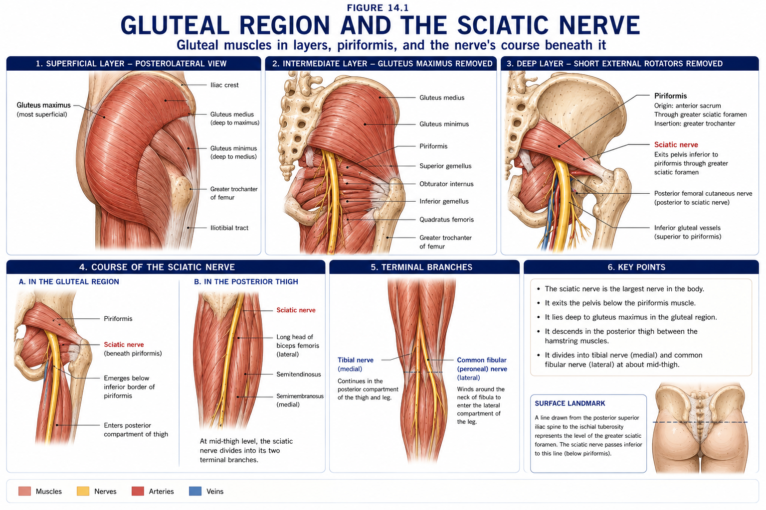
Figure 2 · The abductor mechanism in single leg stance
| Muscle | Origin | Insertion | Nerve | Actions |
|---|---|---|---|---|
| Gluteus maximus | Ilium behind the posterior gluteal line, sacrum, coccyx, sacrotuberous ligament, thoracolumbar fascia | ~75% into the iliotibial tract; ~25% into the gluteal tuberosity | Inferior gluteal (L5, S1, S2) | Powerful hip extensor (used in rising from sitting, climbing, running — not in level walking), external rotator; upper fibres abduct, lower fibres adduct; tensions the ITB |
| Gluteus medius | Ilium between the posterior and anterior gluteal lines | Lateral greater trochanter | Superior gluteal (L4, L5, S1) | Principal abductor; anterior fibres internally rotate, posterior fibres externally rotate. Stabilises the pelvis in single-leg stance |
| Gluteus minimus | Ilium between the anterior and inferior gluteal lines | Anterior greater trochanter | Superior gluteal (L4, L5, S1) | Abduction, internal rotation, pelvic stabilisation; a capsular stabiliser via its deep attachment |
| Tensor fasciae latae | Outer lip of the crest behind the ASIS | Iliotibial tract | Superior gluteal (L4, L5, S1) | Abduction, flexion, internal rotation; tensions the ITB |
| Piriformis | Anterior sacrum, through the greater sciatic foramen | Upper greater trochanter | Nerve to piriformis (S1, S2) | External rotator in extension; becomes an abductor/internal rotator beyond ~60–90° of flexion — a genuinely useful fact for positioning and for testing |
| Obturator internus + superior and inferior gemelli | Inner obturator membrane; ischial spine and tuberosity | Medial greater trochanter | Nerve to obturator internus (L5, S1); nerve to gemellus inferior | External rotation; abduction in flexion; capsular stabilisation |
| Quadratus femoris | Lateral ischial tuberosity | Quadrate tubercle | Nerve to quadratus femoris (L5, S1) | External rotation, adduction |
| Obturator externus | Outer obturator membrane | Trochanteric fossa | Obturator (L3, L4) | External rotation; it is an adductor-compartment muscle by innervation, lying deep in the gluteal region |
The short external rotators from above down: Piriformis, Gemellus superior, Obturator internus, Gemellus inferior, Quadratus femoris, Obturator externus — “Play Golf Or Go On Quaaludes” or simply P-GO-G-Q-O.
| Above piriformis | Below piriformis |
|---|---|
| Superior gluteal nerve and vessels | Sciatic nerve; inferior gluteal nerve and vessels; posterior femoral cutaneous nerve; pudendal nerve and internal pudendal vessels; nerve to obturator internus; nerve to quadratus femoris |
The gluteal region is a standard intramuscular injection site, and the sciatic nerve is the reason precision matters. Inject into the upper outer quadrant of the buttock, dividing the buttock by a vertical and a horizontal line through its centre. Better still, use the ventrogluteal (von Hochstetter) site: palm on the greater trochanter, index finger on the ASIS, middle finger spread toward the iliac crest. Inject into the triangle so formed, which is over gluteus medius and minimus and safely away from the sciatic nerve, superior gluteal vessels and subcutaneous fat.
Injection into the lower medial quadrant risks sciatic nerve injury — a classic, entirely avoidable, and still-occurring cause of foot drop, particularly in children.
In single-leg stance (which occupies ~60% of the gait cycle on each side), body weight acts medial to the hip and would drop the unsupported side of the pelvis. The abductors (gluteus medius and minimus, with TFL) must generate a counterbalancing moment.
The geometry is brutally unequal: the body-weight moment arm is roughly 2.5 times longer than the abductor moment arm. To balance, the abductors must generate about 2.5 times body weight of force. Add that to body weight, and the hip joint reaction force in single-leg stance is approximately 2.5–3 × body weight — rising to 4–5 × in stair climbing and 6–8 × in running.
Three clinical conclusions follow directly:
Causes of a positive Trendelenburg sign: abductor weakness or inhibition (superior gluteal nerve lesion, L5 radiculopathy, post-arthroplasty via a lateral approach, pain inhibition, gluteal tendinopathy), a shortened femoral neck or coxa vara reducing the abductor moment arm, dislocation, and severe pain.
Figure 3 · Compartments of the thigh
Divided by the medial, lateral and posterior intermuscular septa arising from the fascia lata, whose lateral thickening is the iliotibial tract.
| Anterior (extensor) | Medial (adductor) | Posterior (flexor) | |
|---|---|---|---|
| Nerve | Femoral (L2–L4) | Obturator (L2–L4) | Sciatic — tibial division (L5–S2) |
| Artery | Femoral | Profunda femoris / obturator | Perforating branches of profunda femoris |
| Muscles | Quadriceps, sartorius, iliopsoas (in part), pectineus | Adductor longus, brevis, magnus, gracilis, obturator externus | Biceps femoris, semitendinosus, semimembranosus |
| Muscle | Origin | Insertion | Nerve | Action |
|---|---|---|---|---|
| Psoas major | T12–L5 vertebral bodies, discs and transverse processes | Lesser trochanter (with iliacus) | Anterior rami L1–L3 directly | The most powerful hip flexor; lumbar spine stabiliser |
| Iliacus | Iliac fossa | Lesser trochanter | Femoral (L2, L3) | Hip flexion |
| Rectus femoris | Straight head: AIIS. Reflected head: above the acetabulum | Patella → tibial tuberosity via the patellar ligament | Femoral (L2–L4) | The only quadriceps head crossing two joints: hip flexion + knee extension. Prone to strain in kicking, and to adolescent AIIS avulsion |
| Vastus lateralis, medialis, intermedius | Femoral shaft (linea aspera and anterior surface) | Common quadriceps tendon → patella | Femoral (L2–L4) | Knee extension. Vastus medialis obliquus (VMO) — the distal oblique fibres from the adductor magnus tendon — resists lateral patellar tracking |
| Sartorius | ASIS | Medial tibia, via the pes anserinus | Femoral (L2, L3) | The longest muscle in the body; hip flexion, abduction, external rotation + knee flexion — the “tailor’s” cross-legged position |
| Pectineus | Pecten pubis | Pectineal line of femur | Femoral (± obturator) | Hip flexion and adduction; a transitional muscle |
| Articularis genu | Distal anterior femur | Suprapatellar bursa | Femoral | Retracts the bursa during extension |
The pes anserinus (“goose’s foot”) is the conjoined insertion of Sartorius, Gracilis and Semitendinosus — “Say Grace before Tea” — each from a different compartment and a different nerve (femoral, obturator, tibial respectively). It is a common site of bursitis and of medial knee pain that mimics MCL or meniscal pathology.
| Muscle | Origin | Insertion | Nerve | Note |
|---|---|---|---|---|
| Adductor longus | Body of pubis | Middle linea aspera | Obturator | The most commonly strained groin muscle; its palpable tendon is the medial border of the femoral triangle |
| Adductor brevis | Inferior pubic ramus | Upper linea aspera | Obturator | The obturator nerve’s two divisions pass in front of and behind it |
| Adductor magnus | Adductor part: inferior pubic and ischial rami → linea aspera. Hamstring (ischiocondylar) part: ischial tuberosity → adductor tubercle | As above | Adductor part: obturator. Hamstring part: tibial division of sciatic | A composite muscle with two nerve supplies — one of the classic examination points. Its tendon forms the adductor hiatus |
| Gracilis | Body and inferior ramus of pubis | Medial tibia (pes anserinus) | Obturator | The only medial-compartment muscle crossing the knee; commonly harvested for graft |
| Obturator externus | Obturator membrane | Trochanteric fossa | Obturator | Functionally an external rotator |
| Muscle | Origin | Insertion | Nerve | Note |
|---|---|---|---|---|
| Biceps femoris — long head | Ischial tuberosity | Head of fibula | Tibial division of sciatic (L5–S2) | The most commonly strained |
| Biceps femoris — short head | Linea aspera | Head of fibula | Common fibular division (L5–S2) | The only hamstring not crossing the hip, and the only one with common fibular innervation |
| Semitendinosus | Ischial tuberosity | Medial tibia (pes anserinus) | Tibial division | Long tendon distally; a common graft |
| Semimembranosus | Ischial tuberosity | Posteromedial tibial condyle, with expansions to the oblique popliteal ligament and posterior capsule | Tibial division | A posteromedial knee stabiliser |
All hamstrings extend the hip and flex the knee (except the short head of biceps, which only flexes the knee). Because they are biarticular, active and passive insufficiency (Chapter 6) apply — which is why hamstring length is tested with the hip flexed, and why they cramp when the hip is flexed and the knee flexed simultaneously.
Figure 4 · The femoral triangle
| Boundary | Structure |
|---|---|
| Superior (base) | Inguinal ligament |
| Lateral | Medial border of sartorius |
| Medial | Medial border of adductor longus (some texts use its lateral border) |
| Floor | Iliopsoas and pectineus laterally to medially (with adductor longus medially) |
| Roof | Fascia lata, pierced by the saphenous opening for the great saphenous vein; cribriform fascia; superficial inguinal lymph nodes |
Contents, lateral to medial — “NAVEL”: Nerve (femoral) · Artery (femoral) · Vein (femoral) · Empty space (femoral canal) · Lymphatics (deep inguinal nodes, including the node of Cloquet)
The femoral sheath — a prolongation of transversalis and iliac fascia — encloses the artery, vein and canal, but NOT the nerve, which lies outside it. The femoral canal, the medial compartment, permits venous distension and is the site of femoral hernia (below and lateral to the pubic tubercle — versus inguinal hernia, above and medial).
Clinical uses: femoral pulse palpation at the mid-inguinal point; femoral vein cannulation and arterial access (the vein is medial to the artery — remembered by the fact that you aim medially for the vein); femoral nerve block; and lymph node assessment.
A fascial tunnel in the middle third of the thigh, from the apex of the femoral triangle to the adductor hiatus.
| Boundary | Structure |
|---|---|
| Anteromedial (roof) | Fibrous subsartorial fascia, covered by sartorius |
| Anterolateral | Vastus medialis |
| Posterior | Adductor longus and magnus |
Contents: the femoral artery and vein, the saphenous nerve, and the nerve to vastus medialis.
At the adductor hiatus in adductor magnus, the femoral vessels pass into the popliteal fossa and become the popliteal artery and vein. The saphenous nerve does not — it pierces the fascia to become subcutaneous, supplying the medial leg and foot. Adductor canal block, now widely used after knee arthroplasty, targets the saphenous nerve while sparing quadriceps motor function — which is why it has largely replaced the femoral nerve block, whose quadriceps weakness caused falls.
Formed in psoas, emerges at its lateral border, passes under the inguinal ligament lateral to the femoral artery and outside the femoral sheath, then divides.
Descends on the lateral pelvic wall, through the obturator canal, dividing into anterior and posterior divisions on either side of adductor brevis.
The largest nerve in the body, ~2 cm wide at its origin. Leaves the pelvis through the greater sciatic foramen below piriformis (in ~85–90%; variants exist in which the common fibular division pierces piriformis, and these are cited in discussions of piriformis syndrome), descends midway between the ischial tuberosity and the greater trochanter, and runs on adductor magnus deep to the hamstrings.
It is two nerves in one sheath from the outset:
| Division | Supplies | Vulnerability |
|---|---|---|
| Tibial | All hamstrings except the short head of biceps; the hamstring part of adductor magnus; the whole posterior leg and sole | More medial, better protected |
| Common fibular | Short head of biceps; the anterior and lateral leg and dorsum of foot | More lateral, fewer fascicles, less connective tissue, and tethered at the fibular neck — hence preferentially injured in hip dislocation, injection injury and stretch |
It divides at the apex of the popliteal fossa (variably higher).
Sciatic nerve lesions: posterior hip dislocation, misplaced gluteal injection, hip surgery, pelvic tumour, prolonged sitting. A complete lesion produces total loss below the knee plus hamstring weakness — with preserved knee extension and hip flexion (femoral) and preserved sensation over the medial leg (saphenous).
“Sciatica” is a symptom, not a diagnosis, and in the great majority of cases it originates at the lumbar nerve root, not the sciatic nerve itself. Piriformis syndrome as a cause is controversial, over-diagnosed, and best regarded as a diagnosis of exclusion; deep gluteal syndrome is the broader modern term for extrapelvic sciatic entrapment.
Passes under the inguinal ligament near the ASIS. Entrapment causes meralgia paraesthetica — burning, numbness and dysaesthesia over the anterolateral thigh, with no motor deficit. Associated with obesity, pregnancy, tight belts and waistbands, and prolonged hip extension. Recognising it prevents a great deal of unnecessary lumbar imaging.
The femoral artery continues from the external iliac at the inguinal ligament, gives the profunda femoris (the main supply to the thigh, itself giving the medial and lateral circumflex femoral arteries and four perforating arteries), and passes through the adductor hiatus to become the popliteal artery.
The cruciate anastomosis (inferior gluteal, medial and lateral circumflex femoral, first perforating) and the trochanteric anastomosis provide collateral circulation around the hip — clinically important when the femoral or external iliac artery is occluded.
The great saphenous vein — the longest vein in the body — ascends from the anterior to the medial malleolus (a reliable cut-down site), up the medial leg and thigh, to pierce the cribriform fascia at the saphenous opening and join the femoral vein at the saphenofemoral junction, ~3–4 cm inferolateral to the pubic tubercle.
Abnormal contact between the proximal femur and the acetabular rim during movement, producing labral and chondral damage.
| Type | Morphology | Typical patient |
|---|---|---|
| Cam | Aspherical femoral head–neck junction (reduced head–neck offset, “pistol grip” deformity) | Young athletic males; develops during adolescent growth in high-impact sport |
| Pincer | Acetabular over-coverage (global or focal retroversion) | Middle-aged, more often female |
| Mixed | Both | Most cases |
The Warwick Agreement definition requires all three of symptoms, clinical signs and imaging findings — morphology alone is not FAI, and cam morphology is present in a large proportion of asymptomatic athletes. Clinical: groin pain, the FADIR/anterior impingement test (flexion–adduction–internal rotation), and reduced internal rotation in flexion. First-line management is physiotherapist-led rehabilitation; arthroscopy for those who fail it, with trials (FASHIoN, UK FASHIoN) showing a modest advantage for surgery over physiotherapy at 12 months, and continuing debate about longer-term outcomes.
Formerly called “trochanteric bursitis” — the pathology is predominantly tendinopathy of gluteus medius and minimus at the trochanteric insertion, with secondary bursal involvement.
The commonest injury in running and kicking sports.
The Doha agreement classification is now standard, and it is worth using because “groin strain” as a label is useless:
| Condition | Notes |
|---|---|
| Snapping hip (coxa saltans) | External — ITB or gluteus maximus over the greater trochanter; internal — iliopsoas over the iliopectineal eminence or femoral head; intra-articular — labral tear or loose body |
| Piriformis / deep gluteal syndrome | Buttock pain with sciatic distribution symptoms; a diagnosis of exclusion after lumbar causes |
| Meralgia paraesthetica | Anterolateral thigh burning; no motor loss |
| Adductor-related groin pain | The Copenhagen adduction exercise has good preventive evidence |
| Avulsion injuries in adolescents | ASIS (sartorius), AIIS (rectus femoris), ischial tuberosity (hamstrings), lesser trochanter (iliopsoas) — the apophyseal equivalents of adult muscle strains |
15 questions on this chapter. Tap one to see the answer and the reasoning.
Answer: (B) It limits extension and permits relaxed standing.
Answer: (B) The first option is the resting position.
Answer: (C)
Answer: (B) In approximately 85–90% of people.
Answer: (C) Rising to 4–5 × on stairs and 6–8 × in running.
Answer: (B) It provides a long counterbalancing moment arm, reducing joint reaction force by 20–30%.
Answer: (C) Its adductor and hamstring parts have different supplies.
Answer: (B) NAVEL.
Answer: (B)
Answer: (C)
Answer: (B)
Answer: (A) Lateral femoral cutaneous nerve entrapment.
Answer: (B) Because the ITB compresses the tendon in adduction — hence ITB stretching is counterproductive.
Answer: (C)
Answer: (B) It is more lateral, has fewer and larger fascicles, less protective connective tissue and is tethered distally.
Everything on this page, in one screen
| Source | Why it earns its place |
|---|---|
| Standring S (ed.) — Gray’s Anatomy, 42nd edn | Definitive regional anatomy |
| Neumann DA — Kinesiology of the Musculoskeletal System | The definitive account of hip joint reaction forces and the abductor mechanism |
| Palastanga N, Field D, Soames R — Anatomy and Human Movement | Functional anatomy of hip and thigh |
| Snell RS — Clinical Anatomy by Regions | Femoral triangle, adductor canal and gluteal region with clinical notes |
| Griffin DR et al. — “The Warwick Agreement on femoroacetabular impingement syndrome”, Br J Sports Med, 2016 | The consensus definition |
| Griffin DR et al. — “Hip arthroscopy versus best conservative care (UK FASHIoN)”, Lancet, 2018 | The comparative trial |
| Mellor R, Bennell K, Grimaldi A et al. — “Education plus exercise versus corticosteroid injection versus a wait and see approach… gluteal tendinopathy (LEAP trial)”, BMJ, 2018 | The evidence that reframed GTPS management |
| Weir A et al. — “Doha agreement meeting on terminology and definitions in groin pain in athletes”, Br J Sports Med, 2015 | The standard groin pain classification |
| van Dyk N et al. — Nordic hamstring meta-analysis, Br J Sports Med, 2019 | Hamstring injury prevention |
| Chaurasia BD — Human Anatomy, Vol 2 | Indian syllabus-matched descriptive account |
Chapter 14 of 24 · Human Anatomy · Physiotherapist India Next: Chapter 15 — Knee and Leg: the knee joint, menisci and cruciates, and the compartments of the leg.
Reviewed by the Physiotherapist India Team. · Human Anatomy contents