Evidence-based physiotherapy and rehabilitation resources
Editorial & review policy
Evidence-based physiotherapy and rehabilitation resources
Editorial & review policyHuman Anatomy · General anatomy
Tendon, ligament, capsule and fascia are made from the same few ingredients in different proportions and different arrangements. Learn the recipe once and each tissue's behaviour -- how it loads, how it fails, how long it takes to heal -- follows from it.
Part 1 · General anatomy
One recipe behind tendon, ligament and fascia — and why compartments are dangerous
Bone, cartilage, tendon, ligament, fascia, dermis, capsule, aponeurosis and even blood are all connective tissue. They share a single formula:
Connective tissue = cells + fibres + ground substance, with the matrix, not the cells, doing the work.
What distinguishes one from another is the proportion of the three ingredients and the orientation of the fibres. Change the ratio of collagen to elastin and you get a ligament instead of a tendon. Change the fibre orientation from parallel to interwoven and you get fascia instead of tendon. Add hydroxyapatite and you get bone. Add water and aggrecan and you get cartilage.
Learn the recipe, and the tissues stop being a list.
| Class | Subtype | Character | Locations |
|---|---|---|---|
| Connective tissue proper — loose | Areolar | Loosely arranged fibres in abundant ground substance; the general packing tissue | Subcutaneous; around vessels and nerves; deep to epithelia |
| Adipose | Adipocytes dominant | Subcutaneous, perinephric, orbital, buccal fat pad, infrapatellar | |
| Reticular | Type III collagen network | Bone marrow, lymph node, spleen, liver stroma | |
| Connective tissue proper — dense | Dense regular | Parallel collagen bundles; resists tension in one direction | Tendon, ligament, aponeurosis |
| Dense irregular | Interwoven bundles in many directions | Deep fascia, joint capsule, dermis, periosteum, epimysium, dura mater | |
| Dense elastic | High elastin proportion | Ligamenta flava, ligamentum nuchae, walls of elastic arteries, suspensory ligament of the penis | |
| Specialised | Cartilage, bone, blood, lymphoid | See Chapters 2, 3 | — |
The functional rule: parallel fibres resist tension in one direction and are therefore found where load direction is predictable (tendon). Interwoven fibres resist tension in many directions and are found where load direction varies (fascia, capsule, dermis).
Figure 1 · What every connective tissue is made of
| Cell | Function |
|---|---|
| Fibroblast | The workhorse: synthesises collagen, elastin, proteoglycans and glycoproteins. Named tenocyte in tendon, fibrocyte when quiescent. Highly mechanosensitive — matrix synthesis is up-regulated by cyclical load |
| Myofibroblast | A contractile fibroblast expressing α-smooth muscle actin. Responsible for wound contraction; central to scar contracture, Dupuytren’s disease and adhesive capsulitis |
| Macrophage | Phagocytosis, debridement, cytokine signalling, orchestration of repair |
| Mast cell | Histamine, heparin; vascular permeability; a role in fascial and tendon pathology |
| Adipocyte | Energy storage, insulation, cushioning, endocrine (leptin, adiponectin) |
| Plasma cell, lymphocyte, neutrophil, eosinophil | Immune surveillance and inflammation |
| Fibre | Composition | Mechanical property |
|---|---|---|
| Collagen | Triple-helical tropocollagen; the most abundant protein in the body (~30% of total protein) | High tensile strength (~50–100 MPa in tendon), low extensibility (~4–8% before failure) |
| Elastin | Elastin cross-linked by desmosine into a rubber-like network, sheathed by fibrillin microfibrils | Extensible to 150% and fully recoiling; low strength |
| Reticular | Type III collagen with a glycoprotein coat; argyrophilic | Fine supporting mesh |
Collagen types worth knowing:
| Type | Where | Clinical link |
|---|---|---|
| I | Bone, tendon, ligament, dermis, fascia, fibrocartilage, mature scar | ~90% of body collagen. Osteogenesis imperfecta; classical EDS |
| II | Hyaline and elastic cartilage, nucleus pulposus, vitreous | Chondrodysplasias |
| III | Reticular fibres, blood vessels, granulation tissue, early healing tissue and immature scar | Vascular EDS. The ratio of type III to type I is the key index of scar maturity |
| IV | Basement membrane | Alport syndrome; Goodpasture disease |
| V | With type I, regulating fibril diameter | Classical EDS |
| X | Hypertrophic zone of physis; calcified cartilage | Marker of chondrocyte hypertrophy |
Collagen synthesis in outline: transcription → preprocollagen → hydroxylation of proline and lysine (vitamin C-dependent) → glycosylation → triple-helix formation as procollagen → secretion → cleavage of terminal propeptides to tropocollagen → self-assembly into fibrils with quarter-stagger overlap (giving the 67 nm banding) → cross-linking by lysyl oxidase (copper-dependent) → fibrils to fibres.
Two clinical consequences: scurvy (vitamin C deficiency) prevents hydroxylation, producing unstable collagen — poor wound healing, wound breakdown, bleeding gums, subperiosteal haemorrhage; and cross-linking increases with age and with non-enzymatic glycation in diabetes, making tissue stiffer, more brittle and less able to remodel — one reason diabetic patients have stiffer tendons, more frozen shoulders and slower connective tissue healing.
An amorphous, hydrated gel of:
Its functions are to hydrate, to provide the medium for diffusion of nutrients and metabolites, to lubricate the sliding of collagen fibres past one another (the critical interfibrillar distance), and to resist compression.
Immobilisation reduces GAG and water content in dense connective tissue. Collagen fibres consequently lie closer together, and new collagen is deposited randomly across the existing fibres, forming abnormal cross-links at the intersections. The fibres can no longer glide past one another. Add adhesion formation and the result is the stiff, restricted joint seen after even a few weeks of casting.
This is why controlled early motion is not simply “good for morale”. It maintains interfibrillar distance and GAG content, and orients new collagen along lines of stress rather than randomly. The tissue’s organisation is a direct consequence of the loading applied during healing.
Figure 2 · Tendon and ligament compared
Tropocollagen → microfibril → subfibril → fibril → fibre (with tenocytes between) → fascicle (surrounded by endotenon) → tendon (surrounded by epitenon and then paratenon or a synovial sheath)
At rest, collagen fibres in tendon and ligament lie in a regular wavy crimp. Under initial load the crimp straightens before the collagen itself is stretched. This is the structural basis of the toe region of the stress–strain curve, and it is the shock-absorbing buffer that protects the tissue from small loads. Most daily activity operates within the toe region.
| Tendon | Ligament | |
|---|---|---|
| Connects | Muscle to bone | Bone to bone |
| Collagen | ~85–95% type I, dry weight | ~70–80% type I |
| Fibre arrangement | Highly parallel, unidirectional | Less regular, near-parallel with interwoven bundles — because ligaments must resist load from several directions |
| Elastin | ~1–2% | ~5–15% (much higher in ligamentum flavum, ~60–70% elastin) |
| Water | ~55–70% | ~60–70% |
| Cells | Tenocytes in rows | Fibroblasts, more rounded, less regularly arrayed |
| Blood supply | Poor; from musculotendinous junction, enthesis, paratenon/vincula. Watershed zones exist | Poor; better in extra-articular than intra-articular ligaments |
| Innervation | Golgi tendon organs at the MTJ; free nerve endings; sympathetic fibres | Type I–IV receptors (Chapter 4); proprioceptively important |
| Principal function | Transmit muscle force; store and return elastic energy | Restrain joint motion at end range; provide proprioceptive feedback |
Several tendons have a hypovascular segment where degeneration and rupture concentrate:
These are the four tendons that rupture spontaneously in middle age, and their locations are not coincidental.
Figure 3 · How connective tissue behaves over time
| Region | Strain | Behaviour |
|---|---|---|
| Toe region | 0–2% | Crimp straightens. Very compliant; little force for substantial elongation. Normal physiological range |
| Linear (elastic) region | ~2–4% | Collagen fibres themselves stretch. Force rises steeply and linearly; the slope is the Young’s modulus (stiffness). Deformation is fully recoverable |
| Yield point / microfailure | ~4–6% | Individual fibre failure begins; permanent deformation. Clinically, a grade I–II sprain |
| Plastic region and rupture | ~6–8% | Progressive fibre failure to complete rupture, typically at 8–10% strain. Clinically a grade III tear |
Two important derived points. First, the tissue is permanently lengthened once the yield point is passed — which is precisely what a repeatedly sprained ankle ligament has undergone, and why its mechanoreceptor-mediated protective function is impaired even when it “heals”. Second, ligaments fail at their insertion (enthesis) in the immature and in the immobilised, and in mid-substance in the mature, well-conditioned adult — which is why the adolescent sustains a tibial spine avulsion where the adult sustains an ACL mid-substance rupture.
Because connective tissue contains water and a proteoglycan gel, its response is time-dependent, not purely elastic.
| Property | Definition | Clinical application |
|---|---|---|
| Creep | Progressive deformation under a constant load over time | The basis of prolonged low-load stretch, serial casting and dynamic splinting — and the reason a sustained slouched posture progressively lengthens posterior spinal tissues, with measurable loss of protective reflex response |
| Stress relaxation | Progressive fall in the force required to maintain a constant length | Why a sustained stretch feels easier after 30 seconds; the basis of static stretching and of the load reduction seen in a maintained mobilisation hold |
| Hysteresis | Energy lost as heat during a load–unload cycle | Why tissue warms with repeated loading, and why preconditioning (a warm-up of repeated submaximal cycles) reduces stiffness before activity |
| Strain-rate dependence | The tissue is stiffer and stronger at higher loading rates | High-speed injury stores more energy; slow loading permits greater deformation before failure |
| Thixotropy | Reduced resistance with repeated movement | Part of the mechanism of “warming up” and of morning stiffness |
Practical consequence. Low-load, long-duration stretch produces more lasting tissue change than high-load, short-duration stretch, and does so with far less risk of microfailure. This is why serial casting, dynamic splints and 20–30 minute positioning programmes are effective in contracture management, and why brief aggressive stretching is not.
A necessary caveat. The acute range-of-motion gain from a single stretching session is now attributed largely to increased stretch tolerance (a sensory adaptation) rather than to lasting tissue lengthening. Genuine structural lengthening — increased fascicle length, increased sarcomeres in series — is achieved much more reliably by eccentric loading at long muscle lengths and by prolonged positioning, not by conventional stretching.
| Phase | Timing | Events |
|---|---|---|
| Haemorrhage and inflammation | 0–~72 h (up to 7 days) | Haematoma, vasodilatation, neutrophils then macrophages, cytokine release, phagocytosis of debris |
| Proliferation (fibroplasia) | ~3 days – 6 weeks | Fibroblast proliferation; abundant type III collagen, randomly oriented; neovascularisation; a disorganised, weak but voluminous matrix. Tensile strength is at its lowest around days 5–21 |
| Remodelling and maturation | ~6 weeks – 12 months or more | Type III is progressively replaced by type I; fibres realign along lines of stress; cross-links mature; cellularity and vascularity decline; strength rises slowly |
The essential clinical statement: at 6 weeks a healing ligament may have as little as 50–60% of its final strength, and full maturation takes a year or longer. Scar tissue never fully regains normal properties, typically plateauing at ~70–80% of original tensile strength with more type III collagen, smaller fibril diameter and inferior mechanical behaviour.
Immobilised healing produces a weak, disorganised, adhesion-bound scar. Controlled progressive loading during remodelling directly increases collagen alignment, cross-link maturation and ultimate tensile strength — the tissue equivalent of Wolff’s law. This principle, established in animal ligament models and confirmed clinically, is the single strongest justification for early, protected, criteria-progressed rehabilitation rather than prolonged rest.
Intra-articular ligaments heal worst. The ACL is bathed in synovial fluid, which lyses the haematoma and prevents the fibrin clot scaffold from forming. It therefore has essentially no capacity for spontaneous healing — the anatomical reason reconstruction rather than repair is the standard approach.
Tendon heals by both intrinsic (tenocyte-mediated) and extrinsic (surrounding sheath and paratenon) mechanisms. The extrinsic route brings vascularity but also adhesions, which bind the tendon to its sheath and destroy gliding. This is the central problem in flexor tendon repair in the hand, and the reason early controlled mobilisation protocols (Kleinert, Duran, and modern early active motion regimes) exist: they apply enough excursion to prevent adhesion without exceeding repair strength. The window is narrow and the protocols are precise; deviating from them costs the patient hand function.
The old model — “tendinitis”, an inflammatory condition treated with rest, ice and anti-inflammatories — is wrong, and knowing why changes what you do.
Histopathology of chronic tendinopathy shows: collagen disorganisation and fibre separation, increased type III collagen, increased ground substance and proteoglycan, neovascularisation with accompanying neural ingrowth, tenocyte rounding and proliferation, and an absence of the classic inflammatory cell infiltrate. The correct term is tendinosis or, in the umbrella clinical sense, tendinopathy.
The continuum model (Cook and Purdam) describes three overlapping stages that a tendon can move between in either direction:
| Stage | Tissue state | Reversibility | Management |
|---|---|---|---|
| Reactive tendinopathy | Non-inflammatory proliferative cell and matrix response to acute overload; the tendon thickens to reduce stress | Highly reversible | Load reduction and relative rest, then graded return; isometrics for pain relief |
| Tendon disrepair | Greater matrix breakdown, increased proteoglycan and vascularity, separation of collagen | Potentially reversible | Progressive loading; heavy slow resistance or eccentric programmes |
| Degenerative tendinopathy | Areas of cell death, disorganisation, acellular regions, extensive neovascularity | Largely irreversible in the degenerated portion | Load the remaining healthy tendon; capacity building; surgery only for the minority |
What this means for treatment:
Figure 4 · Fascial layers and the compartments of the thigh
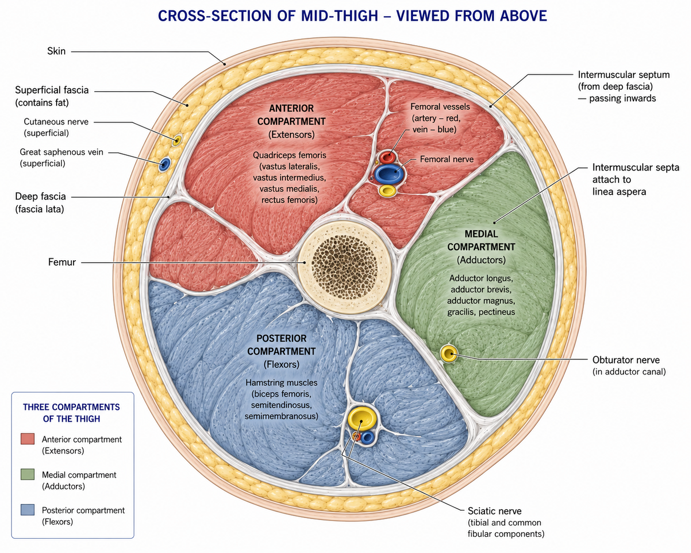
Loose areolar and adipose tissue between the dermis and deep fascia. It contains fat, cutaneous nerves and vessels, superficial veins and lymphatics, and in some regions a membranous layer (Scarpa’s fascia of the abdomen, Colles’ fascia of the perineum, the superficial fascial system of the limbs) and skin ligaments (retinacula cutis) anchoring skin to deep fascia.
Clinical relevance: it is the plane of subcutaneous oedema (hence pitting oedema and the compartment for lymphoedema), the layer mobilised in skin-rolling and soft tissue techniques, and the plane in which superficial veins can be harvested or cannulated. The membranous layer of the perineum (Colles’ fascia) and its attachments determine the direction in which extravasated urine can spread — a classic anatomical point.
A tough, dense irregular sheet investing muscles, and continuous throughout the body. Its several roles:
| Role | Example |
|---|---|
| Investing sheath | Fascia lata of the thigh; crural fascia of the leg; brachial fascia |
| Intermuscular septa | Extend inward from investing fascia to bone, dividing the limb into osteofascial compartments |
| Muscle attachment | Many muscles take origin from deep fascia (e.g. part of gluteus maximus into the iliotibial tract; tensor fasciae latae) |
| Retinacula | Local thickenings holding tendons close to bone at joints, preventing bowstringing: flexor retinaculum (roofing the carpal tunnel), extensor retinaculum of wrist and ankle, superior and inferior fibular retinacula, patellar retinacula |
| Force transmission | Fascia transmits tension between muscles and across regions (myofascial force transmission); the thoracolumbar fascia is the clearest example, linking latissimus dorsi, gluteus maximus, the abdominal wall and the erector spinae |
| Neurovascular sheaths | Carotid sheath; axillary sheath (a prolongation of prevertebral fascia — the basis of brachial plexus block) |
| Pump assistance | Contraction of muscle within a fascial envelope compresses veins and lymphatics — the muscle pump |
Because deep fascia is strong and essentially inextensible, the compartments it forms are closed spaces. Any increase in their contents raises pressure.
| Region | Compartments |
|---|---|
| Arm | Anterior (flexor, musculocutaneous n.), posterior (extensor, radial n.) |
| Forearm | Anterior, posterior, and the mobile wad |
| Thigh | Anterior (femoral n.), medial (obturator n.), posterior (sciatic n.) |
| Leg | Anterior (deep fibular n., anterior tibial a.), lateral (superficial fibular n.), superficial posterior, deep posterior (tibial n., posterior tibial a.) |
Mechanism. Bleeding or oedema within a closed compartment raises intracompartmental pressure above capillary perfusion pressure. Perfusion fails, causing ischaemia, which causes further oedema, which raises pressure further — a vicious cycle. Irreversible muscle necrosis begins at approximately 4–6 hours; nerve damage begins earlier.
Crucially: distal pulses are usually PRESENT. Arterial pressure far exceeds the compartment pressure that occludes capillaries. A palpable pulse does not exclude compartment syndrome, and waiting for pulselessness means waiting until the limb is lost.
The cardinal features — the 5 Ps are late and unreliable. The early signs are:
Common causes: tibial fracture (the commonest), forearm fracture (especially supracondylar humeral fracture in children), crush injury, reperfusion after vascular repair, tight casts and dressings, burns, and prolonged limb compression.
What a physiotherapist must do. Do not elevate above heart level (this reduces perfusion pressure further); do not apply ice or compression; split or remove any constricting cast or dressing; do not “work through” the pain; and escalate immediately. Definitive treatment is emergency fasciotomy. The late consequence of a missed compartment syndrome is Volkmann’s ischaemic contracture — fibrotic replacement of the flexor compartment producing a permanently clawed, useless hand.
Chronic exertional compartment syndrome is a different entity: exercise-induced pressure rise causing predictable, reproducible exertional pain and paraesthesia that resolves within minutes of stopping. It is a diagnosis made on exertional pressure measurement, and it responds to gait retraining in some cases and to elective fasciotomy in others.
Worth naming specifically because of its role in trunk mechanics. Classically described in three layers (posterior, middle and anterior) enclosing the erector spinae and quadratus lumborum. Its posterior layer receives latissimus dorsi, the contralateral gluteus maximus, and the aponeuroses of transversus abdominis and internal oblique. It is therefore the connective tissue node through which trunk and limb forces are transmitted diagonally across the body, and the anatomical basis of the posterior oblique sling.
It is also densely innervated, with free nerve endings and nociceptors, and has been demonstrated to be a plausible source of non-specific low back pain in its own right — a more defensible claim than most made about fascia.
Fascia has attracted a large body of claim, and the honest position is a mixed one:
Supported by evidence:
Not supported:
The defensible synthesis: manual and myofascial techniques are best understood as producing neurophysiological effects — altered mechanosensitivity, descending inhibition, autonomic change, and improved movement tolerance — which create a window in which active loading can be delivered. That is a legitimate and useful role. It is not the same as the mechanical claim usually made for it, and being clear about the difference is part of practising honestly.
15 questions on this chapter. Tap one to see the answer and the reasoning.
Answer: (B) Deep fascia, capsule, dermis and periosteum are dense irregular.
Answer: (B) It provides a compliant buffer for everyday loads.
Answer: (C) It is the mechanism exploited by serial casting and prolonged low-load stretch.
Answer: (C) It is progressively replaced by type I during remodelling.
Answer: (B) Deficiency produces the defective collagen of scurvy.
Answer: (B) Roughly 60–70% elastin, which is why it recoils and does not buckle into the canal in youth.
Answer: (B) Its intra-articular position is the decisive factor.
Answer: (B) Hence tendinosis, not tendinitis.
Answer: (C) Complete rest reduces capacity; heavy loading too early aggravates a reactive tendon.
Answer: (B) Pulses are usually present, and the 5 Ps are late.
Answer: (B) Nerve injury begins earlier still.
Answer: (B) Classically after supracondylar humeral fracture in a child.
Answer: (C) Anterior, lateral, superficial posterior and deep posterior.
Answer: (B) Structural lengthening requires eccentric loading at long lengths or prolonged positioning.
Answer: (B) The forces required exceed what manual contact can deliver.
Everything on this page, in one screen
| Source | Why it earns its place |
|---|---|
| Standring S (ed.) — Gray’s Anatomy, 42nd edn | Reference account of connective tissue and fascial anatomy |
| Ross MH, Pawlina W — Histology: A Text and Atlas | The clearest treatment of connective tissue classification and collagen synthesis |
| Nordin M, Frankel VH — Basic Biomechanics of the Musculoskeletal System | Stress–strain behaviour of tendon and ligament, and viscoelasticity |
| Woo SL-Y et al. — “Injury and repair of ligaments and tendons”, Annu Rev Biomed Eng, 2000 | The reference account of healing and the effect of loading |
| Cook JL, Purdam CR — “Is tendon pathology a continuum?”, Br J Sports Med, 2009 | The model that reorganised tendinopathy management |
| Cook JL, Rio E, Purdam CR, Docking SI — “Revisiting the continuum model”, Br J Sports Med, 2016 | The updated version, including compression and load management |
| Kongsgaard M et al. — “Corticosteroid injections, eccentric decline squat training and heavy slow resistance training in patellar tendinopathy”, Scand J Med Sci Sports, 2009 | The comparative evidence on loading versus injection |
| Schleip R, Findley TW, Chaitow L, Huijing PA — Fascia: The Tensional Network of the Human Body | The most complete fascia text; read alongside the critical literature |
| Willard FH, Vleeming A, Schuenke MD, Danneels L, Schleip R — “The thoracolumbar fascia: anatomy, function and clinical considerations”, J Anat, 2012 | The definitive anatomical account of the TLF |
| Weerakkody N, Taylor CJ, Bulmer CL et al.; and Freitas SR et al. — reviews on stretching mechanisms, Scand J Med Sci Sports | The evidence behind stretch tolerance rather than tissue lengthening |
| Via AG, Oliva F, Maffulli N — compartment syndrome reviews, Muscles Ligaments Tendons J | Clinical presentation and timelines |
Chapter 7 of 24 · Human Anatomy · Physiotherapist India Next: Chapter 8 — Skin, and How the Body Forms: skin layers and repair, the germ layers, and why one nerve root has both a muscle and a skin patch.
Reviewed by the Physiotherapist India Team. · Human Anatomy contents