Evidence-based physiotherapy and rehabilitation resources
Editorial & review policy
Evidence-based physiotherapy and rehabilitation resources
Editorial & review policyHuman Anatomy · General anatomy
Cartilage has no blood vessels, no nerves and no lymphatics. Every difficult thing about it follows from that one sentence: it is fed by movement, it cannot signal its own damage, and when it tears it does not heal the way the tissues around it do.
Part 1 · General anatomy
No vessels, no nerves, no lymphatics — and everything difficult follows from that
Cartilage is avascular, aneural and alymphatic.
Hold that sentence and you can derive almost everything else. No blood vessels means nutrition by diffusion, which means slow metabolism, which means slow healing and a strict dependence on movement to circulate nutrients. No nerves means articular cartilage cannot hurt — so pain in an osteoarthritic joint must be coming from somewhere else, and knowing where changes how you treat it. No lymphatics and no blood supply means immune privilege, which is why osteochondral allografts can be transplanted between unrelated donors without matching.
Bone rebuilds itself; cartilage largely does not. That asymmetry is the reason cartilage injury dominates long-term musculoskeletal disability while bone injury usually resolves.
By the end of this chapter you should be able to:
Cartilage is a specialised connective tissue consisting of cells (chondrocytes) embedded in an extensive extracellular matrix, with the unusual property that the matrix, not the cells, does virtually all of the work.
| Component | Detail |
|---|---|
| Chondroblast | The immature, matrix-secreting cell, derived from mesenchyme. |
| Chondrocyte | The mature cell, occupying a space in the matrix called a lacuna. Often found in groups of 2–8 (isogenous groups / cell nests) representing the progeny of one cell after interstitial growth. Metabolically active but functioning at low oxygen tension — chondrocyte metabolism is largely anaerobic, an adaptation to avascularity. |
| Chondron | The functional unit: the chondrocyte plus its pericellular matrix — a thin shell rich in type VI collagen and perlecan, which acts as a mechanical transducer and protects the cell from direct strain. |
| Territorial matrix | Immediately around the chondron; more proteoglycan-rich, staining more strongly. |
| Interterritorial matrix | The bulk of the tissue between chondrons; more collagen-rich; carries the mechanical load. |
| Perichondrium | A fibrous sheath with an outer fibrous and an inner chondrogenic layer, carrying the blood supply that nourishes cartilage by diffusion. Present around most cartilage — but absent over articular cartilage and over fibrocartilage. This single absence is the reason articular cartilage cannot mount a repair response. |
Figure 1 · The three types compared
Figure 2 · The three types of cartilage under the microscope
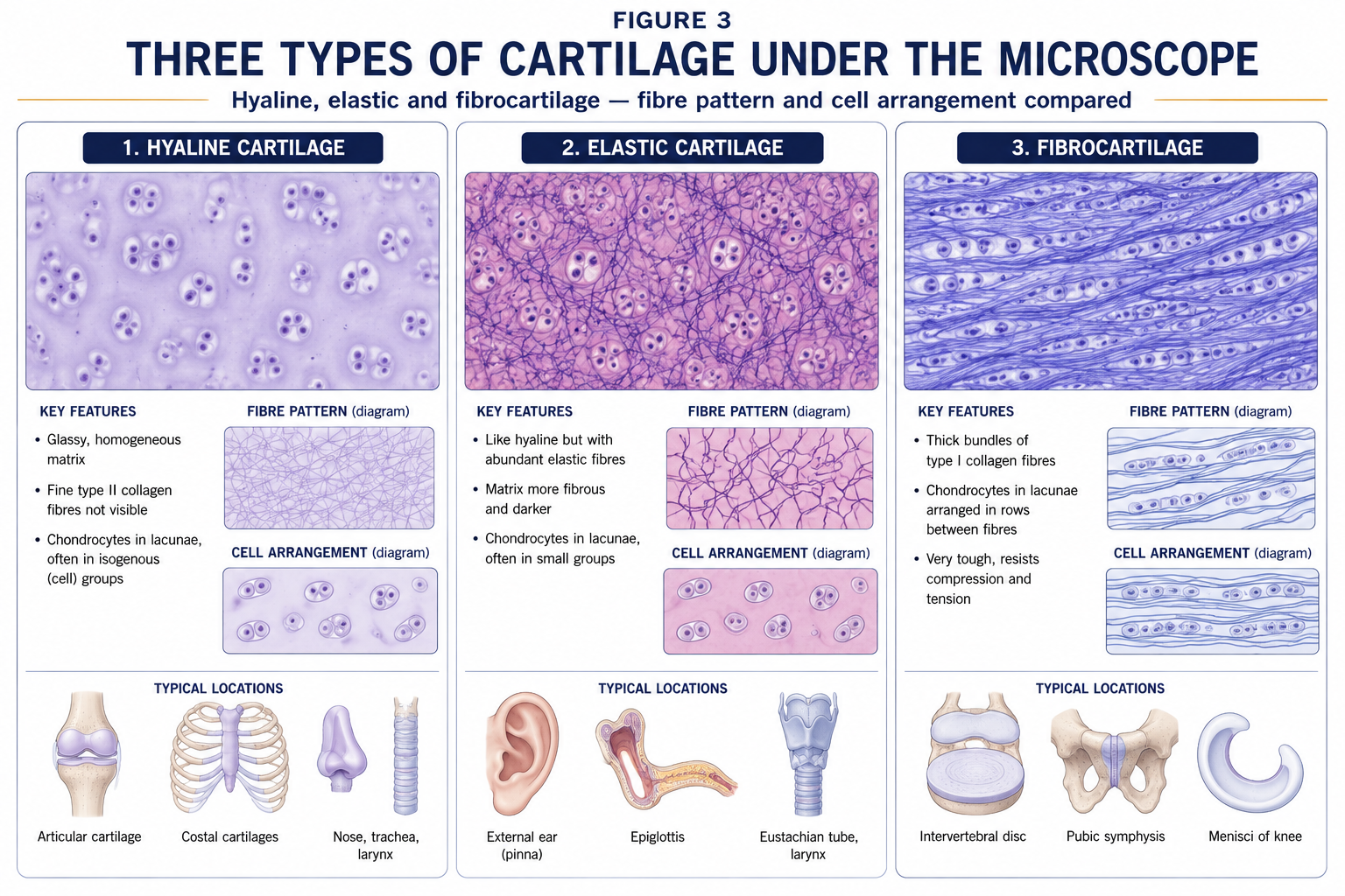
| Hyaline | Elastic | Fibrocartilage | |
|---|---|---|---|
| Principal collagen | Type II (fine fibrils, masked by ground substance — hence the glassy appearance) | Type II plus abundant elastic fibres | Type I predominantly, with some type II |
| Ground substance | Very high proteoglycan (aggrecan) content | High | Lower proteoglycan, higher collagen |
| Appearance | Glassy, bluish-white, translucent | Yellowish, opaque | Dense, white, with visible fibre bundles |
| Perichondrium | Present, except on articular surfaces | Present | Absent |
| Chondrocytes | Rounded, in lacunae, isogenous groups | Rounded, densely packed | Sparse, often in rows between collagen bundles |
| Chief property | Resists compression; low-friction surface | Resists compression and returns to shape after deformation | Resists tension and shear as well as compression; tough |
| Locations | Articular surfaces; costal cartilage; epiphyseal plate; nasal septum; larynx (thyroid, cricoid, most of arytenoid); trachea and bronchi; fetal skeletal model | External ear (auricle); external acoustic meatus; auditory (Eustachian) tube; epiglottis; corniculate and cuneiform cartilages | Intervertebral disc anulus fibrosus; menisci of the knee; glenoid and acetabular labra; triangular fibrocartilage complex (TFCC); articular disc of the sternoclavicular, acromioclavicular and temporomandibular joints; pubic symphysis; fibrocartilaginous entheses (Achilles, supraspinatus, patellar tendon insertions) |
| Calcifies with age? | Yes, readily (costal cartilage, thyroid cartilage) | No — elastic cartilage does not calcify | Rarely |
Two examination points follow: the epiglottis and auricle are elastic (they must spring back), and the intervertebral disc is a hybrid — an outer fibrocartilaginous anulus surrounding a gelatinous nucleus pulposus that is a notochordal remnant and behaves as a fluid-filled pressure vessel rather than as cartilage proper.
By wet weight, articular cartilage is approximately 65–80% water, 10–20% collagen, and 4–7% proteoglycan, with chondrocytes occupying only 1–5% of the volume. The mechanical behaviour is entirely a property of how those first three interact.
Type II collagen forms roughly 90–95% of the collagen, arranged in fine fibrils cross-linked into a three-dimensional mesh. Minor collagens (IX, XI, VI, X) organise, stabilise and anchor it: type IX cross-links fibrils to one another and to proteoglycans, type XI regulates fibril diameter, type VI forms the pericellular shell, and type X appears only in the calcified zone and the hypertrophic zone of the physis — its presence anywhere else indicates chondrocyte hypertrophy and matrix degeneration.
The collagen network provides tensile strength and, crucially, restrains the swelling of the proteoglycans. It is the pressure vessel; the proteoglycans are the pressure.
Aggrecan is the dominant proteoglycan: a core protein carrying about 100 chondroitin sulphate and 30 keratan sulphate glycosaminoglycan (GAG) chains. GAGs are densely sulphated and carboxylated, and therefore carry a very high fixed negative charge density.
Many aggrecan molecules bind non-covalently, via link protein, to a long backbone of hyaluronan, producing a bottle-brush proteoglycan aggregate of enormous molecular size, physically trapped within the collagen mesh.
The mechanics follow directly:
This is exactly the mechanism of a car tyre, and it is why loss of proteoglycan — the earliest measurable change in osteoarthritis, detectable on delayed gadolinium-enhanced MRI of cartilage (dGEMRIC) and T1ρ mapping before any structural loss — softens cartilage long before it thins.
Other matrix molecules: decorin and biglycan (small leucine-rich proteoglycans regulating fibrillogenesis), fibronectin, COMP (cartilage oligomeric matrix protein, a biomarker of turnover), and lubricin / PRG4 (superficial zone protein), secreted by superficial chondrocytes and synoviocytes and responsible for boundary lubrication.
Figure 3 · The zones of articular cartilage
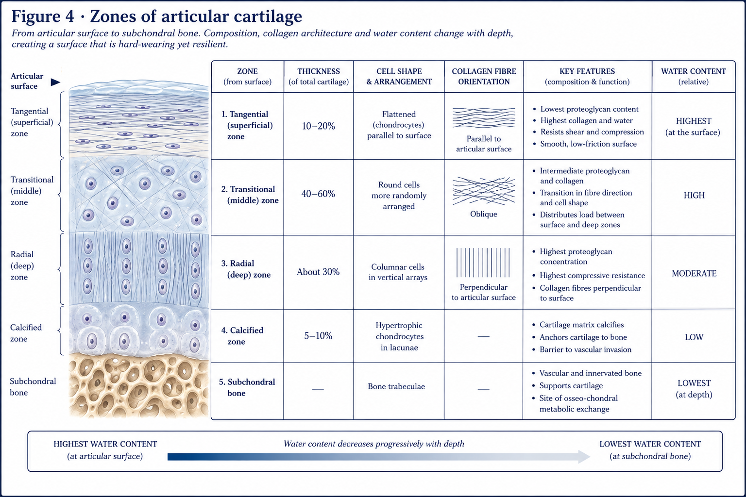
Articular cartilage is only 2–4 mm thick, yet it is organised into four distinct zones. Cartilage is thickest where contact stress is highest — up to 5–7 mm on the patella, the thickest in the body.
| Zone | Depth | Collagen orientation | Chondrocytes | Proteoglycan | Function |
|---|---|---|---|---|---|
| 1. Superficial (tangential) | ~10–20% | Parallel to the surface, densely packed | Flattened, parallel to surface; secrete lubricin | Lowest | Resists shear and tension; the barrier to large molecules; the low-friction bearing surface. Its loss is the first event of osteoarthritis |
| 2. Middle (transitional) | ~40–60% | Obliquely arranged, thicker fibrils | Rounded, sparse | Highest | The transition; the main compressive zone |
| 3. Deep (radial) | ~30% | Perpendicular to the surface, thickest fibrils | Rounded, in vertical columns | High | Greatest resistance to compression; anchors cartilage to bone |
| Tidemark | — | A basophilic line | — | — | The boundary between uncalcified and calcified cartilage; the mechanical transition. It advances with age, thinning the true cartilage |
| 4. Calcified cartilage | Thin | Type II and type X collagen; mineralised | Hypertrophic, few, largely inactive | Low | Anchors cartilage to subchondral bone via interdigitation; collagen fibres cross the tidemark but not the calcified–bone junction |
The classic description of the fibre arrangement is Benninghoff’s arcades: collagen fibres arise perpendicular from the calcified zone, arch through the middle zone, and run parallel at the surface, forming a series of leaf-spring arches. Whether these are literal continuous arcades or a statistical description remains debated, but the model correctly predicts the tissue’s behaviour: vertical fibres resist compression and anchor, horizontal fibres resist shear.
Clinical corollary. Because the deep zone is anchored through the calcified layer to bone but the calcified layer is not crossed by fibres from bone, the osteochondral junction is a plane of mechanical weakness. Shear injury therefore produces either a superficial fibrillation or a full osteochondral flap, but rarely a clean mid-substance tear.
Articular cartilage has no blood supply of its own. Its nutrition comes from two sources:
Diffusion alone is slow across 2–4 mm of dense matrix. What makes it adequate is convection: cyclical loading and unloading squeezes interstitial fluid out of the cartilage and draws it back in with dissolved nutrients — a pumping action driven by joint movement.
This produces the most important clinical statement in the chapter:
Joint movement is the nutritional supply route of articular cartilage. Immobilisation starves it.
The experimental evidence is unambiguous:
It is also the rationale for continuous passive motion after certain cartilage procedures, for early protected range after joint surgery, and for the advice that a patient with knee osteoarthritis should keep moving rather than rest.
Cartilage is modelled as a biphasic material: a porous, permeable solid phase (collagen–proteoglycan matrix) saturated by an interstitial fluid phase (water and ions).
When load is applied:
Two important consequences:
The coefficient of friction of a healthy synovial joint is approximately 0.002–0.02 — lower than ice on ice, and lower than any engineered bearing. Several mechanisms operate together:
| Mode | Mechanism | When it dominates |
|---|---|---|
| Boundary lubrication | A molecular layer of lubricin (PRG4), hyaluronan and surface-active phospholipids bound to the surface prevents direct contact | High load, low speed — e.g. standing, and at the start and end of movement |
| Fluid-film (hydrodynamic / elastohydrodynamic) | A pressurised fluid film separates the surfaces; the surfaces deform elastically, widening the film | Moderate load, higher speed |
| Interstitial fluid pressurisation (“biphasic lubrication”) | Pressurised interstitial fluid carries the load, so very little is borne at the contacting asperities | The dominant mechanism in modern accounts of joint lubrication |
| Weeping / boosted lubrication | Fluid exuded from cartilage under load (weeping); or water driven out of synovial fluid leaving a concentrated hyaluronan gel in surface irregularities (boosted) | Historical models, still taught, now considered contributory rather than primary |
Clinical relevance. In inflammatory arthritis, lubricin production is suppressed by IL-1 and TNF-α, and hyaluronan is depolymerised — friction rises before structural damage appears. This is part of the rationale for intra-articular hyaluronan injection, whose clinical effect size in osteoarthritis nonetheless remains small and contested.
Figure 4 · Why cartilage heals badly
Five reasons, and they compound:
| Partial-thickness (chondral) defect | Full-thickness (osteochondral) defect | |
|---|---|---|
| Depth | Confined to cartilage; does not breach the tidemark or subchondral plate | Penetrates subchondral bone |
| Healing response | Essentially none. A brief local burst of matrix synthesis, then nothing. The defect persists indefinitely and its margins may degenerate | A fibrin clot forms, marrow-derived mesenchymal stem cells enter, and repair tissue develops |
| Repair tissue | — | Fibrocartilage (type I collagen-dominant), not hyaline cartilage |
| Durability | — | Inferior: lower compressive stiffness, poorer wear resistance, tends to degrade over 1–5 years |
This paradox — that the deeper injury heals better — is the entire basis of marrow stimulation techniques, which deliberately convert a non-healing chondral defect into a healing osteochondral one.
| Procedure | Principle | Repair tissue | Typical rehabilitation implication |
|---|---|---|---|
| Debridement / chondroplasty | Remove unstable flaps | None | Symptomatic only; early motion and loading |
| Microfracture / marrow stimulation | Perforate subchondral plate to admit marrow MSCs | Fibrocartilage | Protected weight bearing (commonly 6 weeks), early continuous passive motion, prolonged return to sport (6–12 months). Graft is mechanically weakest at 6–12 weeks — the point at which patients feel well |
| Osteochondral autograft transfer (OATS / mosaicplasty) | Transfer cylindrical plugs of the patient’s own cartilage and bone from a low-load area | Hyaline (transplanted) | Bone-to-bone healing at ~6–8 weeks governs weight bearing; donor-site morbidity is a real consideration |
| Osteochondral allograft | Fresh donor osteochondral plug | Hyaline (donor) | Feasible because cartilage is immune-privileged — no vessels, no lymphatics, and chondrocytes shielded within matrix. Bone incorporation determines timeline |
| ACI / MACI (autologous chondrocyte implantation, matrix-assisted) | Two-stage: harvest chondrocytes, culture, reimplant under a membrane or on a scaffold | Hyaline-like | The longest and most graduated protocol: strict early protection, progressive loading over 6–12 months, return to impact at 12–18 months. Graft maturation phases (proliferation → transition → remodelling → maturation) drive the protocol |
The general rehabilitation principle across all of them: motion early, load late. Movement feeds and organises the repair tissue; premature compressive and shear loading destroys it.
Two semilunar wedges of fibrocartilage, triangular in cross-section, thicker peripherally.
Osteoarthritis is not passive wear and tear of an inert bearing surface. It is an active, cell-mediated disease of the whole joint as an organ — cartilage, subchondral bone, synovium, capsule, ligament, meniscus, fat pad and periarticular muscle.
| System | Basis |
|---|---|
| Kellgren–Lawrence (0–4) | Radiographic: joint space narrowing, osteophytes, sclerosis, deformity |
| Outerbridge (0–4) | Arthroscopic: softening → fissuring <1.25 cm → fissuring >1.25 cm → subchondral bone exposed |
| ICRS (0–4) | The modern arthroscopic/surgical standard for focal chondral defects |
Articular cartilage is aneural. It cannot generate pain. Osteoarthritic pain arises from:
This is the single most useful fact in the chapter for a physiotherapist, because none of those sources is cartilage, and every one of them is modifiable. It is why exercise therapy improves pain and function in osteoarthritis despite doing nothing to reverse cartilage loss, and it is the basis of the GLA:D and comparable structured exercise programmes recommended as first-line care in every major OA guideline (NICE, OARSI, ACR).
Contrary to lay belief and much clinical folklore, moderate loading is protective, not destructive. Long-distance recreational running is not associated with an increased incidence of knee osteoarthritis in the available cohort data, and is associated with a lower incidence than sedentary living; the risk lies with elite-level volume, with previous joint injury (particularly ACL rupture and meniscectomy, which carry a large increase in post-traumatic OA risk), and with obesity, whose effect is both mechanical and metabolic (adipokines, systemic low-grade inflammation). Advising an osteoarthritic patient to “save the joint” by resting is not neutral advice; it removes the nutritional and anabolic stimulus the tissue depends on.
15 questions on this chapter. Tap one to see the answer and the reasoning.
Answer: (B) Type I dominates fibrocartilage and bone; type X marks hypertrophic and calcified cartilage.
Answer: (B) The tyre analogy: proteoglycans supply the pressure, collagen the casing.
Answer: (C) Its absence removes the chondroprogenitor reservoir needed for repair.
Answer: (C) This orientation resists shear; its loss is the first structural event in osteoarthritis.
Answer: (D) This is why cartilage tolerates rapid loading better than sustained loading.
Answer: (C) No blood supply reaches it, so no repair cascade begins.
Answer: (B) Type I collagen-dominant, mechanically inferior, and prone to deterioration over years.
Answer: (C) It is vascularised by the perimeniscal plexus and is the zone in which repair is likely to succeed.
Answer: (B) Hoop stress can no longer be contained and the meniscus extrudes.
Answer: (B) Proteoglycan loss precedes collagen breakdown, which is the point of irreversibility.
Answer: (C) Cartilage is aneural.
Answer: (C) Both are avascular; only elastic cartilage resists calcification.
Answer: (B) Its production falls under IL-1 and TNF-α in inflammatory arthritis.
Answer: (B)
Answer: (C) Movement drives convective nutrient exchange; static compression is catabolic.
Everything on this page, in one screen
| Source | Why it earns its place |
|---|---|
| Standring S (ed.) — Gray’s Anatomy, 42nd edn | Reference descriptive and histological account |
| Ross MH, Pawlina W — Histology: A Text and Atlas | The best diagrams of zonal architecture and matrix organisation |
| Mow VC, Huiskes R — Basic Orthopaedic Biomechanics and Mechano-Biology | The primary source for biphasic theory and cartilage mechanics |
| Nordin M, Frankel VH — Basic Biomechanics of the Musculoskeletal System | Accessible treatment of cartilage viscoelasticity and lubrication |
| Sophia Fox AJ, Bedi A, Rodeo SA — “The basic science of articular cartilage”, Sports Health, 2009 | A concise, widely cited review that maps almost exactly onto this chapter |
| Arnoczky SP, Warren RF — “Microvasculature of the human meniscus”, Am J Sports Med, 1982 | The original description of the red–red / red–white / white–white zones |
| Loeser RF, Goldring SR, Scanzello CR, Goldring MB — “Osteoarthritis: a disease of the joint as an organ”, Arthritis Rheum, 2012 | The paper that reframed OA away from wear and tear |
| Skou ST, Roos EM — “Good Life with osteoArthritis in Denmark (GLA:D)”, BMC Musculoskelet Disord, 2017 | The exercise-therapy model now embedded in international OA guidance |
| NICE NG226 — Osteoarthritis in over 16s: diagnosis and management (2022); OARSI and ACR guidelines | Current first-line management recommendations |
| Benninghoff A (1925) | The original description of the collagen arcades |
| Palastanga N, Field D, Soames R — Anatomy and Human Movement | The physiotherapy-facing synthesis |
Chapter 3 of 24 · Human Anatomy · Physiotherapist India Next: Chapter 4 — Joints: how they are classified, what makes them stable, and what stops them moving.
Reviewed by the Physiotherapist India Team. · Human Anatomy contents