Evidence-based physiotherapy and rehabilitation resources
Editorial & review policy
Evidence-based physiotherapy and rehabilitation resources
Editorial & review policyHuman Anatomy · Upper limb
The upper limb gave up stability to buy reach and dexterity. Its whole bony attachment to the trunk is one small joint at the sternum. Everything that follows — the mobility, the vulnerability, the nerve injuries — comes from that decision.
Part 2 · The upper limb
Clavicle to phalanges, the nerves at risk along the humerus, and the palpable landmarks
The upper limb was freed from weight bearing. Everything about its skeleton follows from that.
The price of that design is written across the trauma list: the clavicle is the most commonly fractured bone in childhood, the shoulder is the most commonly dislocated major joint, and the distal radius is the most commonly fractured bone in adults.
Figure 1 · Bones of the upper limb
| Region | Bones | Number (each side) |
|---|---|---|
| Pectoral (shoulder) girdle | Clavicle, scapula | 2 |
| Arm | Humerus | 1 |
| Forearm | Radius, ulna | 2 |
| Carpus | Scaphoid, lunate, triquetrum, pisiform; trapezium, trapezoid, capitate, hamate | 8 |
| Metacarpus | Metacarpals 1–5 | 5 |
| Phalanges | Proximal, middle, distal (thumb has only proximal and distal) | 14 |
| Total | 32 |
The girdle articulates with the axial skeleton at one point only — the sternoclavicular joint. The scapula has no bony articulation with the thorax; it is held by muscle in the functional scapulothoracic articulation.
An S-shaped strut, subcutaneous throughout its length, convex forwards medially and concave forwards laterally.
| Surface | Structures |
|---|---|
| Superior | Deltoid (lateral third, anterior); trapezius (lateral third, posterior); subcutaneous in the middle |
| Inferior | Subclavius in the subclavian groove; conoid tubercle and trapezoid line for the coracoclavicular ligament laterally; costoclavicular (rhomboid) ligament impression medially |
| Anterior | Pectoralis major (medial two-thirds); deltoid (lateral third) |
| Posterior | Sternocleidomastoid (medial); trapezius (lateral) |
The commonest fracture of childhood and among the commonest overall. Around 80% occur at the junction of the middle and lateral thirds — the weakest point, where the two curvatures meet, the cross-section changes from prismatic to flattened, and no muscle or ligament attaches.
The classic deformity: the medial fragment is pulled up by sternocleidomastoid, and the lateral fragment is pulled down and medially by the weight of the limb and by pectoralis major.
Complications to know: neurovascular injury (subclavian vessels, brachial plexus — uncommon but serious), pneumothorax, malunion with cosmetic deformity, non-union (more common in the middle third with displacement), and medial physeal injury mistaken for sternoclavicular dislocation in adolescents — a posterior displacement here can compress the trachea, oesophagus or great vessels and is an emergency.
A flat triangular bone lying on the posterolateral thorax over ribs 2 to 7, held entirely by muscle.
| Feature | Detail |
|---|---|
| Superior border | Thinnest; interrupted by the suprascapular notch, bridged by the superior transverse scapular ligament. The suprascapular nerve passes under the ligament; the suprascapular artery passes over it — “Army under the bridge, Navy over it.” A site of nerve entrapment |
| Medial (vertebral) border | Long; attachment for rhomboids and levator scapulae; winging is judged from it |
| Lateral (axillary) border | Thickest, buttressing the glenoid; teres major and minor attach |
| Superior angle | At the level of T2; levator scapulae |
| Inferior angle | At the level of T7 and the 7th rib — the standard surface landmark for auscultation and for the lower lung border |
| Lateral angle | Bears the glenoid cavity |
| Costal surface | The subscapular fossa, filled by subscapularis |
| Dorsal surface | Divided by the spine of the scapula (root at the level of T3) into the supraspinous and infraspinous fossae, communicating through the spinoglenoid notch |
| Process | Features and attachments |
|---|---|
| Spine | A shelf continuing laterally as the acromion; deltoid from its lower border, trapezius to its upper |
| Acromion | Articulates with the clavicle; the most lateral bony point of the shoulder, used to measure limb length and shoulder width. Bigliani types I (flat), II (curved), III (hooked) — type III associated with cuff pathology, though causality is contested. An unfused acromial epiphysis is the os acromiale (~1–8% of people), a mimic of fracture and a cause of pain |
| Coracoid | Anterior, hook-shaped. “The Maypole of the shoulder”: three muscles and two ligaments. Muscles — pectoralis minor (medial), coracobrachialis and short head of biceps (conjoint tendon, lateral). Ligaments — coracoacromial and coracoclavicular (conoid and trapezoid). Palpable ~2 cm inferomedial to the anterolateral acromion; tender in almost everyone, so tenderness alone is not a sign |
| Glenoid cavity | Shallow, pear-shaped; accommodates only ~⅓ of the humeral head; deepened by the labrum. Supraglenoid tubercle — long head of biceps; infraglenoid tubercle — long head of triceps. Normal retroversion of ~5–7° and upward tilt contribute to stability |
Scapular position and dyskinesis. Normal resting position:
Figure 2 · Nerves at risk along the humerus
The longest bone of the upper limb, and the single most important bone for a physiotherapist to know in nerve terms — four different nerves are injured at four different levels.
| Feature | Detail |
|---|---|
| Head | Hemispherical, articular; faces superiorly, medially and posteriorly; retroverted ~30° and inclined ~130–140° to the shaft |
| Anatomical neck | The narrow groove immediately around the articular margin; the capsular attachment |
| Surgical neck | The constriction below the tubercles — a common fracture site, and the level at which the axillary nerve and posterior circumflex humeral artery are at risk |
| Greater tubercle | Lateral; three facets from above down for supraspinatus, infraspinatus, teres minor — an examination classic |
| Lesser tubercle | Anterior; subscapularis |
| Intertubercular (bicipital) groove | Between them, holding the tendon of the long head of biceps and its ascending branch of the anterior circumflex humeral artery. Lips: lateral lip — pectoralis major; floor — latissimus dorsi; medial lip — teres major. (Mnemonic: “a lady between two majors”) |
| Feature | Detail |
|---|---|
| Deltoid tuberosity | Mid-lateral; deltoid insertion |
| Radial (spiral) groove | Runs obliquely across the posterior surface, transmitting the radial nerve and profunda brachii artery |
| Nutrient foramen | Directed distally |
| Borders/surfaces | Anteromedial, anterolateral, posterior; medial and lateral supracondylar ridges distally |
| Feature | Detail |
|---|---|
| Capitulum | Lateral, rounded; articulates with the radial head |
| Trochlea | Medial, pulley-shaped; articulates with the trochlear notch of the ulna |
| Medial epicondyle | Prominent, subcutaneous; common flexor origin; the ulnar nerve grooves behind it |
| Lateral epicondyle | Smaller; common extensor origin |
| Coronoid fossa (anterior, medial), radial fossa (anterior, lateral), olecranon fossa (posterior) | Accommodate the corresponding processes in full flexion or extension |
| Carrying angle | The valgus angle between arm and forearm in full extension and supination: ~5–15° in men, 10–20° in women. Increased = cubitus valgus (risk of tardy ulnar nerve palsy); decreased/reversed = cubitus varus (“gunstock deformity”), classically after malunited supracondylar fracture |
| Level of injury | Nerve at risk | Clinical picture |
|---|---|---|
| Surgical neck | Axillary nerve (C5–6) + posterior circumflex humeral artery | Weak abduction (deltoid), loss of sensation over the regimental badge area of the lateral shoulder. Also at risk in anterior shoulder dislocation |
| Mid-shaft / radial groove | Radial nerve (C5–T1) + profunda brachii | Wrist drop, loss of finger and thumb extension, weak grip (Chapter 6), sensory loss over the dorsal first web space. Triceps usually spared (its branches leave proximally) |
| Distal shaft / supracondylar | Median nerve and brachial artery (anterior interosseous nerve in the classic paediatric supracondylar fracture) | Loss of thumb IP and index DIP flexion (inability to make an “OK” sign — anterior interosseous); vascular compromise, Volkmann’s ischaemic contracture if missed |
| Medial epicondyle | Ulnar nerve (C8–T1) | Clawing of the ring and little fingers, loss of intrinsics, sensory loss over the medial 1½ digits. Also injured late in cubitus valgus (tardy ulnar palsy) |
Learn this as a sequence down the bone; it converts a fracture level into a neurological prediction, and it is asked in every clinical examination.
The humerus has one primary centre (shaft, week 8) and up to eight secondary centres. The proximal humeral epiphyses (head at ~1 year, greater tubercle ~3 years, lesser tubercle ~5 years) fuse to each other and then to the shaft at ~18–20 years — the last epiphysis of the upper limb to fuse, and the growing end, hence the proximal humerus is the growing end and the nutrient artery is directed away from it, distally. Distal centres appear in the sequence C-R-I-T-O-E: Capitellum (1 yr), Radial head (3), Internal (medial) epicondyle (5), Trochlea (7), Olecranon (9), External (lateral) epicondyle (11) — a sequence used to interpret paediatric elbow radiographs and detect a displaced medial epicondyle.
| Region | Features |
|---|---|
| Head | Disc-shaped, concave superiorly for the capitulum; its circumference articulates with the radial notch of the ulna within the anular ligament. Not attached to the ulna by ligament other than the anular — hence a young child’s radial head can slip out of the ligament (pulled elbow / nursemaid’s elbow, ages 1–4, from axial traction on a pronated extended arm) |
| Neck | Narrow; fracture site |
| Radial (bicipital) tuberosity | Medial, just distal to the neck; biceps brachii insertion — the reason biceps is a powerful supinator |
| Shaft | Triangular, convex laterally; pronator teres on the mid-lateral surface; interosseous border medially |
| Distal end | Broad. Dorsal (Lister’s) tubercle — a pulley for extensor pollicis longus (which is why EPL ruptures after distal radius fracture). Styloid process, projecting ~1 cm distal to the ulnar styloid. Ulnar notch for the head of the ulna. Articulates with scaphoid and lunate |
| Region | Features |
|---|---|
| Olecranon | The point of the elbow; triceps insertion; forms the posterior part of the trochlear notch |
| Coronoid process | Anterior; brachialis insertion on its tuberosity; the anterior buttress against posterior dislocation — hence its fracture in the “terrible triad” (elbow dislocation + radial head fracture + coronoid fracture) |
| Trochlear notch | Between olecranon and coronoid; grips the humeral trochlea — the source of elbow bony stability |
| Radial notch | Lateral, on the coronoid; for the radial head |
| Supinator crest and fossa | Below the radial notch |
| Shaft | Triangular, tapering distally |
| Distal end | Small head with an articular circumference for the ulnar notch of the radius, and a styloid process posteromedially. The ulnar head does not articulate with the carpus directly — the triangular fibrocartilage complex (TFCC) intervenes |
The membrane’s fibres run obliquely, downwards and medially from radius to ulna. This orientation means the membrane is taut when force is transmitted from hand to radius, transferring load medially to the ulna and hence to the humerus.
The functional distribution: approximately 80% of axial load crosses the wrist through the radius and only 20% through the ulnar side (TFCC), but by the elbow the distribution has shifted to about 60% through the radiocapitellar joint — the interosseous membrane having transferred the difference.
Clinical consequences:
| Eponym | Description |
|---|---|
| Colles’ | Distal radius fracture with dorsal displacement and angulation, radial shortening; the “dinner-fork” deformity. The commonest adult fracture; typically a fall on the outstretched hand in osteoporotic bone. Complications: median nerve compression, EPL rupture, malunion, and complex regional pain syndrome |
| Smith’s | The reverse — volar displacement (“garden spade” deformity) |
| Barton’s | Intra-articular marginal fracture of the distal radius with carpal subluxation |
| Chauffeur’s (Hutchinson) | Radial styloid fracture |
| Monteggia | Fracture of the proximal ulna with dislocation of the radial head |
| Galeazzi | Fracture of the distal radius with dislocation of the distal radio-ulnar joint |
| Essex-Lopresti | Radial head fracture + interosseous membrane rupture + DRUJ disruption |
Mnemonic for the two that are always confused: MUGR — Monteggia = Ulna fracture; Galeazzi = Radius fracture.
Figure 3 · The eight carpal bones
Figure 4 · The carpal bones and the carpal tunnel
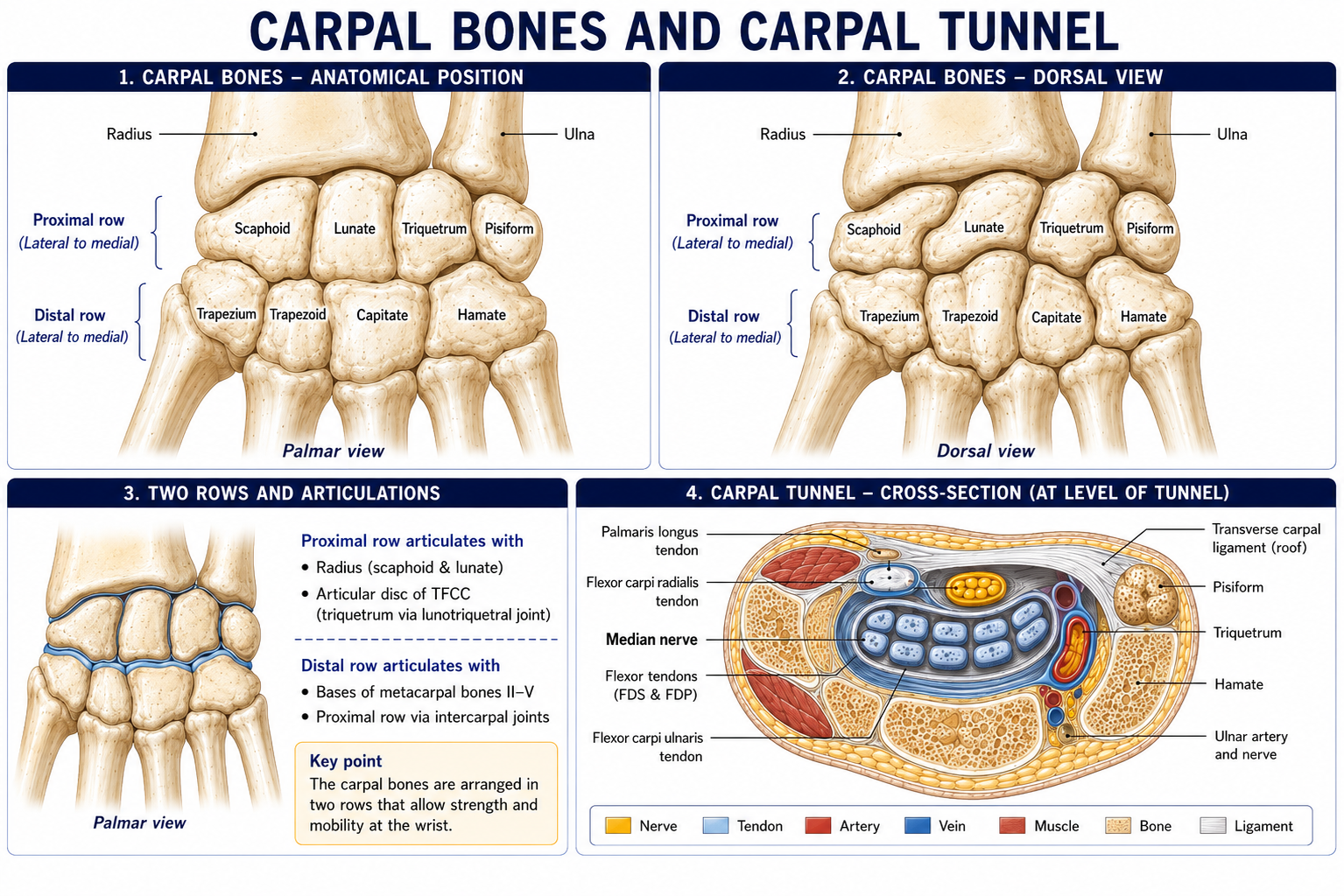
Eight bones in two rows of four, arranged as a transverse arch concave anteriorly, roofed by the flexor retinaculum to form the carpal tunnel.
| Row | Bones (lateral → medial) |
|---|---|
| Proximal | Scaphoid, Lunate, Triquetrum, Pisiform |
| Distal | Trapezium, Trapezoid, Capitate, Hamate |
Mnemonic: So Long To Pinky, Here Comes The Thumb (read proximal row lateral→medial, then distal row medial→lateral).
| Bone | Key facts |
|---|---|
| Scaphoid | The link between the rows; the most commonly fractured carpal bone (~60–70%). Retrograde blood supply entering the distal dorsal ridge from the radial artery — hence proximal pole avascular necrosis and non-union after waist fracture. Tender in the anatomical snuffbox, over the scaphoid tubercle, and on axial compression of the thumb. Initial radiographs are normal in up to 25% — treat clinically and re-image or obtain MRI |
| Lunate | Crescentic; the most commonly dislocated carpal bone (volar dislocation compresses the median nerve). Site of Kienböck’s disease (avascular necrosis, associated with negative ulnar variance) |
| Triquetrum | Articulates with the TFCC, not the ulna directly; the second most commonly fractured carpal (usually a dorsal chip) |
| Pisiform | A sesamoid in the tendon of flexor carpi ulnaris; forms the medial wall of Guyon’s canal |
| Trapezium | Bears the saddle-shaped first carpometacarpal joint — the site of thumb-base osteoarthritis, one of the commonest and most disabling hand conditions. Its tubercle is a flexor retinaculum attachment, and the flexor carpi radialis tendon grooves it |
| Trapezoid | Small, wedge-shaped; least commonly injured |
| Capitate | The largest carpal; the central pivot around which the carpus moves; its head sits in the concavity of the lunate and scaphoid |
| Hamate | Its hook (hamulus) forms the lateral wall of Guyon’s canal and a flexor retinaculum attachment. Hook of hamate fracture occurs in racquet, club and bat sports and can rupture the little finger flexor tendons or injure the ulnar nerve |
Flexor retinaculum attachments (all four are carpal tubercles): laterally the tubercle of the scaphoid and the tubercle of the trapezium; medially the pisiform and the hook of the hamate.
All carpal bones are cartilaginous at birth. They ossify in a broadly anticlockwise sequence starting with the capitate: capitate and hamate (~2–3 months), triquetrum (~2–3 years), lunate (~4), scaphoid, trapezium, trapezoid (~5–6), pisiform (~9–12, the last). A useful rule of thumb: the number of ossified carpal bones roughly equals the child’s age in years, up to about six. This is a standard skeletal-age assessment method (Greulich and Pyle).
Five metacarpals, each with a base (proximal), shaft and head (distal). The heads form the knuckles.
Fourteen phalanges per hand: proximal, middle and distal for digits 2–5; proximal and distal only for the thumb. Distal phalanges bear a terminal tuft (ungual tuberosity) supporting the pulp and nail bed.
Sesamoids are constant at the first MCP joint (two, in the tendons of flexor pollicis brevis and adductor pollicis) and variable elsewhere.
Learn these as a sequence you can perform on a patient, with the reason for each.
| Landmark | How to find it | Why it matters |
|---|---|---|
| 1. Suprasternal notch and sternoclavicular joint | Base of the neck; follow the clavicle medially | The only joint between limb and axial skeleton; palpate for anterior/posterior dislocation |
| 2. Clavicle throughout | Subcutaneous along its whole length | Fracture (junction of middle and lateral thirds), step deformity |
| 3. Acromioclavicular joint | Follow the clavicle laterally to a small step ~2–3 cm medial to the acromial tip | AC joint sprain and osteoarthritis; the step deformity of AC separation |
| 4. Acromion | The flat lateral shelf; its lateral border is the widest point of the shoulder | Reference for limb length and abduction measurement; the “squared-off” shoulder of dislocation |
| 5. Coracoid process | ~2 cm inferomedial to the anterolateral acromion, in the deltopectoral triangle | Attachment of the conjoint tendon and pectoralis minor; a fixed reference for brachial plexus block |
| 6. Greater tubercle of the humerus | Just lateral and inferior to the acromion; best felt with the arm extended and internally rotated | Supraspinatus footprint — the site of tenderness in cuff pathology |
| 7. Spine and inferior angle of the scapula | Spine root at T3; inferior angle at T7 | Vertebral level reference; scapular position and dyskinesis assessment |
| 8. Medial and lateral epicondyles and the olecranon | The three points of the elbow | In extension they are in a straight line; in 90° flexion they form an equilateral triangle. Disruption of this relationship indicates dislocation; preservation with deformity indicates supracondylar fracture — a genuinely useful bedside discriminator |
| 9. Ulnar nerve | Rolled in the groove behind the medial epicondyle | Tinel’s sign; subluxation of the nerve on flexion |
| 10. Ulnar shaft and styloid | Subcutaneous along the whole posteromedial forearm | The subcutaneous border is the reference for splinting |
| 11. Radial styloid and Lister’s tubercle | Distal radius, lateral and dorsal | Radial length assessment; EPL runs around Lister’s tubercle |
| 12. Anatomical snuffbox | The hollow between the tendons of EPL (posteriorly) and APL/EPB (anteriorly), with the thumb extended | Floor contains the scaphoid and the radial artery crosses it |
| 13. Pisiform and hook of hamate | Pisiform at the base of the hypothenar eminence; the hook ~1–2 cm distal and radial to it | The boundaries of Guyon’s canal |
| 14. Scaphoid tubercle | At the base of the thenar eminence, wrist in radial deviation | The volar half of the scaphoid examination |
| 15. Metacarpal heads and the distal palmar crease | The knuckles; the distal palmar crease overlies the MCP joints, not the finger web | Splinting position — the MCP joints must be free to flex to 70–90° |
Two clinically important length measurements:
15 questions on this chapter. Tap one to see the answer and the reasoning.
Answer: (B) It is also the first bone to begin ossifying.
Answer: (B) Around 80% of cases.
Answer: (B) The artery passes over.
Answer: (B) Subscapularis inserts on the lesser tubercle.
Answer: (B) With loss of deltoid function and regimental-badge sensation.
Answer: (C)
Answer: (C) Capitellum 1, radial head 3, internal epicondyle 5, trochlea 7, olecranon 9, external epicondyle 11.
Answer: (B) MUGR.
Answer: (B)
Answer: (B) Scaphoid tubercle and trapezium are the lateral attachments.
Answer: (B) They form a triangle at 90° of flexion; loss of the relationship suggests dislocation.
Answer: (B) Negative variance is associated with Kienböck’s.
Answer: (B) Rolando’s is its comminuted equivalent.
Answer: (C) Normal is 30–60°.
Answer: (B) Metacarpals 2–5 have theirs at the head; all phalanges have theirs at the base.
Everything on this page, in one screen
| Source | Why it earns its place |
|---|---|
| Standring S (ed.) — Gray’s Anatomy, 42nd edn | Definitive osteology and attachments |
| Palastanga N, Field D, Soames R — Anatomy and Human Movement | Osteology with functional interpretation for physiotherapists |
| Snell RS — Clinical Anatomy by Regions | Bone-by-bone clinical correlation |
| Field D, Hutchinson JO — Field’s Anatomy, Palpation and Surface Markings | The reference for palpation technique |
| Chaurasia BD — Human Anatomy, Vol 1: Upper Limb and Thorax | The standard Indian regional text, matched to syllabus |
| McRae R, Esser M — Practical Fracture Treatment | Fracture patterns, eponyms and their functional consequences |
| Netter FH — Atlas of Human Anatomy; Drake, Vogl & Mitchell — Gray’s Atlas | Keep one open alongside this chapter |
| Greulich WW, Pyle SI — Radiographic Atlas of Skeletal Development of the Hand and Wrist | The standard for carpal skeletal age |
| Gilula LA — “Carpal injuries: analytic approach and case exercises”, AJR, 1979 | The arcs |
Chapter 9 of 24 · Human Anatomy · Physiotherapist India Next: Chapter 10 — The Shoulder Region: four joints, the rotator cuff, and why stability here is muscular rather than bony.
Reviewed by the Physiotherapist India Team. · Human Anatomy contents