Evidence-based physiotherapy and rehabilitation resources
Editorial & review policy
Evidence-based physiotherapy and rehabilitation resources
Editorial & review policyHuman Anatomy · Lower limb
The upper limb was built for reach. The lower limb was built to carry you, and every difference between them follows from that. Heavier bones, deeper sockets, stronger ligaments, and a whole architecture devoted to getting weight safely to the ground.
Part 3 · The lower limb
Pelvis to phalanges, the weight-bearing chain, and the palpable landmarks
Figure 1 · How body weight reaches the ground
The upper limb traded stability for reach. The lower limb made the opposite bargain, and every feature of its skeleton follows from it:
The price is written in the pattern of pathology:
Figure 2 · Bones of the lower limb
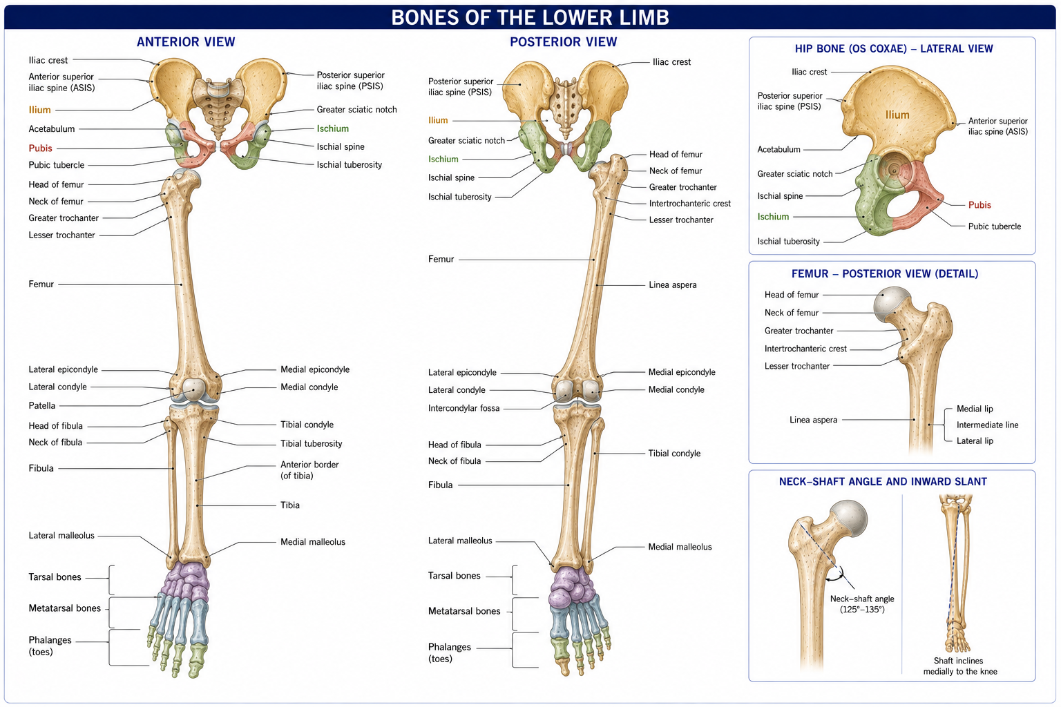
| Region | Bones | Number (each side) |
|---|---|---|
| Pelvic girdle | Hip bone (ilium + ischium + pubis, fused) | 1 |
| Thigh | Femur, patella | 2 |
| Leg | Tibia, fibula | 2 |
| Tarsus | Talus, calcaneus, navicular, cuboid, three cuneiforms | 7 |
| Metatarsus | Metatarsals 1–5 | 5 |
| Phalanges | Proximal, middle, distal (hallux has two) | 14 |
| Total | 31 (+ 2 constant hallucal sesamoids) |
Compare with the upper limb’s 32: the counts are nearly identical, but the carpus has eight bones in two rows for mobility while the tarsus has seven, arranged in a longitudinal column for load transmission.
Three bones fused at the triradiate cartilage in the acetabulum, which ossifies at 15–17 years — an important radiographic landmark in paediatric hip disease.
| Bone | Parts and features |
|---|---|
| Ilium | Iliac crest (running from ASIS to PSIS; the highest point is the supracristal plane at L4, the landmark for lumbar puncture). ASIS (sartorius, inguinal ligament), AIIS (rectus femoris straight head), PSIS (marked by the skin dimples of Venus; overlies S2, the level of the dural sac termination), PIIS. Iliac fossa (iliacus) medially; gluteal surface with posterior, anterior and inferior gluteal lines laterally; auricular surface for the sacroiliac joint; iliac tuberosity for the interosseous sacroiliac ligament |
| Ischium | Ischial tuberosity — the weight-bearing point in sitting; origin of the hamstrings and adductor magnus (ischiocondylar part). Ischial spine, separating the greater and lesser sciatic notches; attachment of the sacrospinous ligament; the landmark for a pudendal nerve block and the reference point for assessing the pelvic outlet in labour. Ischial ramus |
| Pubis | Body with the pubic symphysis medially and the pubic tubercle (the inguinal ligament’s medial attachment, and the landmark distinguishing inguinal from femoral hernia). Superior and inferior rami; the superior ramus bears the pecten pubis (pectineal line) |
Obturator foramen: bounded by the pubic and ischial rami, closed by the obturator membrane except for the obturator canal, which transmits the obturator nerve and vessels.
Converted from notches by two ligaments — sacrospinous (to the ischial spine) and sacrotuberous (to the ischial tuberosity) — which also prevent the sacrum tilting backwards under load.
| Foramen | Contents |
|---|---|
| Greater sciatic foramen | Piriformis, dividing it into suprapiriform and infrapiriform parts. Above: superior gluteal nerve and vessels. Below: sciatic nerve, posterior femoral cutaneous nerve, inferior gluteal nerve and vessels, nerve to quadratus femoris, nerve to obturator internus, pudendal nerve and internal pudendal vessels |
| Lesser sciatic foramen | Tendon of obturator internus, nerve to obturator internus, pudendal nerve and internal pudendal vessels re-entering the pelvis |
The pudendal nerve’s course — out of the greater foramen, around the ischial spine, back in through the lesser foramen — is the reason it can be blocked at the ischial spine and the reason it is compressed in pudendal neuralgia, an under-recognised cause of pelvic pain that physiotherapists working in pelvic health will meet.
| Feature | Male | Female |
|---|---|---|
| General build | Thicker, heavier, more prominent markings | Lighter, thinner, smoother |
| Pelvic inlet | Heart-shaped | Oval / rounded |
| Pelvic cavity | Narrow, funnel-shaped, deep | Wide, cylindrical, shallow |
| Subpubic angle | 50–60° (acute, V-shaped) | 80–85° (rounded, U-shaped) |
| Greater sciatic notch | Narrow (~70°) | Wide (~90°) |
| Ischial spines | Inturned | Everted |
| Sacrum | Long, narrow, more curved | Short, wide, less curved |
| Acetabulum | Larger, faces laterally | Smaller, faces more anteriorly |
| Obturator foramen | Oval | Triangular |
Caldwell–Moloy types: gynaecoid (~50%, the classic female form, most favourable for delivery), android (~20%, male-type), anthropoid (~25%, oval anteroposteriorly), platypelloid (~5%, flattened).
Clinically for physiotherapy, the wider female pelvis produces a greater Q angle at the knee, more pronounced femoral anteversion in some individuals, and a different hip abductor moment arm — factors that contribute (alongside neuromuscular and hormonal ones) to the higher female incidence of patellofemoral pain and ACL injury.
Pelvic diameters worth knowing: at the inlet, the anteroposterior (true conjugate ~11 cm) is the smallest and the transverse (~13 cm) the largest; at the outlet, the reverse — anteroposterior (~12.5 cm) largest and the interspinous transverse (~10.5 cm) smallest. Hence the fetal head engages transversely and delivers anteroposteriorly, rotating in between.
Figure 3 · The hip joint and the femoral neck
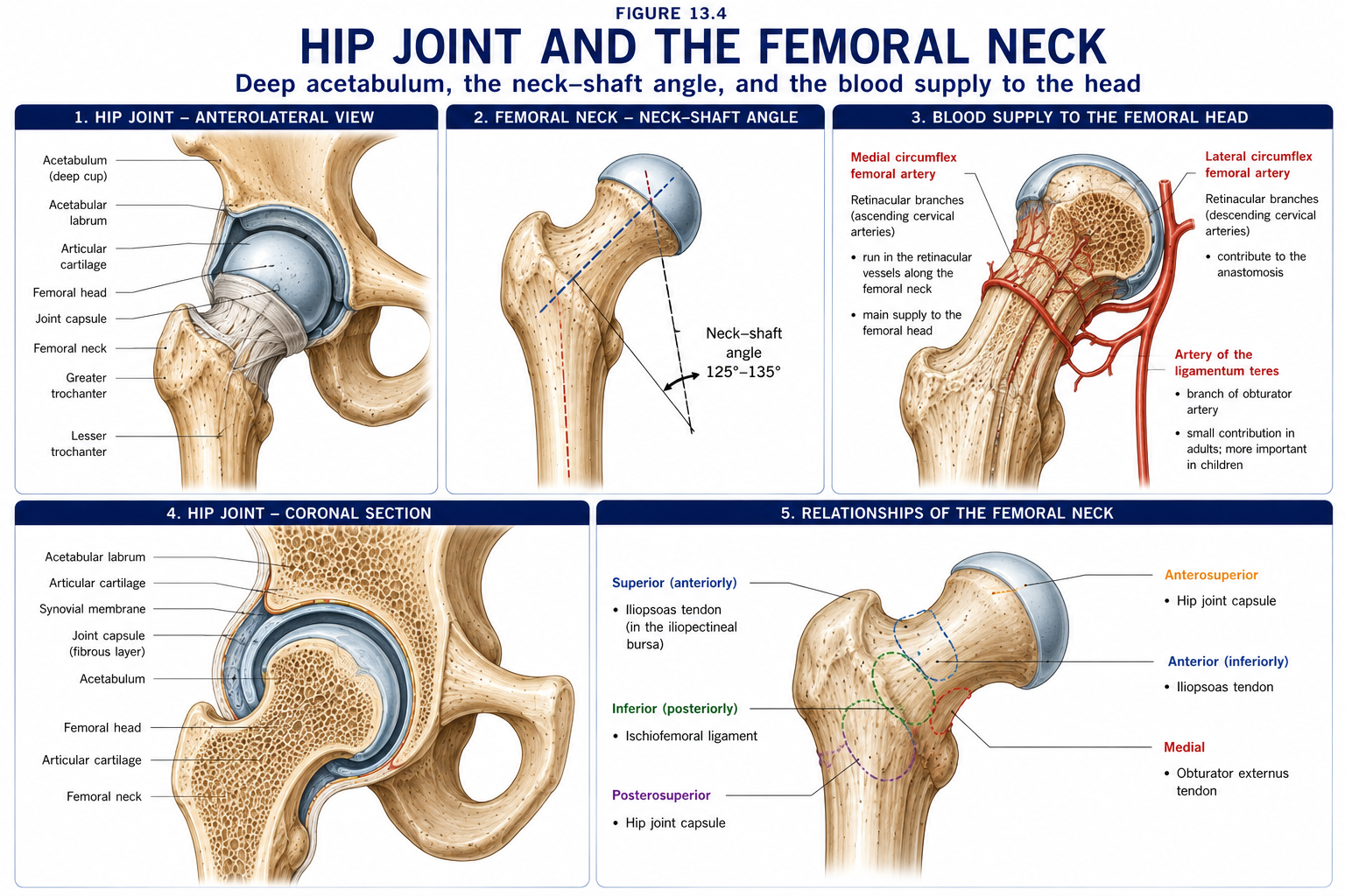
The longest, strongest and heaviest bone in the body — about 26–27% of standing height, which is the basis of stature estimation from a single bone in forensic work.
| Feature | Detail |
|---|---|
| Head | Two-thirds of a sphere, articular except at the fovea capitis, where the ligamentum teres attaches |
| Neck | ~5 cm; intracapsular (the capsule attaches to the intertrochanteric line anteriorly but only to the medial two-thirds of the neck posteriorly) |
| Greater trochanter | Gluteus medius and minimus, piriformis, obturator internus and the gemelli, obturator externus (into the trochanteric fossa) |
| Lesser trochanter | Iliopsoas |
| Intertrochanteric line (anterior) and crest (posterior, with the quadrate tubercle for quadratus femoris) | Capsular attachment anteriorly |
| Calcar femorale | A dense vertical plate of bone from the posteromedial neck into the shaft — the principal internal buttress transmitting load from head to shaft |
| Angle | Normal adult | Increased | Decreased |
|---|---|---|---|
| Neck–shaft (inclination) angle | **125°** (150° at birth, decreasing with weight bearing) | Coxa valga — reduced abductor moment arm, increased joint reaction force, associated with hip instability and cerebral palsy | Coxa vara — increased shear across the neck, Trendelenburg gait, risk of neck fracture; seen in SCFE, Perthes, rickets |
| Femoral torsion (anteversion) | ~10–15° in adults (~30–40° at birth) | Excessive anteversion — in-toeing gait, “W-sitting”, increased internal rotation and reduced external rotation, compensatory external tibial torsion, patellofemoral pain | Retroversion — out-toeing, associated with SCFE and with femoroacetabular impingement |
Craig’s (Ryder’s) test estimates anteversion clinically: with the patient prone and knee flexed 90°, rotate the hip until the greater trochanter is most laterally prominent; the angle of the tibia from vertical approximates the anteversion.
The combined effect of coxa vara or valga on the abductor moment arm is not academic. A varus neck lengthens the abductor moment arm and reduces the force needed; a valgus neck shortens it and increases both the abductor force required and the joint reaction force — which is one reason valgus hips wear.
Two principal systems cross in the femoral neck, mirroring the theoretical stress trajectories (Chapter 2):
Singh index grades osteoporosis radiographically by the progressive loss of these trabecular groups — a useful concept even where DEXA is unavailable.
The femoral head’s supply is precarious and age-dependent:
| Source | Contribution |
|---|---|
| Retinacular vessels from the medial circumflex femoral artery | The dominant adult supply — ascending in the retinacula of Weitbrecht along the neck |
| Lateral circumflex femoral artery | Smaller anterior contribution |
| Artery of the ligamentum teres (from the obturator) | Significant in children; negligible in adults |
Fracture
Location
Consequence
Intracapsular (subcapital, transcervical)
Within the capsule
Disrupts the retinacular vessels → high risk of avascular necrosis and non-union. Usually treated by replacement (hemiarthroplasty or THR) in the elderly
Extracapsular (intertrochanteric, subtrochanteric)
Outside the capsule
Good blood supply; heals well; treated by fixation (DHS or intramedullary nail)
The clinical picture of a displaced neck of femur fracture — the limb shortened, adducted and externally rotated — is produced by the pull of iliopsoas, the short external rotators and the adductors on the distal fragment.
Slipped capital femoral epiphysis (SCFE) — displacement through the hypertrophic zone of the proximal femoral physis in the adolescent (typically 10–16 years, often overweight); presents with hip, thigh or knee pain and an externally rotated limb with obligatory external rotation on hip flexion (Drehmann’s sign). Physiotherapists must know this: an adolescent with knee pain and a limp needs the hip examined and imaged.
The largest sesamoid, within the quadriceps tendon.
| Region | Features |
|---|---|
| Proximal | Medial and lateral condyles with the tibial plateau (medial concave and larger, lateral convex and smaller — hence lateral meniscal mobility). Intercondylar eminence with medial and lateral tubercles; anterior and posterior intercondylar areas for the cruciates and meniscal horns. Gerdy’s tubercle anterolaterally — iliotibial tract insertion. Tibial tuberosity — patellar ligament; the site of Osgood–Schlatter disease. Posterior tibial slope ~7–10°, a factor in ACL strain |
| Shaft | Triangular; anterior border (shin) and medial surface are subcutaneous — hence open fractures and poor soft-tissue cover. Soleal line posteriorly. The nutrient foramen is in the upper posterior shaft, directed distally (“from the knee I flee”) |
| Distal | Medial malleolus; fibular notch laterally for the syndesmosis; the inferior articular surface (plafond) forms the roof of the ankle mortise |
The tibia transmits ~85–90% of the axial load through the leg; the fibula carries only ~10–15% — which is why a fibular shaft can be harvested for grafting with little functional loss, while a tibial shaft cannot.
Named leg fracture patterns:
| Pattern | Description |
|---|---|
| Maisonneuve fracture | Proximal fibular fracture with a syndesmotic and deltoid ligament injury from an external rotation force at the ankle. Palpate the proximal fibula in every ankle injury — this fracture is missed when only the ankle is imaged |
| Pilon (plafond) fracture | Axial-load fracture of the distal tibial articular surface; high-energy, poor prognosis |
| Tibial plateau fracture | Schatzker I–VI; the lateral plateau is most often involved; associated meniscal and ligament injury is common |
| Tibial shaft fracture | The commonest long bone fracture; high rate of open injury and compartment syndrome (Chapter 7) |
| Segond fracture | A small avulsion of the lateral tibial rim — pathognomonic of ACL rupture |
Figure 4 · The foot skeleton and its arches
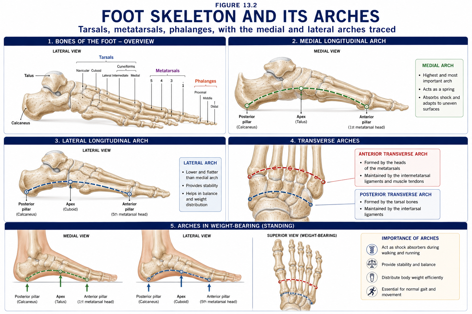
| Bone | Key features |
|---|---|
| Talus | No muscle attaches to it — a unique feature. Body (with the trochlea, wider anteriorly — hence ankle close-packing in dorsiflexion), neck and head. Articulates with tibia, fibula, calcaneus and navicular. Retrograde blood supply (artery of the tarsal canal from the posterior tibial, plus branches from the dorsalis pedis and fibular arteries) → AVN after talar neck fracture (Hawkins classification). Its lateral tubercle may persist as a separate os trigonum (~10%), a cause of posterior ankle impingement in dancers and footballers |
| Calcaneus | The largest tarsal; the calcaneal tuberosity takes the Achilles tendon and transmits body weight to the ground. Sustentaculum tali medially supports the talar head and grooves the flexor hallucis longus tendon below. Three facets for the talus, separated by the sulcus calcanei which with the sulcus tali forms the sinus tarsi. Böhler’s angle (normally 20–40°) is reduced in compression fractures |
| Navicular | Between the talar head and the cuneiforms. Tuberosity medially — tibialis posterior insertion, the keystone attachment of the medial arch. Accessory navicular in ~10% |
| Cuboid | Lateral column; grooved inferiorly for fibularis longus |
| Cuneiforms (medial, intermediate, lateral) | Wedge-shaped, wide dorsally and narrow plantarly — the wedge shape that creates the transverse arch |
Mnemonic: Tall Californian Navy Medics In Long Coats — talus, calcaneus, navicular, medial/intermediate/lateral cuneiforms, cuboid.
| Arch | Components | Keystone | Support |
|---|---|---|---|
| Medial longitudinal | Calcaneus, talus, navicular, three cuneiforms, first three metatarsals | Talar head | Plantar (spring) calcaneonavicular ligament, plantar aponeurosis, tibialis posterior, short and long plantar ligaments, intrinsic muscles |
| Lateral longitudinal | Calcaneus, cuboid, 4th and 5th metatarsals | Cuboid | Long and short plantar ligaments, fibularis longus, plantar aponeurosis |
| Transverse | Cuneiforms, cuboid, metatarsal bases | Intermediate cuneiform | Fibularis longus (its tendon crosses the sole obliquely), tibialis posterior, adductor hallucis (transverse head) |
The passive supports (bone shape and ligaments) do most of the work in quiet standing; the muscles contribute increasingly with load and during propulsion. This is examined constantly and is treated functionally in Chapter 16.
Load descends from the fifth lumbar vertebra to the ground along a defined path:
L5 → sacrum → sacroiliac joints → ilium → acetabulum → femoral head → femoral neck (calcar) → femoral shaft → femoral condyles → tibial plateau → tibial shaft → tibial plafond → talus → calcaneus (posteriorly) and the forefoot (anteriorly) → ground.
Approximate weight distribution in the standing foot: 50% through the calcaneus, 50% through the forefoot, with the medial forefoot (first metatarsal head and sesamoids) carrying roughly twice the load of each lesser metatarsal head.
| Angle | Definition | Normal | Clinical significance |
|---|---|---|---|
| Neck–shaft angle | Femoral neck to shaft | ~125° | Coxa vara / valga |
| Femoral anteversion | Neck axis relative to the transcondylar axis | 10–15° | In-toeing, patellofemoral pain |
| Anatomical femorotibial angle | Femoral shaft to tibial shaft | ~6–7° valgus | The reason the femoral shaft is oblique |
| Mechanical axis | Centre of femoral head → centre of ankle | Passes just medial to the centre of the knee | Deviation medially = genu varum, loading the medial compartment; laterally = genu valgum, loading the lateral compartment |
| Q angle | ASIS → patellar centre → tibial tuberosity | ~13° men, ~18° women | A lateral vector on the patella; >20° historically implicated in patellofemoral pain, though the relationship is weaker than once taught |
| Tibiofemoral (Mikulicz) line | The weight-bearing line | — | The basis of high tibial osteotomy planning |
| Tibial torsion | Proximal to distal tibial axis | ~20–30° external in adults (~5° at birth) | Out-toeing; compensatory external torsion accompanies excessive femoral anteversion — the “miserable malalignment syndrome” |
The developmental sequence of knee alignment is worth knowing so that normal is not treated as pathology: infants are varus until about 18–24 months, become maximally valgus at about 3–4 years, and settle into the adult mild valgus by about 7 years. Parents frequently present a physiologically bow-legged toddler or knock-kneed four-year-old; recognising the normal trajectory prevents unnecessary intervention, while asymmetry, progression, short stature or pain warrants investigation (Blount disease, rickets, skeletal dysplasia).
| Landmark | How to find it | Why it matters |
|---|---|---|
| 1. Iliac crest | Hands on the hips; highest point at the supracristal plane, L4 | Level for lumbar puncture and for counting lumbar segments |
| 2. ASIS | The prominent anterior point of the crest | Leg length measurement; Q angle apex; sartorius and inguinal ligament; AIIS avulsion by rectus femoris in adolescent athletes |
| 3. PSIS | The skin dimples of Venus; overlies S2 | Sacroiliac assessment; the level at which the dural sac ends |
| 4. Pubic tubercle | ~2.5 cm lateral to the symphysis | Inguinal ligament attachment; distinguishes inguinal (above and medial) from femoral (below and lateral) hernia |
| 5. Ischial tuberosity | Palpable in hip flexion, deep to gluteus maximus | Hamstring origin; proximal hamstring tendinopathy and adolescent avulsion; sitting pressure point |
| 6. Greater trochanter | The lateral prominence ~10 cm below the crest | Trendelenburg testing; gluteal tendinopathy / greater trochanteric pain syndrome; leg-length assessment; the reference for femoral anteversion testing |
| 7. Femoral pulse and femoral triangle | Mid-inguinal point, midway between ASIS and symphysis | NAVEL from lateral to medial: nerve, artery, vein, empty space, lymphatics |
| 8. Adductor tubercle | On the medial femoral condyle, at the end of the adductor magnus tendon | Landmark for the MCL and the adductor hiatus |
| 9. Patella, patellar ligament and tibial tuberosity | Anterior knee | Patellar tracking; Osgood–Schlatter at the tuberosity; Sinding-Larsen–Johansson at the inferior pole |
| 10. Joint line of the knee | With the knee flexed 90°, the horizontal cleft either side of the patellar ligament | Meniscal tenderness; the reference for MCL and LCL palpation |
| 11. Gerdy’s tubercle | Anterolateral tibia, lateral to the tuberosity | Iliotibial tract insertion |
| 12. Head and neck of fibula | Lateral, ~2 cm distal to the joint line | The common fibular nerve is rolled against the neck — Tinel’s sign, and the site to protect in casting and positioning |
| 13. Medial and lateral malleoli | The ankle prominences | Lateral is more distal and posterior; the reference for the ankle mortise, for the Ottawa ankle rules, and for oedema assessment |
| 14. Sustentaculum tali | ~2 cm below the medial malleolus | Supports the talus; FHL groove; a landmark for the spring ligament |
| 15. Navicular tuberosity | The prominence on the medial midfoot | Tibialis posterior insertion; the navicular drop test for arch mobility |
| 16. Base of the 5th metatarsal | The prominence on the lateral border of the foot | Fibularis brevis; the Jones fracture zone; an Ottawa ankle rule point |
| 17. Dorsalis pedis and posterior tibial pulses | Lateral to the EHL tendon; behind the medial malleolus | Vascular screening — mandatory before any foot intervention in a diabetic patient |
Leg length measurement:
15 questions on this chapter. Tap one to see the answer and the reasoning.
Answer: (B) Fusion occurs at 15–17 years.
Answer: (C) The level of the dural sac termination. The iliac crest marks L4.
Answer: (B)
Answer: (C) It is ~150° at birth and decreases with weight bearing.
Answer: (B) 30–40° at birth; excessive anteversion causes in-toeing.
Answer: (B)
Answer: (C) Because of the high rate of avascular necrosis and non-union.
Answer: (B)
Answer: (C) The fibula carries only 10–15%.
Answer: (B)
Answer: (C) It also has a retrograde blood supply, hence AVN after neck fracture.
Answer: (B) The cuboid is the keystone of the lateral arch, the intermediate cuneiform of the transverse.
Answer: (B) A watershed zone with a high non-union rate.
Answer: (C) Varus in infancy, maximal valgus at 3–4 years, adult alignment by ~7 years.
Answer: (B) Producing foot drop.
Everything on this page, in one screen
| Source | Why it earns its place |
|---|---|
| Standring S (ed.) — Gray’s Anatomy, 42nd edn | Definitive osteology and attachments |
| Palastanga N, Field D, Soames R — Anatomy and Human Movement | Osteology with functional interpretation |
| Neumann DA — Kinesiology of the Musculoskeletal System | Alignment angles, moment arms and the weight-bearing chain |
| Snell RS — Clinical Anatomy by Regions | Regional clinical correlation |
| Field D, Hutchinson JO — Field’s Anatomy, Palpation and Surface Markings | Palpation technique |
| Chaurasia BD — Human Anatomy, Vol 2: Lower Limb, Abdomen and Pelvis | Indian syllabus-matched descriptive account |
| McRae R, Esser M — Practical Fracture Treatment | Fracture patterns and their consequences |
| Staheli LT — Fundamentals of Pediatric Orthopedics | Developmental alignment, torsional profiles and when to worry |
| Magee DJ — Orthopedic Physical Assessment | Leg length measurement, Craig’s test, alignment assessment |
Chapter 13 of 24 · Human Anatomy · Physiotherapist India Next: Chapter 14 — Gluteal Region, Hip and Thigh: the hip as the shoulder’s opposite, the abductor mechanism, and the femoral triangle.
Reviewed by the Physiotherapist India Team. · Human Anatomy contents