Evidence-based physiotherapy and rehabilitation resources
Editorial & review policy
Evidence-based physiotherapy and rehabilitation resources
Editorial & review policyHuman Anatomy · General anatomy
Bone is not the dry scaffold it looks like in a specimen jar. It is living tissue that rebuilds itself along the lines of force you put through it, stores the body's calcium, makes its blood, and heals by regenerating rather than scarring. Everything a physiotherapist does to a skeleton depends on those four facts.
Part 1 · General anatomy
Living tissue that rebuilds along the lines of force: structure, cells, ossification, blood supply, remodelling and healing
Bone is the only tissue in the body that continuously destroys and rebuilds itself in response to the mechanical demands placed on it. That single property is why bone matters to a physiotherapist more than to almost any other clinician: the load you prescribe is a biological signal. A rehabilitation programme is, at the level of bone, a set of instructions to the osteocyte network about how much skeleton the patient is going to need.
Everything in this chapter serves three practical questions:
By the end of this chapter you should be able to:
Figure 1 · What bone is for
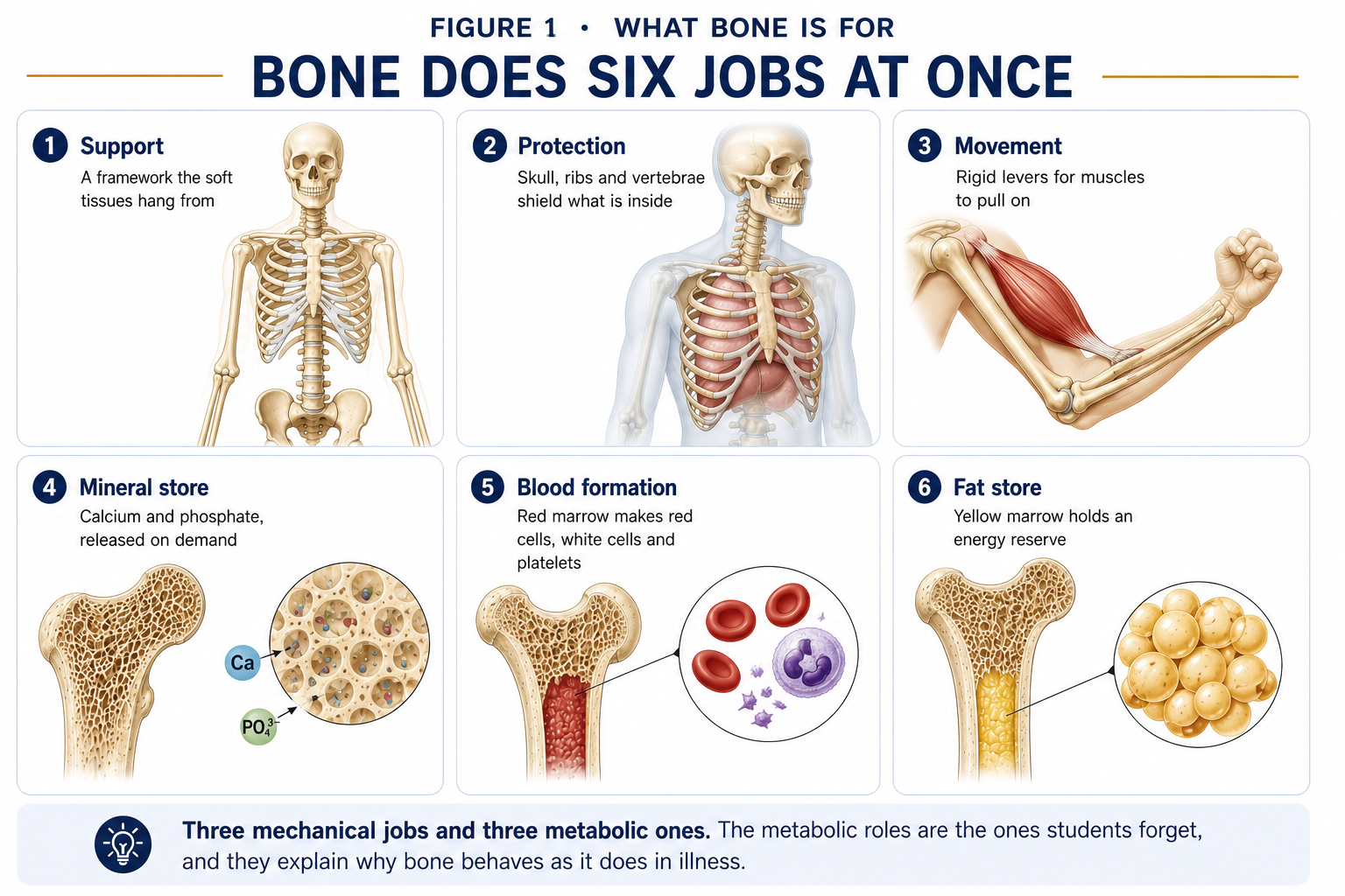
| Function | Detail |
|---|---|
| Support | The rigid framework that maintains body form and provides the levers on which muscles act. |
| Protection | Cranium (brain), vertebral canal (cord), thoracic cage (heart, lungs, great vessels), pelvis (pelvic viscera). |
| Movement | Bones act as levers; joints as fulcrums; muscles as effort. Without a rigid lever, muscle shortening produces no useful displacement. |
| Mineral homeostasis | The skeleton stores approximately 99% of body calcium and 85% of phosphate, plus magnesium and sodium. It is the buffer that keeps serum calcium within its very narrow range. |
| Haemopoiesis | Red marrow produces erythrocytes, leucocytes and platelets. In the adult it is confined largely to the axial skeleton — sternum, ribs, vertebrae, ilium — and the proximal femur and humerus. |
| Fat storage | Yellow marrow is a triglyceride depot; marrow adiposity increases with age and disuse and is inversely related to bone mass. |
| Endocrine organ | Osteocytes secrete FGF-23 (phosphate regulation) and sclerostin; osteoblasts secrete osteocalcin, which influences insulin sensitivity and energy metabolism. Bone is not inert storage. |
| Acid–base buffering | Carbonate and phosphate salts buffer chronic metabolic acidosis — at the cost of bone mineral. |
The adult skeleton has 206 bones: 80 axial (skull 22, hyoid 1, auditory ossicles 6, vertebrae 26, sternum 1, ribs 24) and 126 appendicular. The newborn has around 270 ossification-derived elements, which fuse during growth.
Figure 2 · Classification of bones by shape
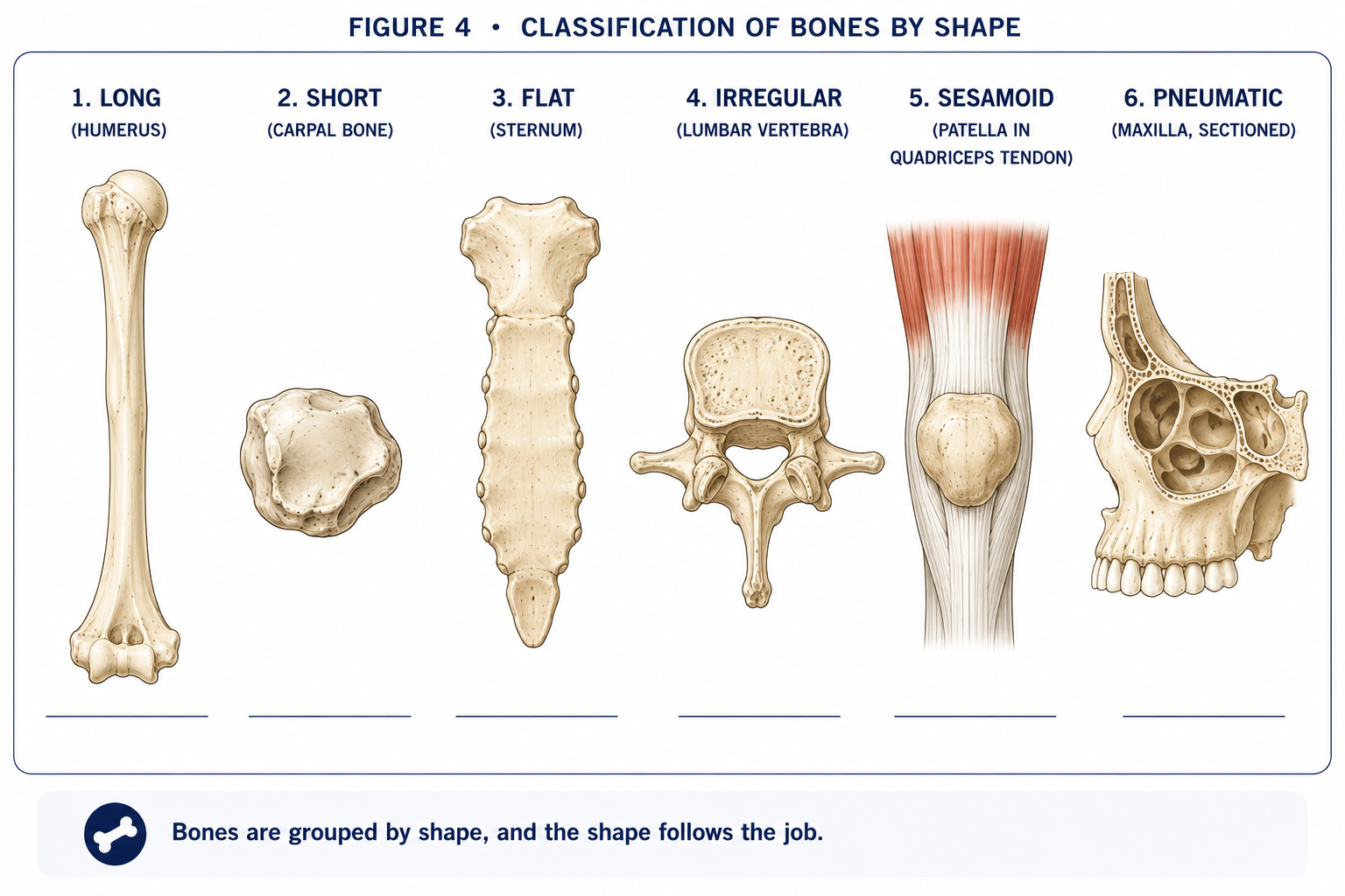
| Type | Structure | Function | Examples |
|---|---|---|---|
| Long | Tubular shaft (diaphysis) with two expanded ends (epiphyses); medullary cavity | Levers for movement; the classic weight-bearing and haemopoietic bones | Femur, tibia, humerus, radius, ulna, metacarpals, phalanges |
| Short | Roughly cuboidal; cancellous core with a thin cortical shell | Stability and shock absorption with limited movement | Carpals, tarsals |
| Flat | Two plates of compact bone with cancellous diploë between | Protection; broad muscle attachment; major haemopoietic sites | Skull vault, sternum, ribs, scapula, ilium |
| Irregular | Complex shapes not fitting other categories | Protection, attachment, articulation | Vertebrae, sacrum, mandible, sphenoid, ethmoid |
| Sesamoid | Develops within a tendon | Alters the tendon’s line of pull; increases the moment arm; protects the tendon from friction | Patella (largest), pisiform, the two hallucal sesamoids, fabella (variable) |
| Pneumatic | Contains air-filled spaces | Reduces weight; resonates the voice; humidifies air | Maxilla, frontal, sphenoid, ethmoid, mastoid |
| Accessory (sutural / supernumerary) | Variable extra ossicles | None — but they are misread as fractures | Wormian bones of the skull; os trigonum; os naviculare accessorium |
Clinical note. The patella increases the quadriceps moment arm at the knee by roughly 30–50% depending on angle. After patellectomy, quadriceps force must rise by a comparable margin to generate the same extension torque — which is why patellar-tendon and extensor-mechanism injuries dominate the difficulty of knee rehabilitation.
Figure 3 · The structure of a long bone
| Region | Description | Clinical significance |
|---|---|---|
| Diaphysis | The shaft. Thick cortical wall enclosing a medullary cavity containing marrow. | Site of the nutrient foramen; the region that heals by callus. |
| Metaphysis | The flared region between shaft and growth plate; predominantly cancellous with a thin cortex. | Richly vascular with sluggish sinusoidal flow — the classic site of acute haematogenous osteomyelitis in children, and of most childhood fractures. |
| Physis (epiphyseal plate) | The cartilaginous growth plate. Present until skeletal maturity, then replaced by the epiphyseal line. | Mechanically the weakest link in the growing skeleton — an injury that would sprain an adult ligament fractures a child’s physis. |
| Epiphysis | The expanded end, largely cancellous, capped by articular cartilage. | Pressure epiphysis where it transmits joint load; traction epiphysis (apophysis) where it exists only for a tendon pull. |
| Articular cartilage | Hyaline cartilage covering the articular surface. No periosteum over it. | Its avascularity governs everything in Chapter 3. |
| Periosteum | Two layers: an outer fibrous layer (dense collagen, richly innervated and vascular) and an inner cambium (osteogenic) layer of osteoprogenitor cells. Anchored by Sharpey’s fibres. | Thick, active and loosely attached in children — hence the periosteal sleeve that produces greenstick fractures and rapid, abundant callus. Thin and less osteogenic in adults. The periosteum is the pain-sensitive part of bone; it is why a shin knock hurts so much and why periosteal elevation is agonising. |
| Endosteum | A thin cellular membrane lining the medullary cavity, Haversian canals and trabecular surfaces. | The principal remodelling surface; endosteal resorption widens the medullary cavity with age. |
| Marrow | Red (haemopoietic) converts to yellow (fatty) from distal to proximal through childhood. | Yellow marrow can reconvert to red under haemopoietic stress (chronic anaemia). Marrow oedema on MRI is a key early sign of bone stress injury. |
Bone is a two-phase composite material, and every mechanical property it has is explained by that fact.
| Phase | Proportion (dry weight) | Constituents | Mechanical contribution |
|---|---|---|---|
| Organic (osteoid) | ~35% | Type I collagen (≈90% of organic matrix); non-collagenous proteins: osteocalcin, osteonectin, osteopontin, bone sialoprotein, proteoglycans; growth factors (BMPs, TGF-β, IGF) sequestered in matrix | Tensile strength, flexibility, toughness — the capacity to absorb energy before failing |
| Inorganic (mineral) | ~65% | Hydroxyapatite, Ca₁₀(PO₄)₆(OH)₂, as plate-like nanocrystals aligned with collagen fibrils; plus carbonate, magnesium, fluoride, citrate | Compressive strength, hardness, stiffness |
| Water | ~10–20% of wet weight | Bound and free | Nutrient transport, viscoelasticity, poroelastic damping |
Decalcify a bone (soak in dilute acid) and the organic matrix remains: the bone becomes rubbery and can be tied in a knot — flexible but unable to bear compression. Deproteinise it (heat or hypochlorite) and the mineral remains: the bone keeps its exact shape but is chalky and shatters under minimal load. The living bone is neither: it is a composite whose toughness exceeds that of either phase alone.
The clinical corollaries follow directly: osteogenesis imperfecta is a type I collagen defect — the organic phase fails, so bones are brittle in a tensile sense and fracture with trivial trauma. Rickets and osteomalacia are mineralisation defects — the organic phase is present but soft, so bones bend (genu varum, coxa vara) rather than snap. Osteoporosis is a loss of both phases in normal proportion — there is simply less bone, of normal quality.
| Cell | Lineage | Location | Function |
|---|---|---|---|
| Osteoprogenitor (osteogenic) cell | Mesenchymal stem cell | Periosteal cambium, endosteum, Haversian canals | The reserve population; proliferates and differentiates into osteoblasts under Runx2/Cbfa1 and Osterix control |
| Osteoblast | Mesenchymal | Bone-forming surfaces, in a cuboidal layer | Synthesises osteoid (type I collagen + NCPs), initiates mineralisation via matrix vesicles and alkaline phosphatase; secretes RANKL and OPG, thereby controlling osteoclasts |
| Osteocyte | Terminally differentiated osteoblast, entombed in matrix | Within lacunae, communicating through canaliculi via gap junctions | 90–95% of all bone cells. The mechanosensor of bone. Detects fluid flow in the lacunocanaliculic system, secretes sclerostin (inhibits formation) and RANKL, performs osteocytic osteolysis, and has a half-life of years to decades |
| Osteoclast | Haemopoietic — monocyte/macrophage lineage; multinucleated by fusion | Resorption surfaces, in Howship’s lacunae | Resorbs bone. Seals to the surface via an actin ring / sealing zone, forms a ruffled border, acidifies the compartment with a proton pump and carbonic anhydrase II (dissolving mineral) and secretes cathepsin K and MMPs (digesting collagen) |
| Bone lining cell | Quiescent osteoblast | Covering inactive surfaces | Forms a barrier controlling ion flux; retracts to expose bone for a new remodelling cycle |
This is the central control system of bone resorption and the target of modern osteoporosis drugs, so it must be understood rather than memorised.
This single ratio explains an entire pharmacology and pathology:
| Influence | Effect on RANKL:OPG | Net effect |
|---|---|---|
| Parathyroid hormone (continuous, as in hyperparathyroidism) | ↑ | Resorption |
| PTH (intermittent, as with teriparatide) | Net anabolic via osteoblast activation | Formation |
| Oestrogen | ↓ RANKL, ↑ OPG | Protective — hence post-menopausal bone loss |
| Glucocorticoids | ↑ RANKL, ↓ OPG, plus direct osteoblast apoptosis | Rapid, severe bone loss |
| IL-1, IL-6, TNF-α (inflammation, RA) | ↑ | Peri-articular and systemic bone loss |
| Mechanical loading | ↓ sclerostin, favourable ratio | Formation |
| Denosumab (a monoclonal antibody) | Acts as a pharmacological OPG | Potent anti-resorptive |
| Bisphosphonates | Taken up in mineral, ingested by osteoclasts, cause their apoptosis | Anti-resorptive |
Figure 4 · Microscopic structure of compact bone
| Woven (immature, primary) | Lamellar (mature, secondary) | |
|---|---|---|
| Collagen | Randomly oriented | Parallel within each lamella, alternating between lamellae |
| Cell density | High, irregularly arranged osteocytes | Lower, regularly arranged |
| Mineralisation | Irregular | Ordered |
| Strength | Weaker, more flexible, isotropic | Stronger, anisotropic |
| Formation | Rapid | Slow |
| Where found | Fetal skeleton; fracture callus; Paget’s disease; bone tumours; the fibrous dysplasia lesion | The entire normal adult skeleton |
Woven bone is always the first bone laid down and is always abnormal in the adult skeleton outside healing. It is subsequently remodelled into lamellar bone.
The structural unit of compact bone: a cylinder 200–300 µm across running roughly parallel to the long axis.
Cancellous bone has no osteons in the classical sense: its trabeculae are thin enough (typically <200 µm) to be nourished by diffusion from marrow, and are built of parallel lamellae organised into hemiosteons (trabecular packets).
Trabeculae are not randomly arranged. In the proximal femur they form two clearly defined systems — a principal compressive group running from the medial cortex (calcar femorale) up into the head, and a principal tensile group arching from the lateral cortex to the head — crossing at approximately right angles and mirroring the theoretical principal stress trajectories of a loaded cantilever. Between them lies a relatively bone-poor region, Ward’s triangle, which is the first area to rarefy in osteoporosis and a standard DEXA region of interest.
This is the strongest visible evidence for Wolff’s law (§2.11): the internal architecture of bone is an engineered response to habitual loading.
Figure 5 · Intramembranous and endochondral ossification
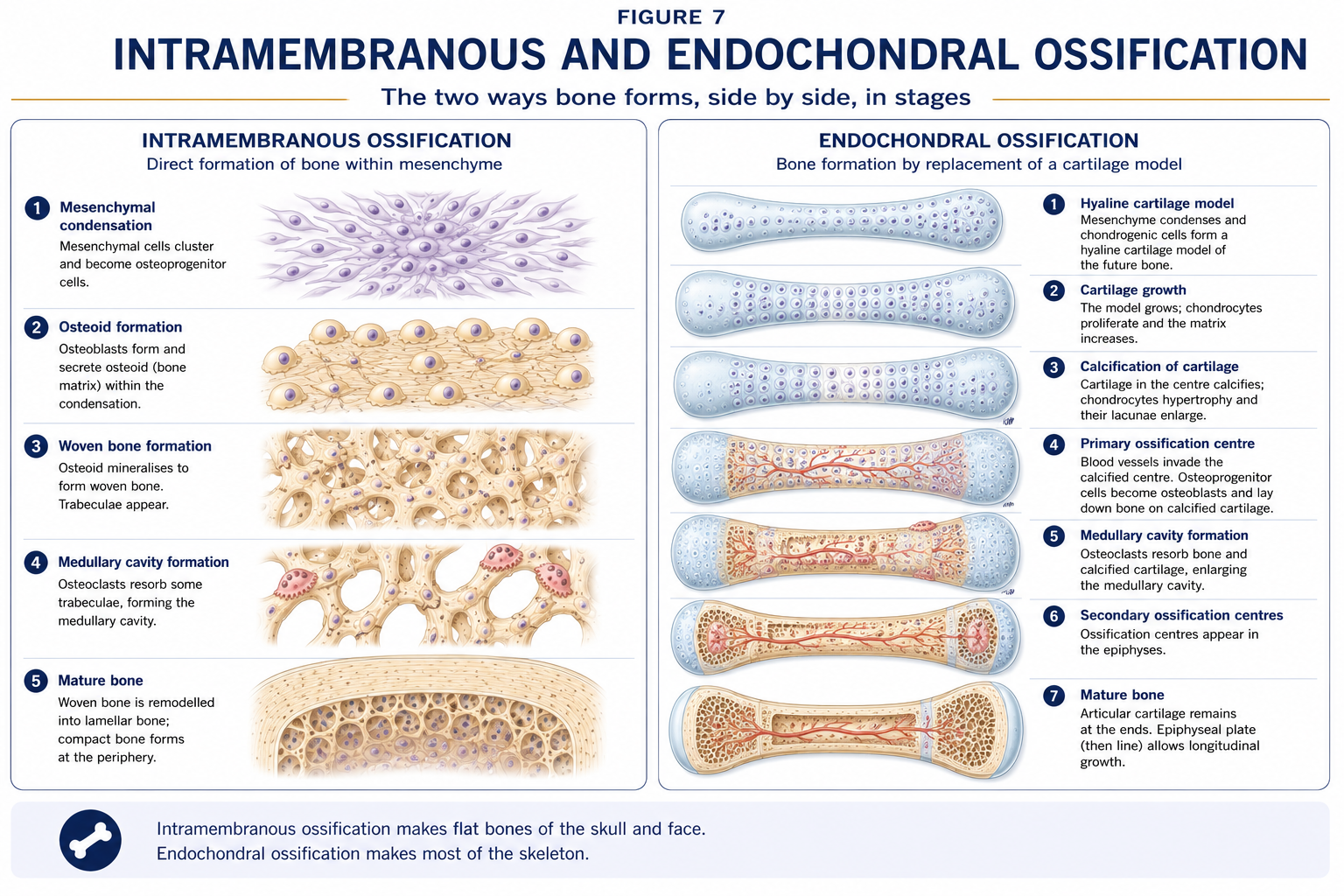
Bone forms in only two ways, and every bone in the body uses one or both.
Direct conversion of mesenchyme into bone, without a cartilage intermediate.
Products: skull vault, most facial bones, mandible, medial clavicle. Because these bones grow by sutural and appositional growth rather than through a physis, cranial vault growth follows brain growth — hence the fontanelles (anterior closing at ~18 months, posterior at ~2–3 months) and the clinical significance of premature sutural fusion (craniosynostosis).
Replacement of a hyaline cartilage model. This is the mechanism for all long bones and is examined constantly.
Learn these in order from epiphysis to metaphysis, with the pathology attached to each — this is the highest-yield table in the chapter.
| Zone | Cell activity | Associated pathology |
|---|---|---|
| 1. Reserve (resting) | Quiescent chondrocytes; matrix storage | Diastrophic dysplasia; Gaucher disease |
| 2. Proliferative | Rapid mitosis; cells stack into longitudinal columns; the zone of longitudinal growth | Achondroplasia (FGFR3 gain-of-function inhibits proliferation → the classic rhizomelic short stature with normal trunk and skull) |
| 3. Hypertrophic | Chondrocytes enlarge 5–10×, accumulate glycogen, then apoptose; matrix is thin here | Mechanically the weakest zone — where physeal fractures and SCFE occur. Also rickets and mucopolysaccharidoses |
| 4. Calcification (provisional calcification) | Matrix mineralises around the empty lacunae | Rickets/osteomalacia (failure to calcify → widened, cupped, frayed physis on radiograph) |
| 5. Ossification (primary spongiosa) | Vascular invasion; osteoblasts deposit bone on calcified cartilage bars | Metaphyseal osteomyelitis; scurvy (defective collagen → subperiosteal haemorrhage) |
Physeal closure is hormonally driven, principally by oestrogen in both sexes (which is why aromatase deficiency causes continued growth, and why girls, with earlier oestrogen exposure, fuse earlier — typically 14–16 years versus 16–18 in boys). Growth hormone acts via IGF-1 on the proliferative zone; thyroid hormone is required for hypertrophic differentiation.
Physeal fractures account for roughly 15–20% of paediatric fractures. The classification predicts growth disturbance.
| Type | Line of injury | Mnemonic | Prognosis |
|---|---|---|---|
| I | Straight through the physis (separation) | S — Slipped | Usually excellent. Includes SCFE |
| II | Through physis + a metaphyseal fragment (Thurston-Holland sign) | A — Above | The commonest (~75%); usually good |
| III | Through physis + epiphysis, into the joint | L — Lower | Intra-articular; needs anatomical reduction; growth arrest risk |
| IV | Through metaphysis, physis and epiphysis | T — Through / Two | Intra-articular; high risk of bony bridge and angular deformity |
| V | Crush injury of the physis | R — Rammed | Worst; often diagnosed retrospectively when growth arrests |
Physiotherapy implications. In a skeletally immature patient, a mechanism that would produce a ligament sprain in an adult far more often produces a physeal injury, because the physis is weaker than the ligament. A child with a “sprained ankle” and tenderness precisely over the distal fibular physis should be treated as a Salter–Harris I until proved otherwise, regardless of normal radiographs. Similarly, apophyseal traction injuries (Osgood–Schlatter at the tibial tuberosity, Sinding-Larsen–Johansson at the inferior patellar pole, Sever’s at the calcaneal apophysis, and avulsions at the ASIS, AIIS and ischial tuberosity) are the adolescent equivalents of adult tendinopathy and require load modification rather than aggressive stretching into the painful apophysis.
Figure 6 · Blood supply of a long bone
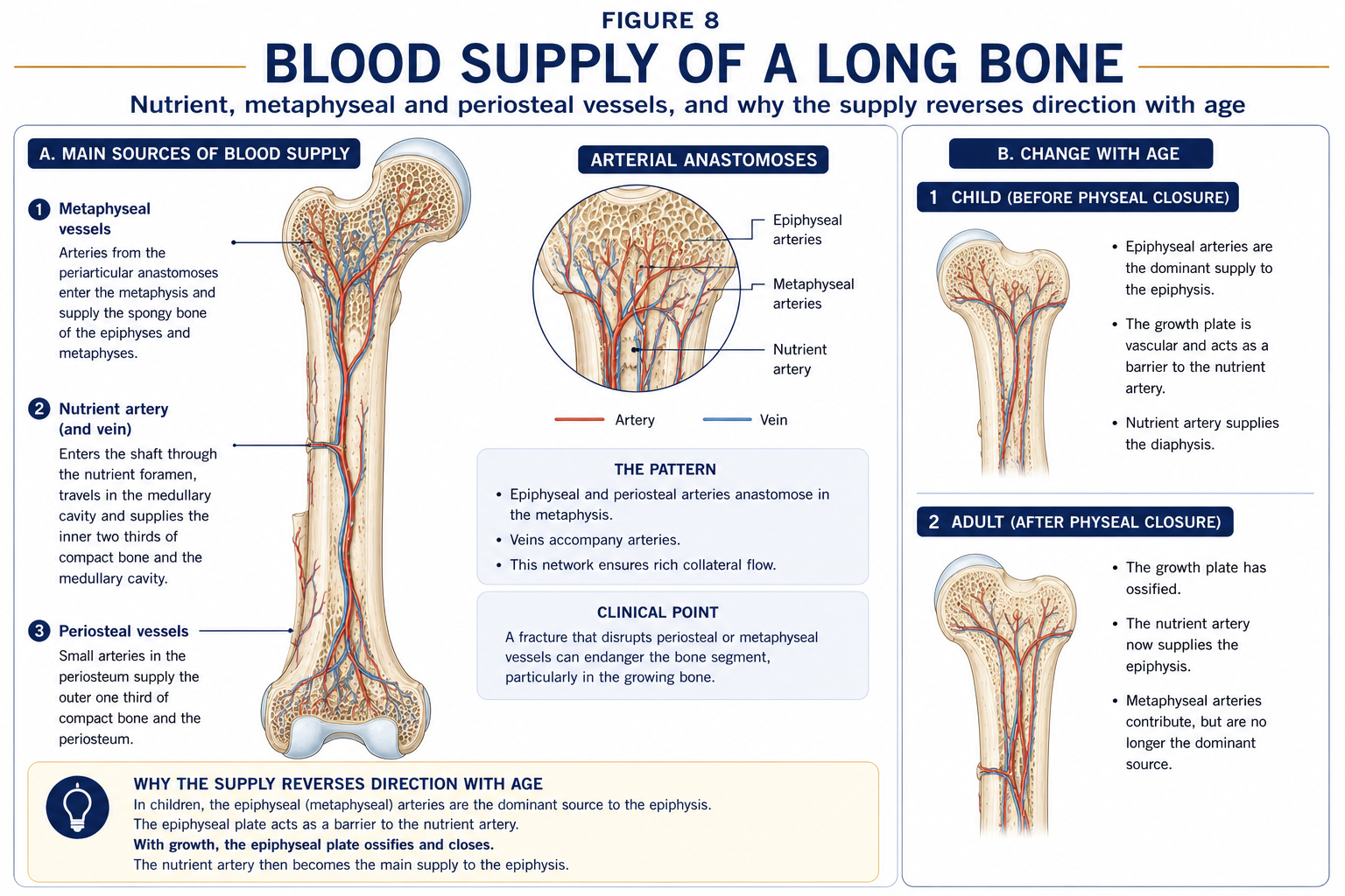
Four sources, and the way they overlap determines which bones die when their supply is interrupted.
| Source | Course | Supplies |
|---|---|---|
| Nutrient artery | Enters through the oblique nutrient foramen of the diaphysis, divides into ascending and descending branches within the medullary cavity | Marrow and the inner two-thirds of the cortex |
| Metaphyseal arteries | Numerous, from adjacent periarticular vessels | Metaphysis; anastomose with the nutrient system |
| Epiphyseal arteries | From the periarticular anastomosis | Epiphysis. Before physeal fusion, the physis is a barrier — epiphyseal and metaphyseal circulations do not communicate, which is why childhood osteomyelitis stays metaphyseal and why epiphyseal blood supply is precarious |
| Periosteal arteries | Numerous, entering where muscle and fascia attach | Outer one-third of the cortex. Their contribution rises dramatically after a fracture disrupts the nutrient supply |
Blood flows centrifugally in the normal diaphysis (medulla → cortex → periosteal capillaries). After the medullary supply is disrupted, flow reverses to centripetal, driven by the periosteum. This is why stripping the periosteum during surgery is far more damaging than reaming the canal, and why periosteal preservation is a principle of modern fracture fixation.
Wherever a bone segment has a single, retrograde, intracapsular supply with poor collateral, interruption causes necrosis:
| Site | Vessel at risk | Consequence |
|---|---|---|
| Head of femur | Medial and lateral circumflex femoral → retinacular vessels; the artery of the ligamentum teres is negligible in adults | AVN after subcapital fracture or dislocation; Perthes disease in children |
| Proximal pole of scaphoid | Retrograde supply entering distally through the dorsal ridge | AVN and non-union after waist fracture — the classic missed injury |
| Body of talus | Largely retrograde from the artery of the tarsal canal | AVN after talar neck fracture (Hawkins classification) |
| Head of humerus | Anterolateral ascending branch of the anterior circumflex humeral (arcuate artery of Laing) | AVN after 4-part proximal humeral fracture |
| Lunate | Variable, sometimes single-vessel | Kienböck’s disease |
| Second metatarsal head | — | Freiberg’s infraction |
| Femoral condyle | — | Spontaneous osteonecrosis of the knee (SONK) |
Nerve supply. Periosteum is richly supplied with sensory (nociceptive) fibres — the most pain-sensitive part of bone. Articular branches, medullary nerves accompanying the nutrient artery, and sympathetic vasomotor fibres complete the picture. This explains periosteal pain from a direct blow, the deep boring pain of intraosseous pathology, and the effectiveness of periosteal anaesthesia.
Figure 7 · The remodelling cycle and the cells that run it
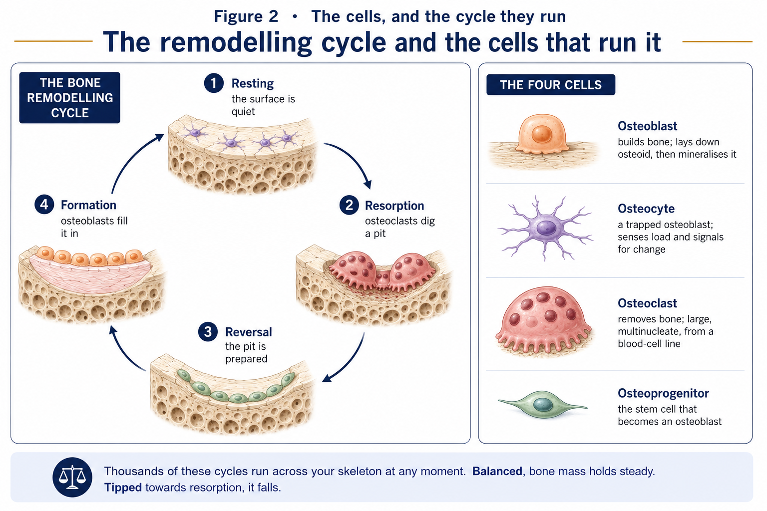
Bone turnover in the adult is not random; it occurs in discrete, coupled cycles executed by a basic multicellular unit (BMU) — a travelling team of osteoclasts followed by osteoblasts, working within a canal in cortical bone or across a trench on a trabecular surface.
| Phase | Duration | Events |
|---|---|---|
| 1. Activation | Days | Lining cells retract; osteoclast precursors are recruited under M-CSF and RANKL; microdamage and osteocyte apoptosis are the usual local triggers |
| 2. Resorption | ~2–3 weeks | Osteoclasts excavate a cutting cone (cortical) or Howship’s lacuna (trabecular); mineral dissolved by acid, collagen by cathepsin K |
| 3. Reversal | ~1–2 weeks | Mononuclear cells prepare the surface; a cement line is laid; TGF-β and IGF released from resorbed matrix recruit osteoblasts — the molecular basis of coupling |
| 4. Formation | ~3–4 months | Osteoblasts deposit osteoid, which mineralises after a ~10–15 day lag; some osteoblasts become osteocytes, some become lining cells, most apoptose |
| 5. Quiescence | Until next cycle | Lining cells re-cover the surface |
Two consequences of this timetable matter clinically:
| Regulator | Action |
|---|---|
| Mechanical strain | The dominant physiological regulator. Detected by osteocytes via canalicular fluid flow; loading ↓ sclerostin → ↑ Wnt/β-catenin signalling → ↑ osteoblast activity |
| PTH | Raises serum calcium: ↑ osteoclastic resorption (indirectly, via osteoblast RANKL), ↑ renal reabsorption, ↑ 1α-hydroxylation of vitamin D. Continuous exposure is catabolic; intermittent is anabolic |
| Calcitriol (1,25-(OH)₂ vitamin D) | ↑ intestinal calcium and phosphate absorption; permits mineralisation. Deficiency → rickets/osteomalacia |
| Calcitonin | ↓ osteoclast activity; physiologically minor in adult humans |
| Oestrogen / testosterone | Restrain resorption, promote physeal closure. Deficiency (menopause, hypogonadism, RED-S) → accelerated loss |
| Growth hormone / IGF-1 | Longitudinal growth and periosteal apposition |
| Thyroid hormone | Necessary for growth; excess accelerates turnover and causes bone loss |
| Glucocorticoids | Reduce formation, increase resorption and apoptosis, reduce calcium absorption — the commonest cause of secondary osteoporosis |
| Cytokines (IL-1, IL-6, TNF-α, prostaglandins) | Drive resorption in inflammatory disease |
| Vitamin C | Cofactor for collagen hydroxylation; deficiency (scurvy) → defective osteoid |
| Vitamin A (excess) | Stimulates resorption |
Figure 8 · How bone answers to load
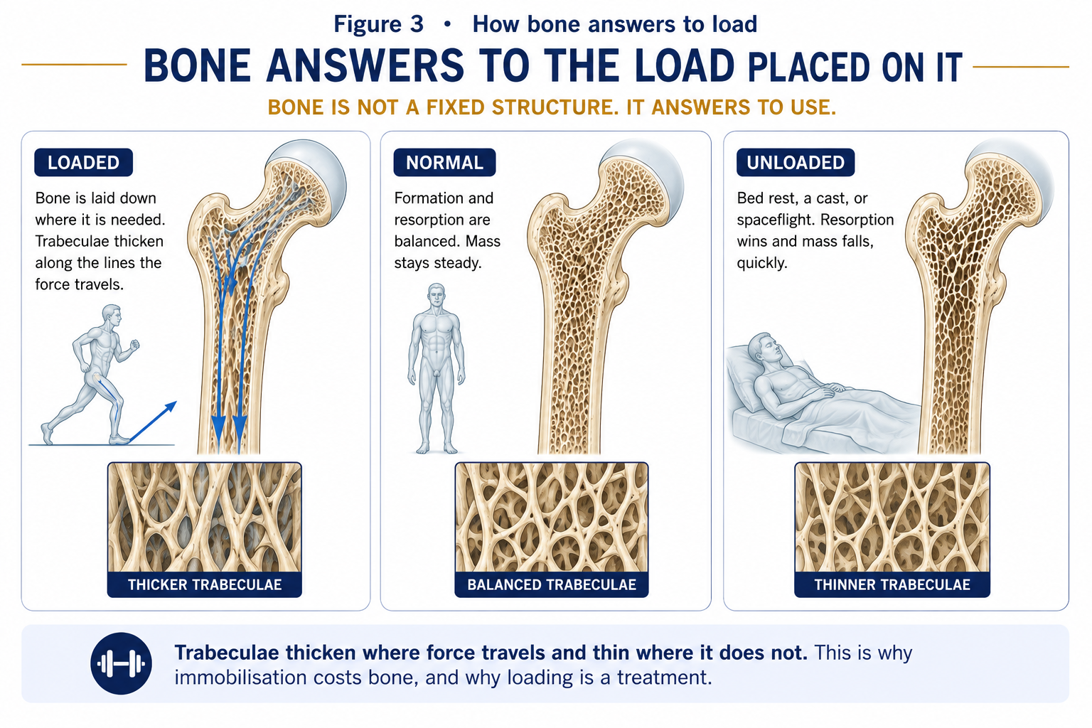
Julius Wolff (1892): bone adapts its internal architecture and external form to the loads habitually placed upon it. Increase the load and bone is laid down; remove it and bone is resorbed. The trabecular systems of the femoral neck are the visual proof.
Harold Frost reframed Wolff’s law as a negative-feedback control system with strain thresholds. Bone “measures” peak strain (in microstrain, µε) and responds:
| Strain window | Approximate range | Bone response |
|---|---|---|
| Disuse (trivial loading) | < ~50–100 µε | Net resorption — remodelling removes bone |
| Adapted (physiological) | ~100–1500 µε | Maintenance — remodelling replaces what is lost |
| Overload (mild) | ~1500–3000 µε | Modelling on — net bone formation |
| Pathological overload | > ~3000–4000 µε | Microdamage accumulates faster than repair → bone stress injury; failure at ~25,000 µε |
Three empirical rules follow, and these are what you build a programme from:
The mechanostat runs in both directions, and the losses are fast and clinically severe.
The practical translation. For the ambulant patient at risk of osteoporosis, the osteogenic prescription is weight-bearing, impact-containing, and progressively resisted: jumping, hopping, skipping, stair climbing, and heavy-load resistance training (which loads bone through muscle pull, the largest force bone experiences). Swimming and cycling, whatever their cardiovascular merit, are essentially non-osteogenic at the lower limb. For the patient with established vertebral fragility, this is tempered by fracture risk: avoid loaded end-range spinal flexion and rotation, and progress impact cautiously. For the non-ambulant patient, functional electrical stimulation cycling and standing frames produce modest, site-specific effects at best; expectations should be set honestly.
Occurs when there is relative stability and some interfragmentary motion: cast treatment, intramedullary nailing, bridge plating, external fixation. It proceeds through callus, recapitulating endochondral ossification.
| Stage | Timing | Events | Rehabilitation implication |
|---|---|---|---|
| 1. Haematoma and inflammation | 0 – ~5 days (peaks 24–48 h) | Vessel disruption, haematoma, clot; platelets and inflammatory cells release TNF-α, IL-1, IL-6, BMPs, PDGF, VEGF; MSCs recruited | Protect. Control pain and oedema. Begin adjacent-joint motion and the unaffected limb immediately. NSAIDs are relevant here — COX-2 inhibition impairs this phase, and while the human evidence is mixed, most units avoid prolonged NSAID use in high-risk or non-union-prone fractures |
| 2. Soft callus (fibrocartilaginous) | ~5 days – 3 weeks | Angiogenesis; MSCs differentiate into chondrocytes and fibroblasts; a cartilaginous bridge unites the fragments; clinically the fracture becomes “sticky” and pain on movement falls | The fracture resists bending but not torsion or shear. Controlled, protected loading in the axial direction is beneficial; rotation is not |
| 3. Hard callus (bony) | ~3 – 12 weeks | Soft callus calcifies and is replaced by woven bone by endochondral ossification, proceeding from the periphery inward; clinical union — painless, non-tender, no movement at the site | Progressive weight bearing per surgical instruction; restore range; begin loading the muscle envelope in earnest |
| 4. Remodelling | 12 weeks – years | Woven bone is replaced by lamellar bone; the callus is resculpted along stress lines by BMUs; the medullary cavity is re-established | The Wolff’s-law phase. This is where physiotherapy has its longest-lasting effect: the remodelled architecture is determined by the loads applied. Full return to sport is a bone-remodelling question, not only a symptom question |
Occurs only with absolute stability and anatomical reduction — compression plating, lag screws. There is no callus. Cutting cones of osteoclasts cross the fracture line directly and osteons are re-established across it (contact healing where the gap is <0.01 mm; gap healing where it is <1 mm, with lamellar bone deposited first and then remodelled). It is slower to achieve mechanical strength and is unforgiving of any residual motion: interfragmentary movement in a construct designed for primary healing produces non-union rather than callus.
Clinically this matters: the absence of visible callus on a radiograph after rigid internal fixation is expected, not a sign of failure. The appearance of callus around a compression plate, conversely, indicates unwanted motion.
| Local | Systemic |
|---|---|
| Inadequate blood supply / soft-tissue stripping | Age |
| Excessive motion at the site | Smoking (a major, dose-dependent, modifiable risk) |
| Distraction / gap at the fracture | Diabetes mellitus |
| Interposed soft tissue | Malnutrition; low protein, calcium, vitamin D |
| Infection | Corticosteroids; chemotherapy; some NSAIDs |
| Comminution; segmental fracture | Peripheral vascular disease |
| Intra-articular location (synovial fluid lyses the haematoma) | Endocrine: hyperparathyroidism, hypothyroidism |
| Bone loss; pathological fracture | Alcohol excess |
Delayed union = healing slower than expected for that site. Non-union = healing has ceased, conventionally by 6–9 months, with no radiographic progress over 3 consecutive months. Non-unions are hypertrophic (abundant callus, “elephant foot” — good biology, insufficient stability; treat mechanically) or atrophic (no callus — poor biology; treat biologically, with bone graft, and address vascularity and systemic factors). Recognising which type you are looking at explains why one patient is told to load more and another less.
| Condition | Mechanism | What the physiotherapist must know |
|---|---|---|
| Osteoporosis | Reduced bone mass with normal mineralisation; microarchitectural deterioration. Post-menopausal (type I, trabecular, vertebral and Colles’ fractures) and senile (type II, cortical and trabecular, hip fractures); secondary causes include glucocorticoids, hypogonadism, hyperthyroidism, malabsorption | Diagnosed by DEXA T-score ≤ −2.5 (osteopenia −1 to −2.5); fracture risk estimated by FRAX. Exercise must be progressive resistance plus impact plus balance/fall prevention; avoid loaded spinal flexion and rotation in established vertebral disease; posture and back-extensor strengthening reduce kyphosis progression |
| Osteomalacia / rickets | Defective mineralisation of osteoid, usually from vitamin D deficiency | Bone pain, proximal myopathy with waddling gait, bowing deformity, Looser’s zones. The proximal weakness is often mistaken for deconditioning; correcting the deficiency is the treatment |
| Osteogenesis imperfecta | Type I collagen mutation | Fragility, blue sclerae, dentinogenesis imperfecta, hearing loss. Rehabilitation aims at safe strengthening, positioning and mobility aids, and at avoiding the immobilisation spiral, since disuse compounds fragility |
| Paget’s disease | Disordered, greatly accelerated remodelling producing structurally weak woven bone in a mosaic pattern | Bone pain, deformity (sabre tibia), pathological fracture, high-output cardiac failure, nerve compression, rare sarcomatous change. Raised alkaline phosphatase with normal calcium |
| Bone stress injury (stress fracture) | Microdamage accumulating faster than repair — the pathological end of the mechanostat curve | The continuum: stress reaction (marrow oedema on MRI) → stress fracture. High-risk sites (femoral neck tension side, anterior tibial cortex, navicular, base of 5th metatarsal, sesamoids, pars interarticularis) demand unloading; low-risk sites tolerate relative rest. Always screen for RED-S / the female athlete triad: low energy availability, menstrual dysfunction, low bone density |
| Heterotopic ossification / myositis ossificans | Lamellar bone forming in soft tissue after trauma, burns, SCI, TBI or hip arthroplasty | Presents as a firm, warm, painful mass with progressively reducing range 1–4 weeks after the insult. Aggressive passive stretching into pain is contraindicated in the acute phase. Maintain gentle active range within comfort; alkaline phosphatase rises; excision, if needed, awaits maturity |
| Osteomyelitis | Infection, haematogenous in children (metaphyseal) or contiguous/post-traumatic in adults | Sequestrum, involucrum, sinus formation. Suspect in the child with fever, refusal to weight-bear and metaphyseal tenderness |
| Avascular necrosis | Interruption of a precarious blood supply (§2.9) | Insidious groin/joint pain, preserved early radiographs, MRI-positive. Weight-bearing restriction may be prescribed pre-collapse |
15 questions on this chapter. Tap one to see the answer and the reasoning.
Answer: (C) Osteocytes detect fluid flow in the lacunocanalicular system and regulate formation through sclerostin.
Answer: (B) They are haemopoietic in origin and become multinucleate by fusion.
Answer: (B) The collagenous organic phase remains, providing tensile flexibility without compressive strength.
Answer: (C) Hence physeal fractures and SCFE occur here.
Answer: (B) It is the commonest type, with a metaphyseal (Thurston-Holland) fragment, and generally has a good prognosis.
Answer: (B) Retrograde supply explains proximal pole avascular necrosis after waist fracture.
Answer: (B) RANKL activates osteoclasts; OPG is a decoy receptor that blocks it.
Answer: (C) Resorption is quick, formation is slow — hence the remodelling transient and the long timescale of bone adaptation.
Answer: (B) Magnitude and strain rate matter more than volume, and the response saturates within 20–40 cycles.
Answer: (B) Absolute stability produces primary healing without callus; callus implies the construct is moving.
Answer: (B) Abundant callus indicates good biology but insufficient stability.
Answer: (B) Remodelling is a surface event; hence metabolic bone disease shows first in the vertebrae and femoral neck.
Answer: (C) Articular cartilage is aneural entirely.
Answer: (B) Aggressive passive stretching in this phase is contraindicated.
Answer: (C) The physis is a vascular barrier, which is why acute haematogenous osteomyelitis remains metaphyseal in children.
Everything on this page, in one screen
| Source | Why it earns its place |
|---|---|
| Standring S (ed.) — Gray’s Anatomy, 42nd edn, chapters on functional anatomy of the musculoskeletal system | The reference account of bone structure, ossification and blood supply |
| Ross MH, Pawlina W — Histology: A Text and Atlas | The clearest treatment of osteon architecture, bone cells and the remodelling cycle |
| Nordin M, Frankel VH — Basic Biomechanics of the Musculoskeletal System | Bone as a material: anisotropy, viscoelasticity, stress–strain behaviour |
| Frost HM — “Bone’s mechanostat: a 2003 update”, Anat Rec | The primary source for the strain-threshold model you prescribe from |
| Turner CH, Robling AG — “Designing exercise regimens to increase bone strength”, Exerc Sport Sci Rev, 2003 | The evidence for magnitude, rate, saturation and rest-insertion |
| Beck BR, Daly RM, Singh MAF, Taaffe DR — “Exercise and Sports Science Australia (ESSA) position statement on exercise prescription for the prevention and management of osteoporosis” | A directly usable clinical prescription framework |
| Einhorn TA, Gerstenfeld LC — “Fracture healing: mechanisms and interventions”, Nat Rev Rheumatol, 2015 | The modern account of the healing cascade |
| Salter RB, Harris WR — J Bone Joint Surg Am, 1963 | The original physeal injury classification |
| Mountjoy M et al. — IOC consensus statement on Relative Energy Deficiency in Sport (RED-S), updates to 2023 | Essential background to bone stress injury in athletes |
| Palastanga N, Field D, Soames R — Anatomy and Human Movement | The physiotherapy-facing synthesis |
| Chaurasia BD — Handbook of General Anatomy | Matched to Indian university syllabi for the descriptive content |
Chapter 2 of 24 · Human Anatomy · Physiotherapist India Next: Chapter 3 — Cartilage: no vessels, no nerves, no lymphatics — and everything difficult follows from that.
Reviewed by the Physiotherapist India Team. · Human Anatomy contents