Evidence-based physiotherapy and rehabilitation resources
Editorial & review policy
Evidence-based physiotherapy and rehabilitation resources
Editorial & review policyHuman Anatomy · Upper limb
The elbow does two entirely different jobs at once. It hinges, so the hand can be brought to the body, and it rotates, so the hand can be turned over without moving the shoulder. Two separate joints share one capsule to make that possible.
Part 2 · The upper limb
Compartments, the cubital fossa, pronation and supination, and the two epicondyles
The shoulder points the arm; the hand does the work; the elbow and forearm position the hand in space and set its orientation. Two functions, two mechanisms:
Because of that second function, the forearm is functionally a joint, not a segment: two bones rotating about each other, connected proximally, distally and throughout by the interosseous membrane. Treat it as a single mechanical unit and its injuries make sense.
Figure 1 · Compartments of the arm
The brachial fascia, with the medial and lateral intermuscular septa running from it to the humeral supracondylar ridges, divides the arm into two.
| Anterior (flexor) compartment | Posterior (extensor) compartment | |
|---|---|---|
| Muscles | Biceps brachii, brachialis, coracobrachialis | Triceps brachii (+ anconeus, distally) |
| Nerve | Musculocutaneous (C5–C7) | Radial (C5–T1) |
| Artery | Brachial | Profunda brachii |
| Muscle | Origin | Insertion | Nerve | Actions |
|---|---|---|---|---|
| Biceps brachii | Long head: supraglenoid tubercle (intracapsular, extrasynovial, running in the intertubercular groove). Short head: coracoid, with coracobrachialis | Radial tuberosity; bicipital aponeurosis (lacertus fibrosus) into deep fascia of the medial forearm | Musculocutaneous (C5, C6) | Powerful supinator (strongest with the elbow at 90°); flexor of the elbow (chiefly when supinated); weak shoulder flexor; the long head contributes to humeral head depression |
| Brachialis | Distal half of the anterior humerus | Coronoid process and ulnar tuberosity | Musculocutaneous (C5, C6), plus a small radial contribution to the lateral part | The workhorse elbow flexor — it flexes the elbow in any forearm position, because it inserts on the ulna, which does not rotate |
| Coracobrachialis | Coracoid | Middle of the medial humeral shaft | Musculocutaneous (C5–C7) — pierced by it | Shoulder flexion and adduction |
Biceps inserts on the radial tuberosity. In supination that tuberosity faces medially and biceps has a good flexion moment arm; in pronation the tuberosity has rotated to face laterally and posteriorly, and biceps’ line of pull first has to unwind the pronation. Test elbow flexion in full pronation and you are testing brachialis and brachioradialis; test it in supination and biceps contributes fully. This is exactly why brachialis, not biceps, is the muscle that never fails you.
| Muscle | Origin | Insertion | Nerve | Actions |
|---|---|---|---|---|
| Triceps brachii | Long head: infraglenoid tubercle (the only head crossing the shoulder). Lateral head: above the radial groove. Medial (deep) head: below the radial groove | Olecranon, via a common tendon | Radial (C6–C8) | Elbow extension (medial head is the workhorse in unresisted extension; the long and lateral heads recruit against resistance); the long head extends and adducts the shoulder |
| Anconeus | Lateral epicondyle | Lateral olecranon and proximal ulna | Radial (C7, C8) | Assists extension; stabilises the elbow; may abduct the ulna in pronation |
Figure 2 · The elbow and radioulnar joints
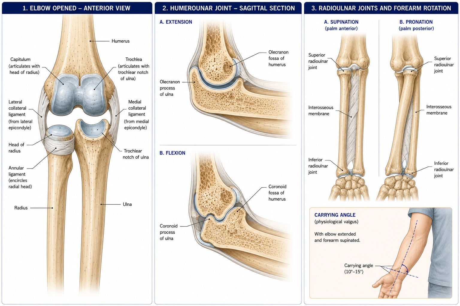
Three articulations within one joint capsule and one synovial cavity — which is why an effusion here distends all three and why septic arthritis of the elbow involves the whole complex.
| Articulation | Surfaces | Type | Movements |
|---|---|---|---|
| Humero-ulnar | Trochlea of humerus ↔ trochlear notch of ulna | Synovial hinge (ginglymus) | Flexion–extension |
| Humeroradial | Capitulum ↔ head of radius | Synovial, functionally hinge + pivot | Flexion–extension and rotation |
| Proximal (superior) radio-ulnar | Head of radius ↔ radial notch of ulna, within the anular ligament | Synovial pivot (trochoid) | Pronation–supination |
| Ligament | Bands | Function |
|---|---|---|
| Ulnar (medial) collateral ligament (UCL/MCL) | Anterior band (medial epicondyle → sublime tubercle of the coronoid) — the primary restraint to valgus stress from 20–120° of flexion; posterior band (→ olecranon), taut in flexion; transverse (Cooper’s) band | Valgus stability. The band injured by repetitive overhead throwing (“Tommy John” injury) |
| Radial (lateral) collateral ligament complex | Radial collateral (lateral epicondyle → anular ligament); lateral ulnar collateral ligament (LUCL) (lateral epicondyle → supinator crest of the ulna); accessory collateral; anular ligament | Varus stability; the LUCL is the key restraint against posterolateral rotatory instability (PLRI) |
| Anular ligament | Encircles the radial head, attached to the front and back of the radial notch; funnel-shaped, narrower below | Holds the radial head against the ulna throughout rotation |
| Quadrate ligament | Radial notch → radial neck | Limits rotation |
| Oblique cord | Ulnar tuberosity → below the radial tuberosity; fibres run opposite to the interosseous membrane | Limits excessive supination |
The elbow is often quoted as the most inherently stable joint in the upper limb. Its stability comes from three primary and several secondary restraints:
Primary: the humero-ulnar (trochlear notch) articulation, the anterior band of the UCL, and the LUCL. Secondary: the radial head (a critical valgus stabiliser when the UCL is deficient — the reason a radial head is now preserved or replaced rather than excised), the common flexor and extensor origins, and the capsule.
Close-packed position: full extension with supination. Resting position: ~70° flexion, 10° supination. Capsular pattern: flexion more limited than extension.
| Movement | Range | Limited by |
|---|---|---|
| Flexion | 0–140/150° | Soft-tissue apposition of the anterior arm and forearm (soft end-feel) |
| Extension | 0° (5–15° hyperextension is common, especially in women) | Olecranon in the olecranon fossa — a bony end-feel |
| Pronation | 0–80–90° | Ligaments and soft tissue |
| Supination | 0–80–90° | Oblique cord, ligaments |
The functional range for most activities of daily living is approximately 30–130° of flexion and 50° each of pronation and supination — a useful target when full range is not achievable.
Carrying angle: 5–15° valgus in men, 10–20° in women, present in full extension and supination and disappearing in flexion and pronation. Increased = cubitus valgus (risk of tardy ulnar nerve palsy); reduced or reversed = cubitus varus / gunstock deformity, the classic sequel of a malunited supracondylar fracture.
The only movement in the body in which one bone rotates around another along its length.
The mechanism, precisely:
| Muscle | Attachments | Nerve | Notes |
|---|---|---|---|
| Pronator teres | Medial epicondyle (humeral head) + coronoid (ulnar head) → mid-lateral radius | Median (C6, C7) | The median nerve passes between its two heads — a site of compression |
| Pronator quadratus | Distal anterior ulna → distal anterior radius | Anterior interosseous (C7, C8) | The primary and constant pronator; also a key DRUJ stabiliser |
| Biceps brachii | See above | Musculocutaneous | The most powerful supinator, especially with the elbow flexed to 90° |
| Supinator | Lateral epicondyle, radial collateral and anular ligaments, supinator crest → proximal radius | Posterior interosseous (C5–C7) | The primary supinator in slow, unresisted movement and with the elbow extended. The PIN passes between its two heads through the arcade of Frohse |
Test it yourself: turning a screw clockwise (supination for a right-handed person) is powerful; anticlockwise is weak. Right-hand screw threads are designed around this asymmetry, which exists because biceps — a large arm muscle — is a supinator, while pronation depends on two smaller forearm muscles.
Figure 3 · Compartments of the forearm
The antebrachial fascia, the interosseous membrane and two intermuscular septa divide the forearm into anterior (flexor–pronator) and posterior (extensor–supinator) compartments, with a lateral “mobile wad” often described separately.
| Layer | Muscle | Origin | Insertion | Nerve | Action |
|---|---|---|---|---|---|
| Superficial (all from the common flexor origin, medial epicondyle) | Pronator teres | + coronoid | Mid-lateral radius | Median | Pronation, elbow flexion |
| Flexor carpi radialis | Base of 2nd (and 3rd) metacarpal | Median | Wrist flexion and radial deviation | ||
| Palmaris longus | Flexor retinaculum and palmar aponeurosis | Median | Weak wrist flexion; tenses the aponeurosis. Absent in ~10–15% — bilaterally in ~half of those; used as a tendon graft | ||
| Flexor carpi ulnaris | + olecranon and posterior ulna | Pisiform, hook of hamate, 5th metacarpal | Ulnar | Wrist flexion and ulnar deviation. The only anterior forearm muscle supplied by the ulnar nerve, apart from half of FDP | |
| Intermediate | Flexor digitorum superficialis | Medial epicondyle, coronoid, radius | Sides of the middle phalanges, digits 2–5, after splitting to let FDP through (the chiasma of Camper) | Median | Flexes PIP joints (and MCP, wrist) |
| Deep | Flexor digitorum profundus | Proximal ¾ anterior and medial ulna, interosseous membrane | Bases of the distal phalanges, digits 2–5 | Lateral half (index, middle): anterior interosseous (median). Medial half (ring, little): ulnar | The only muscle that flexes the DIP joints |
| Flexor pollicis longus | Anterior radius, interosseous membrane | Base of distal phalanx of thumb | Anterior interosseous (median) | Flexes thumb IP joint | |
| Pronator quadratus | Distal ulna | Distal radius | Anterior interosseous (median) | Pronation; DRUJ stability |
The rule: the anterior compartment is median territory, with two ulnar exceptions — FCU and the medial half of FDP.
| Group | Muscle | Insertion | Nerve | Action |
|---|---|---|---|---|
| Mobile wad / lateral | Brachioradialis | Styloid process of radius | Radial (C5, C6) | Elbow flexion, strongest in mid-prone position; the “shaving muscle” |
| Extensor carpi radialis longus | Base of 2nd metacarpal | Radial (C6, C7) | Wrist extension and radial deviation | |
| Extensor carpi radialis brevis | Base of 3rd metacarpal | Posterior interosseous | Wrist extension. The muscle implicated in lateral epicondylalgia | |
| Superficial (common extensor origin, lateral epicondyle) | Extensor digitorum | Extensor expansions, digits 2–5 | PIN | Extends MCP joints principally |
| Extensor digiti minimi | Extensor expansion, little finger | PIN | Extends the little finger | |
| Extensor carpi ulnaris | Base of 5th metacarpal | PIN | Wrist extension and ulnar deviation; important DRUJ stabiliser | |
| Deep | Supinator | Proximal radius | PIN | Supination |
| Abductor pollicis longus | Base of 1st metacarpal | PIN | Abducts and extends the thumb | |
| Extensor pollicis brevis | Base of proximal phalanx of thumb | PIN | Extends thumb MCP | |
| Extensor pollicis longus | Base of distal phalanx of thumb; hooks around Lister’s tubercle | PIN | Extends thumb IP; retropulsion | |
| Extensor indicis | Extensor expansion, index | PIN | Independent index extension |
The rule: the posterior compartment is entirely radial nerve territory — brachioradialis, ECRL and (usually) anconeus from the radial nerve proper, and everything else from the posterior interosseous nerve.
Anatomical snuffbox: anterior/lateral border = APL and EPB; posterior/medial border = EPL; floor = scaphoid and trapezium; crossed by the radial artery and the superficial branch of the radial nerve.
Figure 4 · The cubital fossa
A triangular hollow in front of the elbow — the region you palpate, inject into and read fractures across.
| Boundary | Structure |
|---|---|
| Superior (base) | An imaginary line between the two epicondyles |
| Lateral | Brachioradialis |
| Medial | Pronator teres |
| Floor | Brachialis and supinator |
| Roof | Deep fascia reinforced by the bicipital aponeurosis, superficial fascia and skin; containing the median cubital vein and the medial and lateral cutaneous nerves of the forearm |
Contents, lateral to medial — mnemonic “TAN” (Tendon, Artery, Nerve):
The radial nerve lies just outside the fossa, deep to brachioradialis, where it divides into its superficial (sensory) and deep (posterior interosseous) branches.
Clinical significance:
Course: runs on the lateral side of the brachial artery in the arm, then crosses to lie medial to it in the mid-arm. It passes through the cubital fossa medial to the artery, then between the two heads of pronator teres, then deep to the fibrous arch of flexor digitorum superficialis. It descends between FDS and FDP to reach the wrist between the tendons of FDS and FCR, before entering the carpal tunnel. in the arm, crosses to lie medial to it in the mid-arm, passes through the cubital fossa medial to the artery, then between the two heads of pronator teres, then deep to the fibrous arch of flexor digitorum superficialis, and descends between FDS and FDP to reach the wrist between the tendons of FDS and FCR, before entering the carpal tunnel.
Branches in the forearm:
Lesions:
| Site | Syndrome | Features |
|---|---|---|
| Pronator teres | Pronator syndrome | Forearm pain, weakness, and altered sensation in the palmar cutaneous distribution — the feature distinguishing it from carpal tunnel syndrome |
| Anterior interosseous | Anterior interosseous syndrome (Kiloh–Nevin) | Purely motor: cannot flex the thumb IP or index DIP — the inability to make an “OK” sign, making a triangle instead. No sensory loss |
| Supracondylar fracture | — | Median + brachial artery; the classic paediatric emergency |
| Wrist (carpal tunnel) | See Chapter 12 |
High median nerve palsy produces the “hand of benediction” on attempted fist-making: the index and middle fingers cannot flex (FDP lateral half and FDS), while the ring and little fingers do.
Course: descends medially in the anterior compartment, pierces the medial intermuscular septum in the mid-arm (the arcade of Struthers) to enter the posterior compartment, passes behind the medial epicondyle in the ulnar (cubital) groove — subcutaneous, palpable, and the “funny bone” — then between the two heads of FCU, and descends on FDP, becoming superficial in the distal forearm before entering Guyon’s canal.
Branches in the forearm: FCU, the medial half of FDP, and the dorsal cutaneous branch, which arises ~5–7 cm proximal to the wrist and supplies the dorsomedial hand — spared in Guyon’s canal lesions but lost in cubital tunnel lesions, a useful localising sign.
Cubital tunnel syndrome — the second commonest upper-limb entrapment after carpal tunnel. Compression sites: the arcade of Struthers, the medial epicondyle, the cubital tunnel retinaculum (Osborne’s ligament), and the two heads of FCU. Symptoms worsen with elbow flexion, which stretches and compresses the nerve (tunnel volume falls markedly beyond 90°). Sleeping with the elbow flexed is a classic aggravator; night splinting in extension is a first-line intervention.
A distal ulnar lesion (at the wrist) produces more marked clawing of the ring and little fingers than a proximal lesion (at the elbow), even though the proximal lesion is more extensive. The reason: a proximal lesion also paralyses the medial half of FDP, so the distal interphalangeal joints cannot flex, and the claw is less pronounced. As a proximal lesion recovers and FDP reinnervates before the intrinsics, the claw worsens — a sign of recovery that patients find alarming and must have explained.
Course: passes through the triangular interval into the posterior compartment, spirals around the humerus in the radial groove with the profunda brachii, pierces the lateral intermuscular septum ~10 cm above the lateral epicondyle to re-enter the anterior compartment, lies deep to brachioradialis anterior to the lateral epicondyle, and divides into:
Lesions:
| Site | Features |
|---|---|
| Axilla (crutch palsy, “Saturday night palsy” from sleeping over a chair back) | Triceps weakness plus wrist drop and sensory loss |
| Radial groove (mid-shaft humeral fracture) | Wrist drop, triceps spared, sensory loss over the dorsal first web space |
| Posterior interosseous (arcade of Frohse, ganglion, rheumatoid synovitis) | Finger drop without wrist drop — because ECRL (radial nerve proper) is spared, the wrist extends but deviates radially. No sensory loss |
| Superficial branch (tight watch straps, handcuffs) | Wartenberg’s syndrome — purely sensory, dorsal radial hand |
The brachial artery continues from the axillary artery at the lower border of teres major, lies medial then anterior in the arm on brachialis, and divides in the cubital fossa into radial and ulnar arteries. Branches: profunda brachii (with the radial nerve), nutrient artery to the humerus, superior and inferior ulnar collateral arteries.
The anastomosis around the elbow links the collateral branches (from brachial and profunda brachii) above with the recurrent branches (from radial, ulnar and common interosseous) below. It maintains flow when the brachial artery is occluded or when the elbow is fully flexed and the vessel kinked.
The ulnar artery gives the common interosseous (dividing into anterior and posterior interosseous arteries) and runs with the ulnar nerve deep to FCU. The radial artery runs on the lateral side, is superficial at the wrist between FCR and brachioradialis (the radial pulse), then crosses the anatomical snuffbox to enter the palm and form the deep palmar arch. The ulnar artery forms the superficial palmar arch.
Allen’s test assesses the patency of both arches before radial artery cannulation or harvest — and is worth knowing because a false-negative can cost a hand.
The commonest elbow condition, peaking at 35–55 years, with only a minority of sufferers playing tennis.
Same pathology at the common flexor origin, principally pronator teres and flexor carpi radialis. Roughly a fifth as common as the lateral form. Always examine the ulnar nerve, which is involved in up to 20–50% of cases; and in throwing athletes, assess the UCL for valgus instability.
Covered in §11.7. Note that radial tunnel syndrome produces pain without weakness and without sensory loss (compression of the PIN before its motor branches, or of the nerve to the radial wrist extensors), whereas PIN syndrome produces weakness (finger drop) without pain. They are ends of the same spectrum and both are frequently misdiagnosed as resistant tennis elbow.
The commonest elbow fracture in children (peak 5–8 years), usually from a fall on the outstretched hand with an extension-type displacement (~95%).
The second most commonly dislocated major joint in adults after the shoulder; posterior or posterolateral in ~90%, from a fall on an outstretched hand with valgus, supination and axial load.
| Condition | Features |
|---|---|
| Distal biceps rupture | Usually a sudden eccentric load in a middle-aged man; a “reverse Popeye” deformity (the muscle belly retracts proximally); ~40–50% loss of supination strength and ~30% of flexion strength; positive hook test. Usually repaired surgically |
| Triceps rupture | Rare; loss of active extension against gravity; associated with steroid use and olecranon bursitis |
| Olecranon bursitis | “Student’s elbow”; the swelling is superficial and elbow range is preserved — which distinguishes it from septic arthritis |
| Pulled elbow (nursemaid’s elbow) | Ages 1–4; axial traction on the pronated extended arm slips the radial head out of the anular ligament. The child holds the arm pronated and slightly flexed and refuses to use it; radiographs are normal; reduction by supination-flexion or hyperpronation is immediate and dramatic |
15 questions on this chapter. Tap one to see the answer and the reasoning.
Answer: (B) Its ulnar insertion is why it flexes the elbow in any forearm position.
Answer: (C) The radial head is the important secondary restraint.
Answer: (B) TAN.
Answer: (B)
Answer: (C) FPL and FDP to the index are affected; the lesion is purely motor.
Answer: (B)
Answer: (B)
Answer: (B) ECRL is spared because it is supplied by the radial nerve proper.
Answer: (B) Because a proximal lesion also paralyses the medial half of FDP.
Answer: (B)
Answer: (B)
Answer: (B)
Answer: (B) Disruption indicates dislocation instead.
Answer: (B)
Answer: (C) The radial head has slipped from the anular ligament.
Everything on this page, in one screen
| Source | Why it earns its place |
|---|---|
| Standring S (ed.) — Gray’s Anatomy, 42nd edn | Definitive regional anatomy and attachments |
| Palastanga N, Field D, Soames R — Anatomy and Human Movement | Functional treatment of elbow and forearm mechanics |
| Neumann DA — Kinesiology of the Musculoskeletal System | Pronation–supination mechanics and moment arms |
| Snell RS — Clinical Anatomy by Regions | The cubital fossa, compartments and nerve courses with clinical notes |
| Magee DJ — Orthopedic Physical Assessment | Elbow special tests and their accuracy |
| Nirschl RP, Ashman ES — “Elbow tendinopathy: tennis elbow”, Clin Sports Med, 2003 | The angiofibroblastic tendinosis model |
| Bisset L et al. — “Mobilisation with movement and exercise, corticosteroid injection, or wait and see for tennis elbow”, BMJ, 2006; Coombes BK et al., JAMA, 2013 | The trials on injection versus exercise |
| O’Driscoll SW, Bell DF, Morrey BF — “Posterolateral rotatory instability of the elbow”, JBJS Am, 1991 | The PLRI concept and tests |
| McRae R, Esser M — Practical Fracture Treatment | Supracondylar fracture, dislocation and their complications |
| Chaurasia BD — Human Anatomy, Vol 1 | Indian syllabus-matched descriptive account |
Chapter 11 of 24 · Human Anatomy · Physiotherapist India Next: Chapter 12 — Wrist and Hand: the carpal tunnel, three nerve territories, the arches, and the four grips.
Reviewed by the Physiotherapist India Team. · Human Anatomy contents