Evidence-based physiotherapy and rehabilitation resources
Editorial & review policy
Evidence-based physiotherapy and rehabilitation resources
Editorial & review policyHuman Anatomy · Lower limb
Twenty-six bones, thirty-three joints, and the whole weight of the body landing on them thousands of times a day. The foot has to be a soft shock absorber the moment it lands and a rigid lever the moment you push off. This chapter is about how one structure manages to be both.
Part 3 · The lower limb
The ankle mortise, its ligaments, and the arches of the foot
Figure 1 · One foot, two jobs
The foot has to be two different things within the same stride, roughly half a second apart:
The mechanism that switches between the two is the coupling of subtalar pronation and supination with midtarsal joint mobility — and understanding that mechanism is the single most useful thing in this chapter. Almost every foot condition you will treat is a failure of it.
Over a lifetime the foot performs this transformation something like 150 million times. That repetition, in a structure of 26 bones, 33 joints and only 2 mm of skin between bone and ground, is why the foot carries the highest overuse pathology burden in the body.
Figure 2 · Bones of the foot
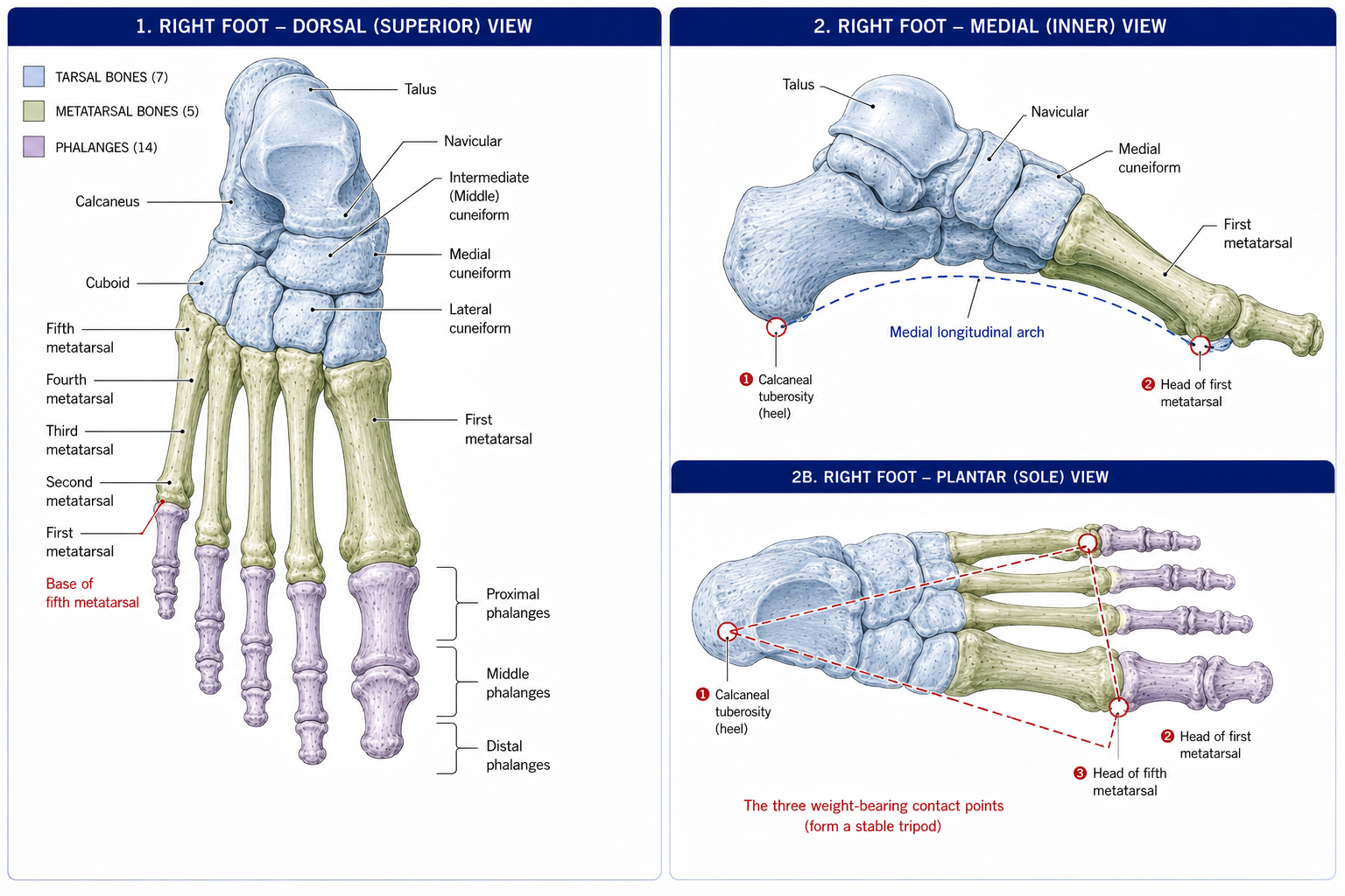
Figure 3 · The ankle mortise and its ligaments
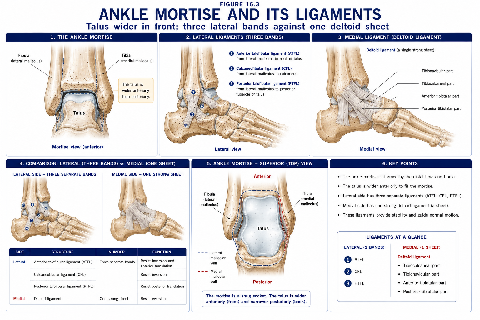
A synovial hinge joint with one degree of freedom (though with a slightly oblique axis, so its motion is not purely sagittal).
The consequence of that wedge shape is the single most examinable fact about the ankle:
| Position | Talar segment in the mortise | Stability |
|---|---|---|
| Dorsiflexion | The wide anterior part; the malleoli splay slightly and the syndesmosis tightens | Close-packed — maximally stable |
| Plantarflexion | The narrow posterior part | Loose-packed — vulnerable |
Hence the classic inversion sprain occurs in plantarflexion and inversion, and hence full dorsiflexion is the safest position to test and to weight-bear in.
Resting position: ~10° plantarflexion, midway between inversion and eversion. Capsular pattern: plantarflexion more limited than dorsiflexion.
| Ligament | Bands | Function |
|---|---|---|
| Lateral collateral complex | Anterior talofibular ligament (ATFL) — the weakest and first injured; taut in plantarflexion. Calcaneofibular ligament (CFL) — crosses both the talocrural and subtalar joints; taut in dorsiflexion. Posterior talofibular ligament (PTFL) — the strongest, rarely injured | Resist inversion. The order of injury is ATFL → CFL → PTFL |
| Medial (deltoid) ligament | Superficial: tibionavicular, tibiocalcaneal, superficial tibiotalar. Deep: anterior and posterior tibiotalar | Very strong, triangular; resists eversion. So strong that eversion force usually avulses the medial malleolus rather than tearing the ligament |
| Syndesmosis (inferior tibiofibular) | Anterior inferior tibiofibular ligament (AITFL), posterior inferior tibiofibular ligament (PITFL), transverse tibiofibular ligament, interosseous ligament | Binds tibia to fibula, maintaining the mortise. Injured by external rotation and dorsiflexion |
| Movement | Range | Note |
|---|---|---|
| Dorsiflexion | 10–20° | Requires at least ~10° for normal gait, and reduced dorsiflexion is one of the most consistent findings associated with lower-limb injury — it forces compensatory pronation, knee valgus and early heel rise |
| Plantarflexion | 40–55° |
The talocrural axis runs obliquely, from just below the medial malleolus to just below the lateral malleolus — angled about 8° from the frontal plane and 20–30° externally rotated in the transverse plane. Because it is oblique, dorsiflexion is accompanied by slight abduction and eversion, and plantarflexion by adduction and inversion. This is why “pure” sagittal ankle motion does not exist.
The Silfverskiöld test distinguishes gastrocnemius from soleus restriction: measure dorsiflexion with the knee extended (loading the biarticular gastrocnemius) and then flexed (unloading it). More dorsiflexion with the knee flexed implicates gastrocnemius.
The articulation between talus and calcaneus, in anterior, middle and posterior facets, with the sinus tarsi and the interosseous talocalcaneal ligament between the middle and posterior facets.
Its axis is oblique in all three planes — running approximately 42° from the horizontal and 16° from the sagittal — which means every motion at this joint is triplanar:
| Motion | Components |
|---|---|
| Pronation (open chain) | Eversion + abduction + dorsiflexion |
| Supination (open chain) | Inversion + adduction + plantarflexion |
In closed chain (weight bearing), the same joint motions appear differently because the calcaneus is fixed to the ground: pronation appears as calcaneal eversion with talar adduction and plantarflexion, and therefore obligatory internal rotation of the tibia and femur. Supination produces the reverse.
This coupling is the mechanical link between the foot and the rest of the limb. Excessive or prolonged pronation drives tibial internal rotation, which drives femoral internal rotation and dynamic knee valgus — the pathway connecting foot mechanics to patellofemoral pain, ITB syndrome and ACL risk. It is also why foot orthoses can influence proximal symptoms and why hip strength can influence foot symptoms.
Talonavicular + calcaneocuboid, forming an S-shaped line across the foot — the surgical amputation level of the same name.
Its behaviour is the key to the transformation:
That is the mechanism the whole chapter turns on. Heel strike → subtalar pronation → midtarsal unlocked → shock absorption and terrain accommodation. Midstance to toe-off → subtalar supination → midtarsal locked → rigid lever for propulsion.
Described by Hicks. The plantar aponeurosis runs from the calcaneal tuberosity to the plantar plates and bases of the proximal phalanges, passing beneath the metatarsal heads.
During toe-off, the MTP joints extend (dorsiflex). The aponeurosis wraps around the metatarsal heads like a cable around a drum, shortening the distance between the calcaneus and the metatarsal heads. This:
It is an elegant purely mechanical solution, requiring no muscular effort, and it is testable at the bedside: passively extend the hallux in a standing patient and watch the arch rise (a positive Jack’s test). Failure of the windlass — from hallux rigidus, plantar fascia rupture, or an incompetent first ray — is a common and under-diagnosed cause of forefoot and arch pain.
Figure 4 · The arches and the windlass
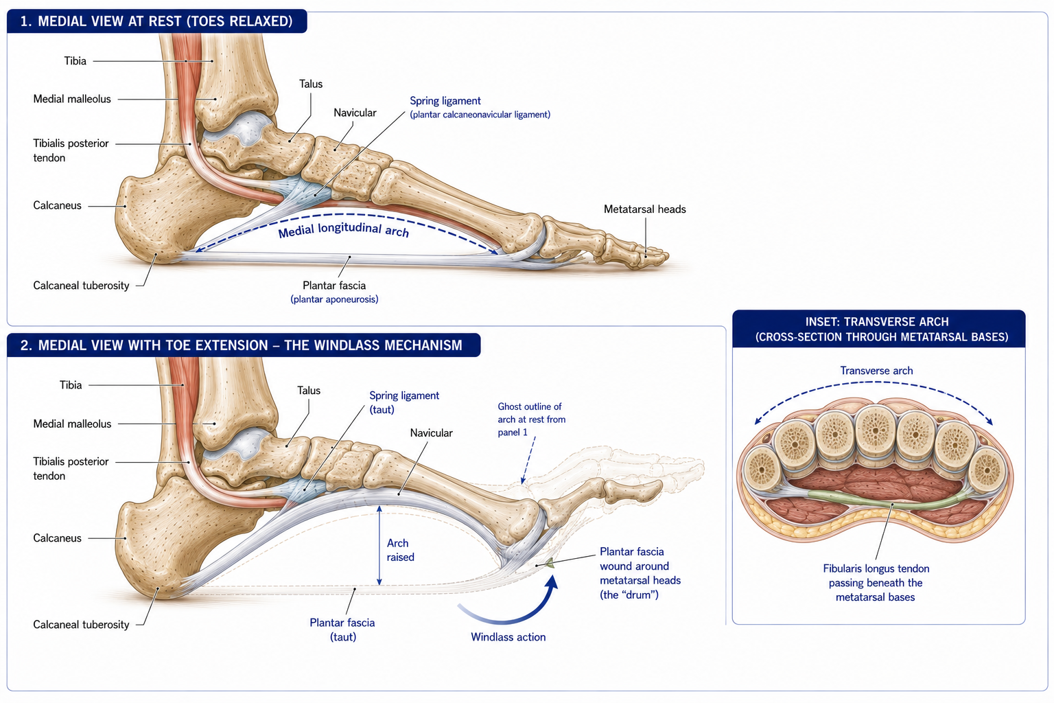
| Arch | Bones | Keystone | Passive supports | Dynamic supports |
|---|---|---|---|---|
| Medial longitudinal | Calcaneus, talus, navicular, three cuneiforms, metatarsals 1–3 | Head of the talus | Plantar calcaneonavicular (spring) ligament, plantar aponeurosis, long and short plantar ligaments, the shapes of the bones themselves | Tibialis posterior (chief), flexor hallucis longus, flexor digitorum longus, abductor hallucis, the intrinsics |
| Lateral longitudinal | Calcaneus, cuboid, metatarsals 4–5 | Cuboid | Long and short plantar ligaments, plantar aponeurosis | Fibularis longus and brevis, abductor digiti minimi |
| Transverse | Cuneiforms, cuboid, metatarsal bases | Intermediate cuneiform (wedge-shaped, wider dorsally) | Deep transverse metatarsal ligaments | Fibularis longus (crossing the sole obliquely), tibialis posterior, adductor hallucis transverse head |
The hierarchy of support — the order matters and is a standard examination point:
This explains why arch collapse follows ligament failure (spring ligament attenuation) or tendon failure (tibialis posterior dysfunction) rather than simple “weak muscles”, and why intrinsic foot muscle training, though useful, is not a complete answer to a collapsing arch.
Pes planus (flatfoot) must be classified before it is treated: flexible (arch present on tiptoe or with Jack’s test — usually asymptomatic and normal in children under 5–6) versus rigid (no arch reformation — consider tarsal coalition, particularly in an adolescent with a stiff, painful flatfoot and recurrent “ankle sprains”). Pes cavus (high arch) should prompt a neurological review — bilateral cavovarus feet with clawed toes suggest Charcot–Marie–Tooth disease until proved otherwise.
| Structure | Contents |
|---|---|
| Superior and inferior extensor retinacula | Tibialis anterior, EHL, EDL, fibularis tertius — with the anterior tibial artery and deep fibular nerve between EHL and EDL |
| Flexor retinaculum (from the medial malleolus to the calcaneus) | The tarsal tunnel |
| Superior and inferior fibular retinacula | Fibularis longus and brevis behind the lateral malleolus |
Contents, from anterior to posterior — “Tom, Dick And Very Nervous Harry”:
Tibialis posterior · Flexor Digitorum longus · Artery (posterior tibial) · Vein · Nerve (tibial) · Flexor Hallucis longus
Tarsal tunnel syndrome produces burning, tingling and aching in the sole, worse on standing and walking, with a positive Tinel’s sign behind the medial malleolus. Causes include space-occupying lesions (ganglion, varicosities), post-traumatic fibrosis, and hindfoot valgus stretching the nerve. It is often confused with plantar fasciopathy, and the distinguishing features are the burning quality, the nocturnal component, and the sensory rather than mechanical pattern.
Note the FHL groove beneath the sustentaculum tali: FHL tenosynovitis here is a classic dancer’s injury, often with posterior ankle impingement from an os trigonum.
Dorsum: extensor digitorum brevis and extensor hallucis brevis — the only intrinsic muscles supplied by the deep fibular nerve. A visible fleshy mass over the lateral dorsum is normal EDB, frequently mistaken for swelling.
Sole — four layers, all supplied by the medial and lateral plantar nerves (tibial):
| Layer | Muscles | Nerve |
|---|---|---|
| 1 (most superficial) | Abductor hallucis, Flexor digitorum brevis, Abductor digiti minimi | Medial plantar (AH, FDB); lateral plantar (ADM) |
| 2 | Quadratus plantae, four lumbricals; plus the tendons of FDL and FHL | Lateral plantar (quadratus, lateral 3 lumbricals); medial plantar (1st lumbrical) |
| 3 | Flexor hallucis brevis (with the two sesamoids), adductor hallucis (oblique and transverse heads), flexor digiti minimi brevis | Medial plantar (FHB); lateral plantar (adductor hallucis, FDMB) |
| 4 (deepest) | Three plantar interossei (PAD), four dorsal interossei (DAB); plus the tendons of fibularis longus and tibialis posterior | Lateral plantar |
The nerve supply rule: the medial plantar nerve supplies only four muscles — abductor hallucis, flexor digitorum brevis, flexor hallucis brevis and the first lumbrical (the exact foot equivalent of the median nerve’s LOAF in the hand). The lateral plantar nerve supplies everything else, exactly as the ulnar nerve does in the hand.
Reference axis: in the foot, abduction and adduction are defined relative to the second toe (compare the middle finger in the hand).
Quadratus plantae is a foot-specific muscle with no hand equivalent: it corrects the oblique line of pull of the FDL tendons, which enter the sole obliquely from the medial side, so that the toes flex straight rather than deviating medially.
Functionally, the intrinsics behave as the foot’s “local stabilisers” — active mainly in the second half of stance, stiffening the arch, controlling toe position, and contributing to balance. They atrophy with prolonged shoe use and immobility, and short-foot and toe-yoga exercises have reasonable evidence for improving arch control and balance, though not for changing static arch height.
| Nerve | Territory |
|---|---|
| Saphenous (femoral) | Medial side of the foot, as far as the first MTP joint |
| Superficial fibular | Most of the dorsum of the foot and toes |
| Deep fibular | First web space only (its autonomous zone) |
| Sural | Lateral border of the foot and little toe |
| Medial and lateral plantar (tibial) | The sole, in a distribution mirroring the median and ulnar nerves of the hand — medial plantar to the medial 3½ toes, lateral plantar to the lateral 1½ |
| Medial calcaneal (tibial) | The heel — and note it arises proximal to the tarsal tunnel, so heel sensation is spared in tarsal tunnel syndrome |
Assessment:
| Phase | % of cycle | What the foot does |
|---|---|---|
| Initial contact (heel strike) | 0% | Heel contacts in slight supination; tibialis anterior works eccentrically to control foot lowering |
| Loading response (foot flat) | 0–10% | Subtalar pronation unlocks the midtarsal joint; the foot becomes a mobile shock absorber; obligatory tibial internal rotation |
| Midstance | 10–30% | The body passes over the foot; subtalar joint begins to supinate; the ankle dorsiflexes over the fixed foot |
| Terminal stance (heel off) | 30–50% | Windlass engages as the MTP joints extend; subtalar supination locks the midtarsal joint; the foot becomes a rigid lever; the calf works concentrically |
| Pre-swing (toe off) | 50–60% | Push-off through the first ray and hallux |
| Swing (initial, mid, terminal) | 60–100% | Dorsiflexors clear the foot; the foot repositions for the next contact |
Stance is ~60% and swing ~40% of the cycle, with two periods of double support totalling ~20% — which disappear in running, the defining difference between the two.
Common gait deviations and their anatomical cause:
| Deviation | Cause |
|---|---|
| Foot slap | Weak eccentric tibialis anterior (deep fibular nerve, L4–5) |
| Steppage (high-stepping) gait | Foot drop — exaggerated hip and knee flexion to clear the toes |
| Antalgic gait | Shortened stance on the painful side |
| Trendelenburg / lurch | Hip abductor weakness (Chapter 14) |
| Circumduction, vaulting, hip hiking | Compensations for a functionally long limb — foot drop, knee stiffness, or true leg-length discrepancy |
| Early heel rise | Reduced ankle dorsiflexion (gastrocnemius or soleus restriction) |
| Absent push-off | Weak plantarflexors (tibial nerve, S1–2); or pain |
The commonest musculoskeletal injury in sport, and the one most often dismissed.
The commonest cause of plantar heel pain.
The commonest cause of progressive adult flatfoot, typically in women over 40, and frequently missed until deformity is fixed.
| Stage (Johnson & Strom) | Features |
|---|---|
| I | Tenosynovitis; tendon length normal; arch normal; pain and swelling behind the medial malleolus |
| II | Tendon elongated; flexible planovalgus deformity; unable to perform a single-leg heel raise; “too many toes” sign |
| III | Rigid hindfoot valgus deformity |
| IV | Deltoid ligament failure with ankle valgus |
The single-leg heel raise is the key test — an inability to perform one, or to invert the heel while doing it, is the earliest reliable sign. Stages I and II are the physiotherapy window: orthoses, eccentric and concentric tibialis posterior strengthening (which has good trial evidence), and activity modification. Once rigid, it is surgical.
| Condition | Notes |
|---|---|
| Hallux valgus | Lateral deviation of the hallux with medial deviation of the first metatarsal; multifactorial (genetic, first-ray hypermobility, footwear); disrupts the windlass and transfers load laterally, causing transfer metatarsalgia. Conservative care manages symptoms; it does not correct the deformity |
| Hallux rigidus | First MTP osteoarthritis; loss of MTP extension destroys the windlass; managed with a rocker sole or rigid orthosis |
| Morton’s neuroma | Perineural fibrosis of the interdigital nerve, most often in the third web space (where the medial and lateral plantar nerves communicate); burning forefoot pain radiating into the toes, relieved by removing the shoe; Mulder’s click |
| Metatarsal stress fracture | Classically the second or third; “march fracture”; consider RED-S and bone health |
| Turf toe / sesamoiditis | Hyperextension injury of the first MTP; loading the sesamoids is the aggravator |
Peripheral neuropathy (loss of protective sensation), peripheral arterial disease, and impaired healing combine to make the diabetic foot the leading non-traumatic cause of lower-limb amputation.
The rules that matter:
15 questions on this chapter. Tap one to see the answer and the reasoning.
Answer: (B) The wide anterior talus wedges into the mortise.
Answer: (C)
Answer: (B)
Answer: (B) The coupling that links foot mechanics to knee and hip.
Answer: (B) The talonavicular and calcaneocuboid axes diverge, creating a rigid lever.
Answer: (B) It raises the arch and supinates the hindfoot passively.
Answer: (B)
Answer: (B) “Tom, Dick And Very Nervous Harry”.
Answer: (B) The exact foot equivalent of LOAF.
Answer: (B)
Answer: (B)
Answer: (C) With swing at 40% and double support totalling ~20%.
Answer: (C) Proprioceptive training substantially reduces recurrence.
Answer: (B) Stages I and II are the physiotherapy window.
Answer: (C) Immediate offloading and urgent specialist referral; continued weight bearing destroys the midfoot.
Everything on this page, in one screen
| Source | Why it earns its place |
|---|---|
| Standring S (ed.) — Gray’s Anatomy, 42nd edn | Definitive regional anatomy |
| Neumann DA — Kinesiology of the Musculoskeletal System | Subtalar and midtarsal mechanics, triplanar motion and gait |
| Palastanga N, Field D, Soames R — Anatomy and Human Movement | Functional anatomy of the foot and ankle |
| Perry J, Burnfield JM — Gait Analysis: Normal and Pathological Function | The reference for the gait cycle |
| Hicks JH — “The mechanics of the foot: the plantar aponeurosis and the arch”, J Anat, 1954 | The original windlass description |
| Vicenzino B, Delahunt E et al. — International Ankle Consortium consensus statements on chronic ankle instability, Br J Sports Med | Current definitions and rehabilitation evidence |
| Rathleff MS et al. — “High-load strength training improves outcome in patients with plantar fasciitis”, Scand J Med Sci Sports, 2015 | The plantar fascia loading protocol |
| Kulig K et al. — tibialis posterior tendon dysfunction exercise trials, Phys Ther | The evidence for eccentric TP strengthening |
| International Working Group on the Diabetic Foot (IWGDF) Guidelines | The standard for screening, offloading and Charcot management |
| Chaurasia BD — Human Anatomy, Vol 2 | Indian syllabus-matched descriptive account |
Chapter 16 of 24 · Human Anatomy · Physiotherapist India End of Part 3 — The lower limb. Next: Chapter 17 — Vertebral Column and Back, opening Part 4.
Reviewed by the Physiotherapist India Team. · Human Anatomy contents