Evidence-based physiotherapy and rehabilitation resources
Editorial & review policy
Evidence-based physiotherapy and rehabilitation resources
Editorial & review policyHuman Anatomy · The trunk
Between the ribs and the pelvis there is no bony wall. The organs are held in by muscle that does three jobs at once: it contains the contents, stiffens the spine, and drives the air out when you cough.
Part 4 · The trunk
The abdominal wall, the peritoneum and the viscera
The first is mechanical. The abdominal wall is not a container; it is a force-transmitting cylinder. It generates intra-abdominal pressure, provides the diaphragm’s fulcrum, powers the cough, transmits load between thorax and pelvis through the thoracolumbar fascia, and is the muscle group most often damaged by pregnancy, surgery and deconditioning.
The second is diagnostic. Abdominal viscera refer pain to the back, the flank, the shoulder and the groin, in patterns that mimic musculoskeletal problems precisely. A physiotherapist who cannot recognise those patterns will eventually treat a kidney stone as a facet joint, an abdominal aortic aneurysm as lumbar pain, or a cholecystitis as a scapular strain. Those are not hypothetical errors; they are documented ones.
Figure 1 · The nine regions and the four quadrants
Four quadrants (by the median and transumbilical planes): right upper, left upper, right lower, left lower — the scheme used in emergency documentation.
Nine regions (by two vertical mid-clavicular planes and two horizontal planes):
| Right | Middle | Left | |
|---|---|---|---|
| Upper | Right hypochondrium | Epigastrium | Left hypochondrium |
| Middle | Right lumbar (flank) | Umbilical | Left lumbar |
| Lower | Right iliac (inguinal) | Hypogastrium (suprapubic) | Left iliac |
| Plane | Level | Marks |
|---|---|---|
| Transpyloric (of Addison) | L1, midway between the jugular notch and pubic symphysis | Pylorus, neck of pancreas, hila of kidneys, origin of the superior mesenteric artery, termination of the spinal cord, 9th costal cartilages, duodenojejunal flexure |
| Subcostal | L3 | Lowest point of the costal margin |
| Umbilical | L3–L4 (variable with age and habitus) | Umbilicus; T10 dermatome |
| Supracristal | L4 | Highest points of the iliac crests; aortic bifurcation; lumbar puncture level |
| Transtubercular | L5 | Iliac tubercles |
| Interspinous | — | ASIS to ASIS |
Dermatome landmarks: T7 epigastrium, T10 umbilicus, L1 groin and inguinal region.
Figure 2 · The layers of the wall and the rectus sheath
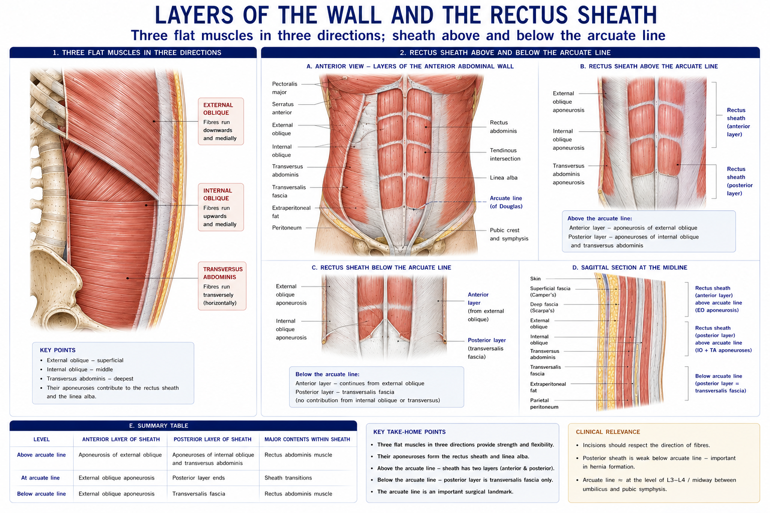
Figure 3 · Three muscles, three fibre directions
| Muscle | Fibre direction | Attachments | Nerve | Action |
|---|---|---|---|---|
| External oblique | Downwards and forwards (“hands in pockets”) | Outer surfaces of ribs 5–12 → iliac crest, pubic tubercle, linea alba; its lower border folds back as the inguinal ligament | T7–T11 (thoracoabdominal), T12 (subcostal) | Flexion; contralateral rotation; ipsilateral lateral flexion; compression |
| Internal oblique | Upwards and forwards (at right angles to external) | Thoracolumbar fascia, iliac crest, lateral inguinal ligament → ribs 10–12, linea alba, conjoint tendon | T7–T12, L1 (iliohypogastric, ilioinguinal) | Flexion; ipsilateral rotation; lateral flexion; compression |
| Transversus abdominis | Transverse | Costal cartilages 7–12, thoracolumbar fascia, iliac crest, lateral inguinal ligament → linea alba, conjoint tendon | T7–T12, L1 | Compression — the principal generator of intra-abdominal pressure; tensions the thoracolumbar fascia |
| Rectus abdominis | Vertical | Pubic crest and symphysis → xiphoid process and costal cartilages 5–7; crossed by three (or more) tendinous intersections | T7–T12 | Trunk flexion; posterior pelvic tilt; compression |
| Pyramidalis | — | Pubis → linea alba; absent in ~20% | T12 | Tenses the linea alba |
Trunk rotation is produced by a force couple: the external oblique of one side with the internal oblique of the other (Chapter 6).
Note the parallel with the intercostals: external oblique continues the direction of external intercostal, internal oblique that of internal intercostal, and transversus abdominis that of innermost intercostal. They are the same three layers, continued into the abdomen — which also explains the segmental thoracoabdominal innervation.
Formed by the aponeuroses of the three flat muscles, and its composition changes at the arcuate line, roughly midway between the umbilicus and the pubis:
| Above the arcuate line | Below the arcuate line | |
|---|---|---|
| Anterior wall | External oblique aponeurosis + anterior lamina of internal oblique | All three aponeuroses |
| Posterior wall | Posterior lamina of internal oblique + transversus abdominis aponeurosis | Absent — only transversalis fascia lies behind rectus |
Consequences:
Contents of the sheath: rectus abdominis, pyramidalis, the superior epigastric (from the internal thoracic) and inferior epigastric (from the external iliac) vessels — which anastomose within it, providing a collateral route between the subclavian and external iliac systems in aortic coarctation — plus the terminal parts of the lower six thoracic nerves.
The linea alba is the midline raphe of interlacing aponeurotic fibres from xiphoid to pubis — relatively avascular, hence the surgical midline incision, and the site of divarication (diastasis) recti.
The linea semilunaris is the lateral border of rectus abdominis, curving from the 9th costal cartilage to the pubic tubercle — the site of the rare Spigelian hernia.
The abdominal cavity behaves as a pressurised cylinder:
| Wall | Structure |
|---|---|
| Roof | Diaphragm |
| Floor | Pelvic floor |
| Front and sides | Transversus abdominis and the obliques |
| Back | Lumbar spine, multifidus and the thoracolumbar fascia |
Co-contraction of these four raises intra-abdominal pressure (IAP), which:
Coordination matters more than strength. The four walls must act together: if the pelvic floor does not co-contract while IAP rises, the pressure is transmitted downwards, which is the mechanism linking heavy lifting, chronic cough and constipation to pelvic organ prolapse and stress urinary incontinence. Conversely, an over-braced abdominal wall with a poorly coordinated pelvic floor is a recognised pattern in pelvic pain.
Forced expiration against a closed glottis raises IAP and intrathoracic pressure markedly, increasing trunk stiffness — which is why powerlifters use it deliberately.
But it also reduces venous return, drops cardiac output and then causes a sharp rebound rise in blood pressure. It is therefore contraindicated in cardiac rehabilitation, uncontrolled hypertension, and after abdominal or ophthalmic surgery, and it must be actively taught against in patients with pelvic floor dysfunction, hernia or recent abdominal repair. “Breathe out on effort” is not folklore; it is this physiology.
Diastasis recti abdominis (DRA) — separation of the recti at the linea alba, present in the great majority of women in late pregnancy and persisting in around a third at 12 months.
The contemporary understanding has shifted in an important way: the inter-recti distance matters less than the ability of the linea alba to generate tension. A narrow but slack linea alba may function worse than a wider taut one. Consequently:
Figure 4 · The inguinal canal and the two inguinal hernias
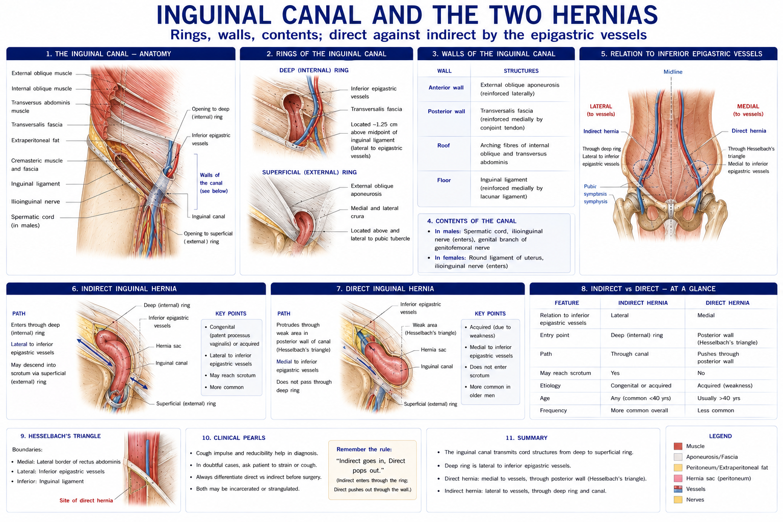
An oblique passage, ~4 cm long, above the medial half of the inguinal ligament — the anatomical price paid for the descent of the testis.
| Boundary | Structure |
|---|---|
| Anterior wall | External oblique aponeurosis throughout; reinforced laterally by internal oblique |
| Posterior wall | Transversalis fascia throughout; reinforced medially by the conjoint tendon |
| Roof | Arching fibres of internal oblique and transversus abdominis |
| Floor | Inguinal ligament, with the lacunar ligament medially |
| Deep ring | An opening in the transversalis fascia, midway between the ASIS and the pubic symphysis (the mid-inguinal point is where the femoral pulse is felt — a different point) |
| Superficial ring | A triangular defect in the external oblique aponeurosis, just above and lateral to the pubic tubercle |
Contents: in the male, the spermatic cord (vas deferens, testicular artery, pampiniform plexus, genital branch of the genitofemoral nerve, and the three fascial coverings); in the female, the round ligament of the uterus. In both, the ilioinguinal nerve enters through the side wall and exits through the superficial ring.
The two protective mechanisms are worth knowing because they explain why the canal usually does not herniate: the obliquity of the canal (so raised IAP presses the anterior and posterior walls together) and the shutter mechanism (contraction of internal oblique and transversus lowers the arching roof onto the floor).
| Type | Relation to the inferior epigastric vessels | Relation to the pubic tubercle | Notes |
|---|---|---|---|
| Indirect inguinal | Lateral | Above and medial | Through the deep ring, along the canal; congenital (patent processus vaginalis); commonest overall, and commoner in the young |
| Direct inguinal | Medial | Above and medial | Through the weak posterior wall (Hesselbach’s triangle: bounded by the inferior epigastric vessels, the lateral border of rectus, and the inguinal ligament); acquired; older men |
| Femoral | — | Below and lateral | Through the femoral canal; commoner in women; highest risk of strangulation because of the sharp lacunar ligament |
The pubic tubercle relationship is the single most useful bedside discriminator, and it is one of the anatomical facts most likely to be tested.
Figure 5 · The peritoneum in section
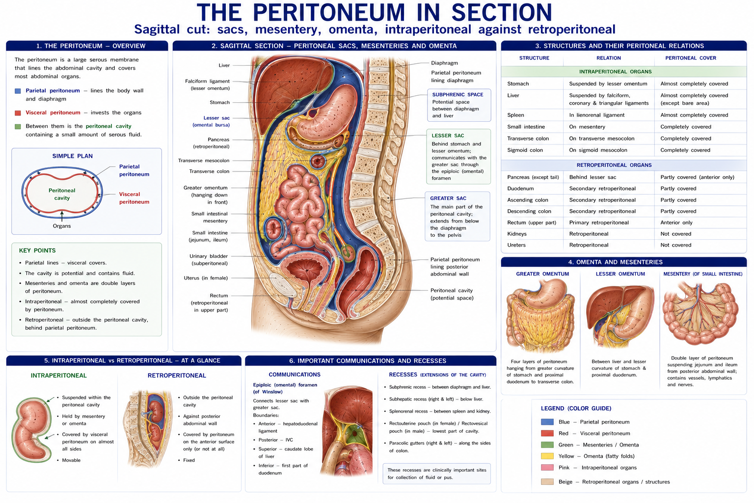
A serous membrane with parietal (lining the wall) and visceral (covering organs) layers, enclosing the peritoneal cavity — the largest serous cavity in the body.
Innervation, and why it matters clinically:
| Layer | Innervation | Pain character |
|---|---|---|
| Parietal peritoneum | Somatic — segmental nerves of the overlying wall; the central diaphragmatic part by the phrenic (C3–C5) | Sharp, severe, WELL LOCALISED; provokes guarding and rebound tenderness |
| Visceral peritoneum | Autonomic (visceral afferents) | Dull, poorly localised, referred to the midline according to embryological gut origin |
This is the anatomy of appendicitis. Early inflammation irritates the visceral peritoneum of a midgut structure, so the pain is felt vaguely in the periumbilical (T10) region. When inflammation reaches the parietal peritoneum of the right iliac fossa, the pain becomes sharp, localised and associated with guarding at McBurney’s point. The migration of the pain is the diagnosis.
| Category | Definition | Examples |
|---|---|---|
| Intraperitoneal | Almost completely covered by visceral peritoneum, suspended by a mesentery | Stomach, jejunum, ileum, transverse colon, sigmoid colon, appendix, spleen, liver (mostly), tail of pancreas |
| Retroperitoneal (primary) | Developed and remained behind the peritoneum | Kidneys, ureters, suprarenal glands, aorta, IVC, lower rectum |
| Retroperitoneal (secondary) | Developed intraperitoneally, then fused to the posterior wall | Duodenum (parts 2–4), pancreas (except tail), ascending and descending colon |
Mnemonic for retroperitoneal structures: SAD PUCKER — Suprarenal, Aorta and IVC, Duodenum (2nd–4th), Pancreas (except tail), Ureters, Colon (ascending and descending), Kidneys, Esophagus (lower), Rectum.
| Organ | Position and surface projection | Key clinical points |
|---|---|---|
| Stomach | Left hypochondrium and epigastrium; cardia at T11, pylorus at the transpyloric plane (L1) | Referred pain: epigastrium (T6–T9) |
| Duodenum | C-shaped around the pancreatic head; parts 2–4 retroperitoneal; duodenojejunal flexure at L2, suspended by the ligament of Treitz | Posterior duodenal ulcer erodes the gastroduodenal artery |
| Jejunum and ileum | Intraperitoneal, ~6 m | Referred pain: periumbilical (T10) |
| Large intestine | Caecum (RIF) → ascending → hepatic flexure → transverse → splenic flexure (higher and more posterior) → descending → sigmoid → rectum | Splenic flexure syndrome — trapped gas causing left upper quadrant and left shoulder pain |
| Appendix | Base at McBurney’s point — one-third along the line from the ASIS to the umbilicus; tip position variable, retrocaecal in ~65%, pelvic in ~30% | A retrocaecal appendix irritates psoas → positive psoas sign, and pain on hip extension. A pelvic appendix irritates the obturator internus → obturator sign |
| Liver | Right hypochondrium and epigastrium, under ribs 7–11; the upper border reaches the 5th intercostal space | Referred pain: right shoulder tip if the diaphragmatic surface is involved |
| Gallbladder | At the tip of the 9th costal cartilage, where the lateral border of rectus crosses the costal margin | Referred pain: right hypochondrium and the inferior angle of the right scapula (T7–T9); Murphy’s sign |
| Pancreas | Retroperitoneal, along the transpyloric plane; head in the duodenal C, tail to the splenic hilum | Pain bores through to the back (T6–T10), classically relieved by leaning forward — a pattern very frequently misread as thoracic spine pain |
| Spleen | Left hypochondrium, along the long axis of the 10th rib, deep to ribs 9–11 | “1, 3, 5, 7, 9, 11” rule: 1 × 3 × 5 inches, 7 oz, ribs 9–11. Not normally palpable — a palpable spleen is enlarged threefold. Rupture → left shoulder-tip pain (Kehr’s sign) |
| Kidneys | Retroperitoneal, T12–L3; the right is lower (liver); hila at L1; move ~2–3 cm with respiration | Referred pain: loin, flank, and radiating to the groin (T10–L1); renal angle tenderness between the 12th rib and the lateral border of erector spinae |
| Ureters | On psoas major, crossing the bifurcation of the common iliac artery at the pelvic brim; three constrictions (pelviureteric junction, pelvic brim, vesicoureteric junction) | Loin-to-groin colic, radiating to the testis or labium (T11–L2) |
| Suprarenal glands | On the upper poles of the kidneys | Cortex mesodermal, medulla neural crest (Chapter 8) |
| Abdominal aorta | T12 to its bifurcation at L4 (supracristal plane) | AAA — see red flags below |
| Type | Branches |
|---|---|
| Unpaired (midline, to the gut) | Coeliac trunk (T12) — foregut; superior mesenteric artery (L1) — midgut; inferior mesenteric artery (L3) — hindgut |
| Paired lateral (visceral) | Suprarenal (L1), renal (L1–L2), gonadal (L2) |
| Paired posterior (parietal) | Inferior phrenic, four lumbar arteries |
| Terminal | Common iliac arteries at L4, plus the median sacral |
The gut is supplied by three arteries corresponding to three embryological divisions, and this determines both the anastomotic watershed areas (the splenic flexure and rectosigmoid junction — the classic sites of ischaemic colitis) and the pattern of referred pain:
| Division | Artery | Extent | Referred pain |
|---|---|---|---|
| Foregut | Coeliac | Oesophagus to the mid-duodenum, plus liver, gallbladder, pancreas, spleen | Epigastrium (T6–T9) |
| Midgut | Superior mesenteric | Mid-duodenum to the proximal two-thirds of the transverse colon | Periumbilical (T10) |
| Hindgut | Inferior mesenteric | Distal transverse colon to the upper rectum | Hypogastrium (T11–L1) |
The hepatic portal vein is formed behind the neck of the pancreas by the union of the superior mesenteric and splenic veins, carrying nutrient-rich blood from the gut to the liver.
Portosystemic anastomoses become clinically important in portal hypertension, when portal blood is forced through them:
| Site | Anastomosis | Clinical result |
|---|---|---|
| Lower oesophagus | Left gastric ↔ oesophageal veins | Oesophageal varices — the major cause of fatal haemorrhage |
| Anal canal | Superior rectal ↔ middle and inferior rectal | Haemorrhoids |
| Umbilicus | Paraumbilical ↔ superficial epigastric | Caput medusae |
| Retroperitoneal | Colic ↔ retroperitoneal veins (of Retzius) | Rarely symptomatic |
| Muscle | Attachments | Nerve | Action |
|---|---|---|---|
| Psoas major | T12–L5 bodies, discs and transverse processes → lesser trochanter | Anterior rami L1–L3 directly | Hip flexion; lumbar spine stabilisation and (with a fixed femur) lumbar flexion or lateral flexion |
| Psoas minor | T12–L1 → pecten pubis; absent in ~40% | L1 | Weak trunk flexion |
| Iliacus | Iliac fossa → lesser trochanter | Femoral (L2, L3) | Hip flexion |
| Quadratus lumborum | Iliac crest and iliolumbar ligament → 12th rib and L1–L4 transverse processes | T12, L1–L4 | Lateral flexion; fixes the 12th rib during inspiration (an accessory respiratory muscle); hip hitching |
| Diaphragm | See Chapter 18 | Phrenic | Respiration |
Psoas is worth pausing on. Its direct segmental innervation from the anterior rami, its attachment to every lumbar vertebra and disc, and its line of pull mean it is simultaneously the strongest hip flexor and a significant lumbar compressive and shear force generator. A psoas abscess (tuberculous or pyogenic — still seen in India) tracks down within the psoas sheath to present as a groin or thigh swelling with a flexion deformity of the hip and pain on passive extension — a presentation that reaches physiotherapy clinics misdiagnosed as a hip flexor strain.
| Nerve | Roots | Supplies |
|---|---|---|
| Iliohypogastric | L1 | Lower abdominal wall; suprapubic skin |
| Ilioinguinal | L1 | Lower abdominal wall; skin of the groin, upper medial thigh, and external genitalia |
| Genitofemoral | L1, L2 | Cremaster (genital branch); skin of the femoral triangle (femoral branch) |
| Lateral femoral cutaneous | L2, L3 | Anterolateral thigh — meralgia paraesthetica |
| Obturator | L2–L4 | Adductor compartment; medial thigh; articular branches to hip AND knee |
| Femoral | L2–L4 | Anterior thigh compartment; saphenous nerve |
| Lumbosacral trunk | L4, L5 | To the sacral plexus |
Iliohypogastric and ilioinguinal nerve entrapment after appendicectomy, hernia repair, Pfannenstiel incision or Caesarean section is a common and under-recognised cause of chronic groin and lower abdominal pain — and it is a genuinely treatable one.
Figure 6 · Referred pain from the three parts of the gut
This is the practical heart of the chapter for a physiotherapist.
| Organ | Refers to | Segments | Features that distinguish it from musculoskeletal pain |
|---|---|---|---|
| Heart | Central chest, left arm and medial forearm, jaw, interscapular | T1–T5 | Exertional, not positional; sweating, nausea, dyspnoea |
| Diaphragm (central) | Shoulder tip | C3–C5 | Not reproducible on shoulder testing |
| Gallbladder | Right hypochondrium, inferior angle of the right scapula | T7–T9 | Relation to fatty meals; Murphy’s sign |
| Liver | Right shoulder, right upper quadrant | C3–C5, T7–T9 | Jaundice, hepatomegaly |
| Stomach / duodenum | Epigastrium, mid-back (T6–T10) | T6–T9 | Relation to meals; night pain relieved by food (duodenal) |
| Pancreas | Epigastrium boring through to the back, left more than right | T6–T10 | Relieved by leaning forward, worse lying flat; often mistaken for thoracic spine pain |
| Spleen | Left shoulder tip (Kehr’s sign) | C3–C5 | Trauma history; hypotension |
| Kidney | Loin and flank, radiating anteriorly | T10–L1 | Renal angle tenderness; haematuria; fever |
| Ureter | Loin to groin, testis or labium | T11–L2 | Colicky, writhing rather than still; unable to find a comfortable position |
| Appendix | Periumbilical → right iliac fossa | T10 → somatic | The migration is the diagnosis |
| Colon | Lower abdomen, sacrum | T11–L1 | Relation to bowel habit |
| Bladder / prostate / uterus | Suprapubic, sacrum, perineum | T11–L2, S2–S4 | Urinary or menstrual relation |
| Abdominal aorta | Low back, deep and boring, flank, abdomen | T12–L2 | See below |
Abdominal aortic aneurysm. Deep, boring, non-mechanical low back or abdominal pain in a patient over 60, usually male, with a smoking history, hypertension or known vascular disease. A pulsatile, expansile abdominal mass (expansile is the key word — a transmitted pulsation is not the same). Pain unrelated to position or movement, often worse at night. A ruptured AAA presents as sudden severe back or abdominal pain with collapse — and it is a not-uncommon cause of a patient dying after presenting to a physiotherapist with “back pain”.
Other red flags:
The general rule again: musculoskeletal pain has a mechanical behaviour. Pain that cannot be provoked or eased by any position, movement or palpation is not musculoskeletal until proved otherwise.
| Consideration | Detail |
|---|---|
| Respiratory | Upper abdominal incisions reduce FRC and vital capacity by 25–50% and impair cough. Early mobilisation, positioning, supported cough (a pillow or rolled towel over the wound) and deep breathing reduce post-operative pulmonary complications |
| Wound and fascial healing | Fascial strength follows the connective tissue timeline (Chapter 7): ~50–60% at 6 weeks, plateauing at 70–80% over months. Graded, not prohibited, loading |
| Log-rolling and transfers | Reduce direct abdominal wall loading in the early phase |
| Adhesions | Peritoneal healing produces fibrous adhesions in most laparotomies; they are the leading cause of small bowel obstruction and of chronic post-surgical pain |
| Progressive loading | Return to lifting is graded by symptoms and tissue timeline, with attention to breathing pattern and avoiding Valsalva |
The abdominal wall lengthens, the linea alba widens, the diaphragm is displaced upwards ~4 cm (with a compensatory increase in the transverse thoracic diameter), lumbar lordosis increases and the centre of mass shifts forwards. Postnatal rehabilitation addresses the entire canister — diaphragm, abdominal wall, pelvic floor and lumbar musculature — with graded load and breathing coordination, rather than isolated exercises or blanket restrictions.
Physiotherapists should recognise the presentations, understand the mechanics (raised IAP, weak posterior wall, chronic cough or constipation as drivers), and be able to teach a safe breathing and loading strategy post-repair. A tender, irreducible, painful hernia with vomiting is a strangulated hernia — a surgical emergency.
15 questions on this chapter. Tap one to see the answer and the reasoning.
Answer: (B) It marks the pylorus, pancreatic neck, renal hila, SMA origin and the end of the spinal cord.
Answer: (C) All three aponeuroses pass in front of rectus there.
Answer: (B) A contralateral force couple.
Answer: (C)
Answer: (B) It carries the highest strangulation risk.
Answer: (B) Through the deep ring.
Answer: (B)
Answer: (B) Foregut → epigastrium; hindgut → hypogastrium.
Answer: (B)
Answer: (B) Via diaphragmatic irritation and C3–C5.
Answer: (B) It is frequently misdiagnosed as thoracic spinal pain.
Answer: (B) The right kidney lies slightly lower than the left.
Answer: (C) Iliacus, by contrast, is femoral.
Answer: (B) Non-mechanical behaviour is the key.
Answer: (C)
Everything on this page, in one screen
| Source | Why it earns its place |
|---|---|
| Standring S (ed.) — Gray’s Anatomy, 42nd edn | Definitive abdominal anatomy |
| Moore KL, Dalley AF, Agur AMR — Clinically Oriented Anatomy | The best clinical treatment of the abdominal wall, inguinal canal and peritoneum |
| Snell RS — Clinical Anatomy by Regions | Regional clinical correlation and surface anatomy |
| Goodman CC, Snyder TEK — Differential Diagnosis for Physical Therapists: Screening for Referral | The single most useful book for the screening content of this chapter |
| Boissonnault WG — Primary Care for the Physical Therapist: Examination and Triage | Red flags and systems screening |
| Hodges PW, Cholewicki J, van Dieën JH — Spinal Control: The Rehabilitation of Back Pain | Intra-abdominal pressure and trunk stiffness |
| Lee D, Hodges PW — “Behavior of the linea alba during a curl-up task in diastasis rectus abdominis”, J Orthop Sports Phys Ther, 2016 | The evidence reframing DRA management |
| Benjamin DR, van de Water ATM, Peiris CL — “Effects of exercise on diastasis of the rectus abdominis muscle”, Physiotherapy, 2014 | Exercise and DRA prevalence |
| Chaurasia BD — Human Anatomy, Vol 2: Lower Limb, Abdomen and Pelvis | Indian syllabus-matched descriptive account |
Chapter 20 of 24 · Human Anatomy · Physiotherapist India Next: Chapter 21 — Pelvis and Endocrine Glands: the pelvic floor, and the glands that regulate the body.
Reviewed by the Physiotherapist India Team. · Human Anatomy contents