Functional Electrical Stimulation (FES): Principles, Clinical Applications, and Rehabilitation Perspectives
Functional Electrical Stimulation (FES) is an evidence-based neuromodulatory technique that uses controlled electrical impulses to elicit muscle contractions in individuals with neurological or musculoskeletal impairments. This article explores the physiological principles, clinical applications, indications, contraindications, procedural considerations, therapeutic benefits, and limitations of FES in the rehabilitation context. The scope encompasses spinal cord injury, stroke, cerebral palsy, and other neurological disorders, supported by current literature and clinical guidelines.
1. Introduction
Functional Electrical Stimulation (FES) is a rehabilitative modality wherein low-energy electrical pulses are applied to motor nerves or muscles to restore or augment voluntary motor function lost due to central nervous system (CNS) damage or disease. It differs from traditional neuromuscular electrical stimulation (NMES) by being task-specific and aiming to produce purposeful movements such as walking, grasping, or standing, thereby integrating motor relearning principles and promoting neuroplasticity (1,2).
2. Physiological Basis of FES
FES operates by delivering brief electrical impulses (typically in the range of 20–50 Hz) to peripheral nerves, leading to depolarization of motor axons and generation of action potentials that result in muscle contractions. The induced contractions mimic voluntary movement and, over time, facilitate neuromuscular re-education through repetitive activation of cortical and subcortical motor circuits (3). This is aligned with Hebbian plasticity principles—“neurons that fire together wire together”—supporting its role in functional recovery after CNS injuries (4).
3. Indications and Clinical Applications
3.1 Neurological Disorders Treated with FES
FES is indicated in various neurological and neuromuscular conditions where voluntary muscle activation is impaired but lower motor neurons remain intact. Major clinical applications include:
Condition | Clinical Use of FES |
Spinal Cord Injury (SCI) | Restoration of hand function, ambulation, bladder/bowel control, and respiratory support (5) |
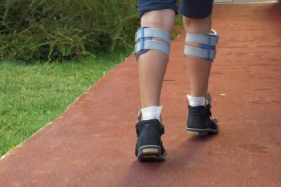
Stroke | Management of foot drop, upper limb functional recovery, shoulder subluxation, and spasticity (6) |
Multiple Sclerosis | Enhancement of ambulation and reduction of fatigue (7) |
Cerebral Palsy | Improvement of gait and upper limb motor control (8) |
Traumatic Brain Injury | Functional restoration in hemiplegia and spastic paresis (9) |
4. Mechanisms of Action and Functional Outcomes
FES promotes motor function through multiple pathways:
- Neuromuscular Re-education: Stimulated muscle contractions facilitate re-learning of movement patterns.
- Spasticity Management: Reciprocal inhibition of antagonistic muscles through repeated stimulation reduces hypertonicity (10).
- Cardiorespiratory Benefits: FES-assisted cycling enhances aerobic fitness and reduces cardiovascular deconditioning in SCI (11).
- Bone Health: Muscle contractions via FES increase mechanical load on bones, mitigating disuse osteoporosis (12).
- Pain Modulation: FES may inhibit nociceptive transmission via gate control theory (13).
5. Components and Types of FES Systems
FES systems may be categorized into surface, percutaneous, or implanted systems based on electrode placement:
Type | Description | Clinical Use |
Surface FES | Electrodes adhered to the skin using conductive gel or adhesive pads | Early rehabilitation; cost-effective and non-invasive |
Percutaneous FES | Needle electrodes inserted through the skin for more focused stimulation | Short-term use in research or specific therapies |
Implanted FES | Electrodes surgically implanted near peripheral nerves or spinal roots | Long-term solutions in chronic SCI or CNS injury (14) |
Core components of an FES system include:
- Stimulator Unit: Delivers controlled electrical pulses.
- Electrodes: Deliver current to target muscles.
- Sensors: Detect limb position or gait phase (in advanced systems).
- Control Interface: May include manual switches, EMG signals, or brain-computer interface (15).
6. Indications and Patient Selection
6.1 Inclusion Criteria
Patients considered for FES typically meet the following criteria:
- Presence of upper motor neuron lesion.
- Intact peripheral motor pathways.
- Residual voluntary control or potential for functional use.
- Motivation and cognitive capacity to participate in therapy.
6.2 Contraindications
Absolute and relative contraindications include (16):
- Implanted cardiac pacemakers or defibrillators.
- Uncontrolled epilepsy.
- Active thrombophlebitis.
- Osteomyelitis or active infection at stimulation site.
- Severe osteoporosis or fractures.
- Pregnancy (relative contraindication).
A comprehensive clinical evaluation including sensory, motor, and cognitive assessments is essential prior to initiation.
7. Procedure and Dosimetry
FES treatment is individualized. Parameters are chosen based on desired outcome, muscle size, and tolerance:
- Pulse Width: 200–400 µs.
- Frequency: 20–50 Hz (higher frequencies may cause fatigue).
- Amplitude: Set to produce functional muscle contraction without pain.
- On/Off Ratio: Adjusted to avoid muscle fatigue (commonly 1:2 to 1:4) (17).
Electrode placement should be optimized for targeted muscle activation and verified through palpation, visual inspection, or electromyographic monitoring.
8. Benefits and Evidence-Based Outcomes
Numerous systematic reviews have demonstrated the effectiveness of FES in restoring motor function and improving quality of life:
Outcome | Evidence Summary |
Improved gait velocity in stroke | Moderate evidence supports FES over conventional therapy for foot drop (18) |
Upper limb function post-stroke | FES combined with task-specific training improves motor scores on Fugl-Meyer and ARAT (19) |
SCI recovery | Long-term use of FES increases muscle bulk, bone density, and functional independence (20) |
Reduced spasticity | FES induces reciprocal inhibition and enhances voluntary control (21) |
9. Risks and Limitations
Despite its benefits, FES is not without potential adverse effects:
- Skin irritation or burns due to improper electrode placement.
- Muscle fatigue with high-frequency stimulation.
- Discomfort or pain in individuals with low tolerance to sensory stimuli.
- Infections (in case of implanted systems).
- Mechanical failure of implanted leads or battery depletion.
Regular follow-up and reassessment mitigate these risks.
10. Future Directions
Recent advancements in neuroengineering are integrating FES with wearable sensors, robotic exoskeletons, and brain-computer interfaces (BCIs), enhancing its precision and functional outcomes. Closed-loop systems using real-time feedback and AI-driven stimulation protocols show promise for individualized therapy and enhanced neuroplasticity (22,23).
Functional Electrical Stimulation represents a powerful modality in neurorehabilitation, harnessing electrical currents to restore purposeful movement and enhance patient independence. While not universally applicable, its clinical value in spinal cord injury, stroke, and other neurological disorders is well-established. Future integration with neurotechnologies may further refine its applications and outcomes.
References
- Sheffler LR, Chae J. Neuromuscular electrical stimulation in neurorehabilitation. Muscle Nerve. 2007;35(5):562–590.
- Peckham PH, Knutson JS. Functional electrical stimulation for neuromuscular applications. Annu Rev Biomed Eng. 2005;7:327–360.
- Rushton DN. Functional electrical stimulation and rehabilitation—an hypothesis. Med Eng Phys. 2003;25(1):75–78.
- Kleim JA, Jones TA. Principles of experience-dependent neural plasticity. J Speech Lang Hear Res. 2008;51(1):S225–239.
- van Middendorp JJ, Hosman AJF, Doi SA, et al. A clinical prediction rule for ambulation outcomes after traumatic spinal cord injury. Lancet. 2011;377(9770):1004–1010.
- Eraifej J, Clark W, France B, Desando S, Moore D. Effectiveness of upper limb functional electrical stimulation after stroke: A systematic review and meta-analysis of the evidence. Clin Rehabil. 2017;31(4):429–440.
- Stein RB, Everaert DG, Thompson AK, et al. Long-term therapeutic and orthotic effects of a foot drop stimulator on walking performance in progressive multiple sclerosis. Neurorehabil Neural Repair. 2010;24(2):152–167.
- Pool D, Valentine J, Bear N, et al. Functional electrical stimulation as an adjunct to botulinum toxin-A treatment in children with cerebral palsy: A randomized controlled trial. Dev Med Child Neurol. 2020;62(2):237–243.
- Daly JJ, Ruff RL. Electrically induced recovery of walking in stroke patients. Arch Phys Med Rehabil. 2000;81(9):1174–1185.
- Thompson AK, Stein RB. Short-term effects of functional electrical stimulation on spinal excitability in spinal cord injury. Exp Brain Res. 2004;159(3):384–393.
- Nash MS, Jacobs PL, Montalvo BM, Klose KJ, Guest RS, Needham-Shropshire BM. Evaluation of a training program for persons with SCI paraplegia using an FES cycle ergometer. Spinal Cord. 2004;42(12):657–665.
- Dudley-Javoroski S, Shields RK. Muscle and bone plasticity after spinal cord injury: Review of adaptations to disuse and to electrical muscle stimulation. J Rehabil Res Dev. 2008;45(2):283–296.
- Johnson MI, Bjordal JM. Transcutaneous electrical nerve stimulation for the management of painful conditions: Focus on neuropathic pain. Expert Rev Neurother. 2011;11(5):735–753.
- Kilgore KL, Hoyen HA, Bryden AM, Hart RL, Keith MW, Peckham PH. An implanted upper-extremity neuroprosthesis using myoelectric control. J Hand Surg Am. 2008;33(4):539–550.
- Bhadra N, Kilgore KL. High-frequency electrical conduction block of mammalian peripheral motor nerve. Muscle Nerve. 2005;32(6):782–790.
- Glanz M, Klawansky S, Stason W, Berkey C, Chalmers TC. Functional electrostimulation in poststroke rehabilitation: A meta-analysis of the randomized controlled trials. Arch Phys Med Rehabil. 1996;77(6):549–553.
- Hara Y. Neurorehabilitation with new functional electrical stimulation for hemiparetic upper extremity in stroke patients. J Neuroeng Rehabil. 2004;1(1):15.
Visit Our Other Resources
You my would like to visit our other resources