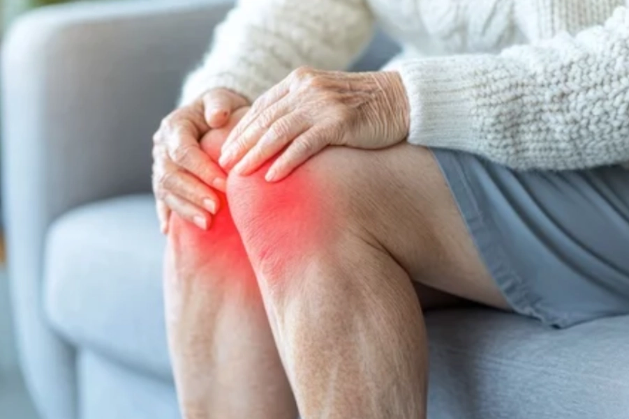
Knee pain can be caused by a variety of conditions with different underlying pathophysiology. For osteoarthritis, it presents with gradual degeneration and functional limitations, while rheumatoid arthritis leads to symmetric inflammation and progressive deformities.
An ACL tear typically follows trauma, and meniscus tears often occur with twisting motions, presenting with mechanical symptoms like locking. Tendinitis involves pain from overuse and repetitive activity, while bursitis may occur due to friction or direct injury.
Treatment for these conditions varies significantly, with more severe cases often requiring surgical interventions, particularly for ACL tears and advanced OA.
Below is a comparative table for the differential diagnosis of Knee Pain in conditions like Osteoarthritis (OA), Rheumatoid Arthritis (RA), Anterior Cruciate Ligament (ACL) Tear, Meniscus Tear, Tendinitis, and Bursitis
| Condition | Osteoarthritis (OA) | Rheumatoid Arthritis (RA) | ACL Tear | Meniscus Tear | Tendinitis | Bursitis |
| Pathophysiology | Degeneration of articular cartilage, osteophyte formation, and synovial inflammation leading to joint space narrowing. | Chronic autoimmune disease causing inflammation in synovial joints, often symmetric. Leads to joint deformity and cartilage damage. | Rupture or sprain of the anterior cruciate ligament, usually caused by traumatic twisting or hyperextension. | Tears in the meniscus (cartilage) of the knee, often caused by twisting or heavy load-bearing. | Inflammation of a tendon, commonly at the knee (e.g., patellar tendinitis), due to overuse or repetitive stress. | Inflammation of the bursae (fluid-filled sacs) around the knee due to friction, injury, or prolonged pressure. |
| Onset | Gradual, often after age 40, due to wear and tear on the joint. | Insidious, often starting in middle age, with gradual and progressive joint swelling and stiffness. | Acute onset following a specific trauma (e.g., sports injury or sudden deceleration). | Acute onset, often after twisting, squatting, or lifting. | Gradual onset, typically from overuse, or repetitive activities (e.g., running or jumping). | Gradual or acute onset, often due to direct trauma or overuse. |
| Pain | Dull, aching pain that worsens with activity and improves with rest. Often worse after prolonged weight-bearing. | Symmetric joint pain, worse in the morning or after inactivity, with stiffness and swelling. | Sudden, sharp pain at the time of injury, especially with twisting or pivoting movements. Pain is often localized to the knee. | Pain with twisting, bending, or squatting. Locking or catching sensations may be felt in the knee. | Aching pain with activity, especially at the front of the knee (for patellar tendinitis) or the back (for hamstring tendinitis). | Pain localized around the bursa, which worsens with pressure or movement. |
| Range of Motion | Limited, especially with flexion and extension. Stiffness and crepitus are common. | Decreased range of motion due to inflammation and joint destruction. | Restricted due to pain, especially with pivoting or weight-bearing activities. | Limited range of motion with bending and twisting. A “locking” sensation may be felt. | Range of motion may be limited by pain, especially with activity or weight-bearing. | Limited range of motion due to pain and swelling around the bursa. |
| Swelling | Mild to moderate, often intermittent and associated with activity. | Significant, often symmetric and associated with morning stiffness. | Swelling after the injury, often accompanied by hemarthrosis (blood in the joint). | Swelling localized to the knee, especially along the joint line. May have joint effusion. | Swelling localized over the affected tendon. | Swelling localized over the bursa, especially around the knee. |
| Tenderness | Tenderness over the joint line, especially medially and laterally. | Tenderness over multiple joints, especially in the morning. | Tenderness over the ACL or at the femoral-tibial joint. | Tenderness along the joint line, especially on the medial or lateral side. | Tenderness localized to the tendon, especially at its origin or insertion. | Tenderness localized over the bursa (prepatellar, infrapatellar, or pes anserine). |
| Imaging | X-rays show joint space narrowing, osteophytes, and subchondral sclerosis. | X-rays may show joint space narrowing, but MRI is required for early detection of joint erosion and inflammation. | MRI is the gold standard to visualize the ACL tear. X-rays may be normal but can show associated fractures. | MRI is the best imaging tool to identify meniscus tears. X-rays may show no abnormalities. | Ultrasound and MRI are helpful for diagnosing tendon inflammation and tears. | Ultrasound and MRI can show inflammation and swelling of the bursa. |
| Special Tests | None specific, but crepitus and positive “knee bending” tests may be observed. | Rheumatoid factor test and anti-CCP antibody test are diagnostic. Physical exam shows joint deformity and synovial thickening. | Positive Lachman’s test, anterior drawer test, or pivot shift test. | Positive McMurray’s test, Apley’s compression test, or joint line tenderness. | Positive pain with resisted movement of the affected tendon. | Positive pain with direct pressure over the bursa (e.g., prepatellar bursitis test). |
| Functional Limitations | Difficulty with weight-bearing activities, stairs, and prolonged standing. | Difficulty with daily activities, including walking, climbing stairs, and prolonged sitting. | Limited ability to pivot, decelerate, or perform cutting movements. | Difficulty with movements that involve twisting, squatting, or bending. | Difficulty with activities that involve knee extension or repetitive movement (e.g., running, jumping). | Limited function due to pain with kneeling or squatting. |
| Treatment | Conservative management: NSAIDs, physical therapy, joint injections, and weight management. Surgical options include arthroscopy or knee replacement. | Disease-modifying antirheumatic drugs (DMARDs), NSAIDs, physical therapy, joint injections, and potentially joint replacement. | Rest, ice, knee brace, and surgery (e.g., ACL reconstruction) if necessary. | Rest, ice, physical therapy, and surgery (e.g., arthroscopy, meniscectomy) if needed. | Rest, ice, NSAIDs, physical therapy, and corticosteroid injections. | Rest, ice, NSAIDs, corticosteroid injections, and possibly surgical drainage if septic. |
| Prognosis | Chronic, progressive disease. Early-stage OA can be managed with conservative measures, but advanced stages may require surgery. | Chronic, progressive disease. Early treatment can control symptoms and slow progression. Joint damage is irreversible. | Good with appropriate rehabilitation. Surgery (ACL reconstruction) may be required for long-term stability. | Generally good with physical therapy and conservative measures. Surgery may be required for severe or irreparable tears. | Good prognosis with rest and conservative treatment. Chronic tendinitis may require more advanced interventions. | Prognosis depends on the cause. Non-septic bursitis typically resolves with conservative treatment. Septic bursitis requires more aggressive treatment. |
Literature References:
- Osteoarthritis (OA):
- Felson, D. T., & Lawrence, R. C. (2023). Osteoarthritis of the knee: Current evidence-based treatment strategies. Current Opinion in Rheumatology, 35(4), 333–340. doi:10.1097/BOR.0000000000000934
- Rheumatoid Arthritis (RA):
- Dumitru, R. L., & Tindall, S. K. (2022). Rheumatoid arthritis and its impact on the knee joint: A review. Journal of Clinical Rheumatology, 28(5), 271–277. doi:10.1097/RHU.0000000000001889
- ACL Tear:
- Donnelly, E. D., & Woods, D. M. (2023). The role of ACL reconstruction in the treatment of anterior cruciate ligament tears: A systematic review. Orthopaedic Journal of Sports Medicine, 11(2), 23259671231127329. doi:10.1177/23259671231127329
- Meniscus Tear:
- Khurana, S., & Verma, A. (2023). Management strategies for meniscus tears: A comprehensive review. International Orthopaedics, 47(6), 1301–1309. doi:10.1007/s00264-023-05854-7
- Tendinitis:
- Miller, S. L., & Davenport, S. E. (2022). Tendinitis and tendonopathy: Pathophysiology and clinical management. Sports Medicine, 52(8), 1835–1844. doi:10.1007/s40279-022-01639-z
- Bursitis:
- Liu, J., & Miller, A. T. (2023). Bursitis: Etiology, diagnosis, and treatment. Journal of Orthopaedic & Sports Physical Therapy, 53(5), 275–282. doi:10.2519/jospt.2023.10223
Disclaimer: This content is for informational purposes only. Always consult with a qualified healthcare provider for diagnosis and treatment recommendations tailored to individual needs.


