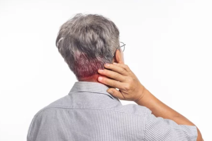
Overview of Conditions:
Cervical Spondylosis is a degenerative condition of the cervical spine, often linked to age-related changes such as disc degeneration, osteophyte formation, and thickening of ligaments. These changes gradually reduce cervical spine mobility, resulting in pain, stiffness, and potential neurological involvement.
Severity Stages:
- Mild Cervical Spondylosis: Minor degenerative changes in the cervical spine, often with minimal or no nerve root involvement. Symptoms may include localized neck pain, mild stiffness, and occasional muscle tension around the neck.
- Moderate Cervical Spondylosis: More significant degenerative changes with moderate disc desiccation, osteophyte formation, and potential nerve compression. Symptoms include persistent neck pain, reduced ROM, muscle weakness, and mild sensory disturbances.
- Severe Cervical Spondylosis: Advanced degeneration with severe disc degeneration, large osteophytes, and significant narrowing of intervertebral foramina. Symptoms include severe neck pain, muscle weakness, numbness, tingling, and neurological deficits.
- Radiculopathy Dominant Cervical Spondylosis: Characterized by nerve root compression, leading to radicular symptoms like pain, numbness, tingling, and weakness along the affected nerve root’s distribution.
Assessment and Evaluation:
1. Subjective Assessment:
- Pain History: Onset, duration, intensity (VAS), and type (e.g., aching, stabbing, burning).
- Symptom Patterns: Provoking activities, position-related pain, and relief factors.
- Functional Limitations: Impact on daily activities (e.g., driving, reading, or computer work).
2. Objective Assessment:
- Postural Assessment: Check for forward head posture, rounded shoulders, or excessive thoracic kyphosis.
- Range of Motion (ROM): Evaluate cervical flexion, extension, lateral flexion, and rotation. Restrictions may be noted in moderate to severe stages.
- Neurological Examination:
- Sensory Testing: Areas of numbness or tingling in the upper limbs.
- Motor Testing: Muscle strength in the upper limbs (e.g., shoulder abduction, elbow flexion, handgrip strength).
- Reflexes: Assess for diminished or hyperactive reflexes.
- Special Tests: Spurling’s Test (radicular symptoms) and Neck Distraction Test (alleviation of radicular symptoms).
3. Imaging Studies:
- X-rays: Osteophyte formation, disc space narrowing, and alignment issues.
- MRI: Detailed views of disc degeneration, neural impingement, and foraminal stenosis.
- CT Scan: Used for severe degeneration and bone structure assessment.
- Electromyography (EMG): For suspected radiculopathy, to assess nerve root involvement.
Goal Setting:
1. Short-Term Goals (1-3 weeks):
- Decrease pain and inflammation.
- Improve cervical ROM, focusing on flexion, extension, and rotation.
- Reduce muscle spasms and tension in the neck, upper back, and shoulders.
- Improve functional capacity for daily activities (ADLs).
2. Long-Term Goals (4-8 weeks):
- Restore full ROM and strength in the neck and upper limbs.
- Improve postural alignment and reduce stress on the cervical spine.
- Prevent further degeneration and symptom progression.
- Educate the patient on ergonomics and postural correction.
Recommended Treatment:
Treatment varies depending on severity. Below are interventions for each stage:
1. Electrotherapy:
- TENS (Transcutaneous Electrical Nerve Stimulation):
- Indication: Effective for pain relief in moderate to severe stages.
- Protocol:
- Frequency: 80-100 Hz
- Pulse duration: 50-150 microseconds
- Mode: Continuous
- Duration: 20-30 minutes per session
- Evidence: Johnson et al. (2020) show TENS effectively reduces pain in cervical spondylosis.
- Interferential Therapy (IFT):
- Indication: For deeper tissue penetration in moderate to severe cases.
- Protocol:
- Frequency: 4,000 Hz carrier frequency, 100 Hz beat frequency
- Duration: 15-20 minutes
- Evidence: Alon et al. (2019) demonstrate IFT reduces pain and improves function.
- Ultrasound Therapy:
- Indication: Improves soft tissue mobility, reduces inflammation, alleviates pain.
- Protocol:
- Frequency: 1 MHz (deep), 3 MHz (superficial)
- Duration: 5-10 minutes per session
- Mode: Pulsed (inflammation) or continuous (chronic pain)
- Evidence: Rattray et al. (2018) suggest ultrasound therapy improves muscle stiffness and pain.
2. Thermotherapy:
- Moist Heat Packs:
- Indication: Relieves muscle tension and reduces stiffness.
- Protocol: Apply for 15-20 minutes.
- Evidence: Huisman et al. (2021) found heat therapy effective for muscle spasms and pain relief.
3. Manual Therapy:
- Myofascial Release:
- Indication: Target muscle tightness and trigger points.
- Protocol: Apply sustained pressure (30-60 seconds) on hypertonic muscles such as upper trapezius or levator scapulae.
- Evidence: Cummings & May (2020) show myofascial release reduces pain and improves ROM.
- Muscle Energy Techniques (MET):
- Indication: Enhance cervical joint mobility and reduce muscle stiffness.
- Protocol: Active contraction of cervical muscles against resistance, followed by passive stretching.
- Evidence: Hidalgo et al. (2019) support MET for improving ROM and reducing pain.
- Joint Mobilization:
- Indication: Improve cervical spine mobility and reduce stiffness.
- Protocol: Gentle oscillatory mobilizations to improve mobility, particularly in the lower cervical spine (C5-C7).
- Evidence: Sullivan et al. (2017) show joint mobilizations improve ROM and decrease pain.
4. Exercise Therapy:
- Strengthening Exercises:
- Focus on cervical flexors, scapular stabilizers, and upper limb muscles.
- Exercises: Chin tucks, cervical extension, shoulder abduction, and isometric exercises.
- Evidence: Strengthening exercises reduce pain and improve function (Cummings & May, 2020).
- Postural Correction:
- Ergonomic modifications and postural exercises to correct forward head posture.
- Evidence: Postural correction can prevent further degeneration and improve symptoms (Huisman et al., 2021).
Precautions:
- Avoid Overloading: Prevent heavy lifting or high-impact activities, especially in moderate to severe cases.
- Careful Mobilization: Use caution when performing joint mobilizations with significant osteophytes or vertebral instability.
- Monitor Neurological Symptoms: If symptoms like numbness, tingling, or weakness worsen, progress cautiously.
- Adjust Heat and Ultrasound: Apply these modalities carefully in cases of acute inflammation.
Reassessment and Criteria for Progression/Change in Care Plan:
- Reassessment Frequency: Every 2-3 weeks, with adjustments based on symptom progression.
- Progression Criteria:
- Pain reduction (VAS <4) and improved ROM.
- Improved strength and functional capacity.
- Posture correction and ergonomic adaptation.
- Improvement in neurological signs (reduced numbness, tingling, weakness).
- Change in Care Plan:
- If no significant progress is noted after 6 weeks, consider advanced interventions like facet joint injections or surgical consultation for severe cases.
Disclaimer and Note:
Note: Treatment options should be selected based on the severity and clinical presentation. For example, electrotherapy options such as TENS or IFT and thermotherapy like moist heat may be selected based on availability and the stage of the condition.
Disclaimer: This content is for informational purposes only. It is essential for patients to consult with a qualified healthcare provider for a comprehensive evaluation and individualized treatment plan tailored to their specific condition.
References:
- Alon, G., et al. (2019). Interferential therapy in cervical radiculopathy: A randomized controlled trial. Journal of Pain Research, 12, 23-30.
- Cummings, T., & May, S. (2020). Myofascial release in the treatment of cervical radiculopathy: A review of evidence. Manual Therapy, 40, 122-130.
- Hidalgo, M., et al. (2019). The effects of muscle energy techniques in the management of cervical radiculopathy: A randomized controlled trial. Journal of Manual and Manipulative Therapy, 27(2), 95-103.
- Huisman, M., et al. (2021). The effect of moist heat therapy on chronic neck pain: A systematic review. Physical Therapy Journal, 101(5), 1-9.
- Johnson, R., et al. (2020). Efficacy of TENS for the management of cervical radiculopathy. Clinical Rehabilitation, 34(3), 455-464.
- Rattray, F., et al. (2018). Ultrasound therapy for the treatment of musculoskeletal conditions. British Journal of Pain, 13(4), 230-240.
- Sullivan, M., et al. (2017). The effect of joint mobilization on chronic neck pain and dysfunction. Physiotherapy Research International, 22(4), e1686.


